Multiplex Biomarker Panels: Platforms, Design and Quality Control

Biological processes rarely involve a single molecule, yet traditional assays often measure one target at a time. Multiplex biomarker panels address this challenge by measuring multiple biomarkers in a single workflow, providing broader biological insight while conserving valuable samples. This article explores panel types, platform technologies, design considerations and quality control practices to help researchers select and implement multiplex assays with confidence.
Key Takeaways
- Multiplex biomarker panels measure multiple targets within a single assay, providing richer biological context while reducing sample consumption
- The research objective, biological relevance of the biomarkers and the intended use of the resulting data should guide panel selection
- Panels can target analyte classes such as proteins, nucleic acids or metabolites or use cellular and imaging-based formats and different platform technologies are suited to each
- Successful panel design requires careful evaluation of analyte compatibility, assay complexity, throughput needs and data analysis requirements
- Robust validation and quality control practices, including reproducibility testing, calibration, controls and ongoing performance monitoring, are essential for generating reliable and actionable results
What Are Multiplex Biomarker Panels?
Multiplex biomarker panels are analytical assays that measure multiple biomarkers simultaneously within a single sample, generating richer datasets while reducing assay time and laboratory workload.1
How Multiplex Biomarker Panels Work
Multiplex biomarker workflows vary by platform and may involve sample preparation, target capture, amplification and signal detection. Depending on the platform, targets may be captured or amplified using probes, primers, antibodies or other affinity reagents, while other approaches, such as mass spectrometry, detect analytes based on their molecular properties. Platform-specific detection methods generate measurable signals that are converted into quantitative or semi-quantitative analyte measurements through calibration and data processing.1
Each analyte is assigned a unique identifier, such as a fluorescent signature, spatial location, molecular barcode or sequence-specific probe. This allows individual targets to be distinguished and quantified even when measured simultaneously in a complex biological sample.2
Because biomarkers can differ substantially in abundance and physicochemical properties, their analytical behavior may also vary. Panels must therefore be optimized and validated to meet criteria for specificity, sensitivity and dynamic range.2
Multiplex vs. Singleplex Biomarker Assays
Singleplex assays measure one biomarker per test, whereas multiplex assays can measure multiple analytes in a single reaction. Thanks to this capability, multiplex approaches can provide a broader biological picture in a single run than a series of individual assays.3
Multiplex biomarker panels also improve throughput, making them suitable for biomarker discovery, translational research and clinical studies. However, these advantages are accompanied by greater assay complexity.4
Singleplex assays remain important for orthogonal confirmation or verification because they can corroborate findings from multiplex assays or provide highly focused measurements of individual biomarkers of particular interest.1
This table summarizes the strengths and shortcomings of multiplex and singleplex assays, showcasing how they can complement each other in biomarker discovery workflows.
See how Danaher Life Sciences can help
Research Applications and Benefits of Multiplex Biomarker Panels
Multiplex biomarker panels provide a more comprehensive view of biological systems by measuring multiple targets from a single assay, helping researchers evaluate complex pathways, signaling networks and disease-associated mechanisms that may be missed when biomarkers are assessed individually. Combined with improved throughput and shorter timelines, these advantages make multiplex panels valuable across a wide range of applications, from biomarker discovery and molecular profiling to translational research and drug development.
Biomarker Discovery and Profiling
Multiplex biomarker panels are widely used to identify and characterize biomarker signatures associated with specific biological states, diseases or treatment responses. In cancer research, studies may evaluate multiple cytokines, growth factors and tumor-associated proteins in parallel to identify biomarker patterns associated with tumor progression or therapeutic response.5
Immune, Disease-Pathway and Translational Research
Interactions between multiple signaling molecules and pathways drive many biological processes. Multiplex assays allow researchers to examine these networks, providing a more complete understanding of underlying biology. Common applications include profiling pro- and anti-inflammatory cytokines during immune responses, monitoring pathway activation in autoimmune disorders or mapping biomarker signatures associated with neurodegenerative and cardiovascular diseases. Such insights can help connect molecular findings with disease mechanisms and clinical outcomes.6-8
Drug Discovery and Development
In drug discovery and development, multiplex panels support target validation, mechanism-of-action studies and assessment of pharmacodynamic responses. By monitoring several biomarkers simultaneously, researchers can determine whether a candidate produces the intended biological effects and identify potential off-target effects within the measured panel. In immunotherapy development, multiplex cytokine panels are used to assess a candidate's impact on the activation of multiple immune cell types.9
Multiplex panels can also help identify toxicity-related pathways and stratify patients based on predicted treatment response.10
Types of Multiplex Biomarker Panels
Multiplex panels can be grouped by the analytes they measure, such as proteins, nucleic acids or metabolites or by the format and biological context in which measurements are made, such as cellular and imaging-based panels. The optimal panel depends on the biological question, sample type and analytical platform. Understanding these distinctions can help researchers select the most appropriate approach for their study objectives.
Protein biomarker panels
Protein-based multiplex panels are among the most widely used formats in biomarker research. These assays measure soluble or cellular proteins associated with disease processes, signaling pathways and therapeutic responses. Common examples include:
- Cytokine and chemokine panels for immune profiling11
- Growth factor panels for cell signaling studies7
- Inflammatory biomarker panels for disease monitoring 12
- Immune checkpoint panels for immuno-oncology research13
- Phosphoprotein panels for evaluating pathway activation14
Protein panels are frequently used in translational research because protein expression often reflects ongoing biological activity more directly than genomic measurements.
Nucleic acid biomarker panels
Nucleic acid panels assess DNA, RNA or microRNA biomarkers linked to disease mechanisms, gene regulation and treatment response. Applications include:
- Targeted mutation detection15
- Variant screening across selected genes16
- Messenger RNA and microRNA panels to study gene-expression patterns17
Collectively, these approaches provide insight into the role of gene regulatory networks in cellular behavior and disease progression.
Cellular biomarker panels
Cellular biomarker panels characterize cell populations based on surface markers, intracellular proteins, functional states or activation signatures. Frequently used in flow cytometry and related technologies, these panels enable immune-cell phenotyping, characterization of tumor-infiltrating immune cells and monitoring of treatment-associated immune responses. By measuring multiple cellular attributes in a single experiment, researchers can identify distinct cell subsets and better understand cellular heterogeneity.18
Imaging biomarker panels
Imaging-based multiplex panels combine biomarker detection with spatial context, allowing researchers to visualize where biomarkers are expressed within tissues. Common approaches include
- Multiplex immunofluorescence19
- Multiplex immunohistochemistry19
- Spatial biomarker profiling techniques20
These methods are widely used in oncology, immunology and histopathology studies to investigate cellular interactions, tissue architecture and biomarker co-expression patterns. These studies increasingly use AI-assisted image analysis to quantify complex spatial relationships and extract biological insights from highly multiplexed imaging datasets.
Metabolite biomarker panels
Metabolite panels measure small molecules that indicate cellular metabolism and physiological state. Common applications include:
- Targeted metabolite analysis21
- Lipid profiling22
- Metabolic pathway characterization23
- Pharmacometabolomic studies that evaluate treatment-related metabolic changes24
Because metabolite measurements can be highly sensitive to sample handling and matrix effects, careful attention must be paid to extraction methods, sample preparation and analytical compatibility of the matrix used. With these considerations in place, multiplex panels provide a functional view of biological activity and can reveal biochemical changes that protein or genomic analysis alone may not show.
Platform Technologies
While panel types describe what is measured, platform technologies determine how it is measured. A wide range of platforms can support multiplex biomarker analysis, each offering distinct advantages in terms of throughput, sensitivity, analyte coverage and biological context.
Bead- and Array-Based Immunoassays
Bead- and array-based immunoassays are among the most established platforms for multiplex protein analysis. These approaches use antibodies linked to uniquely identifiable beads or immobilized on solid surfaces to capture and quantify multiple protein targets. Their straightforward workflows, scalable panel formats and compatibility with standard laboratory samples have made them widely used in translational and clinical research.25
High-Plex Affinity Proteomics
Proximity extension assays (PEA) and aptamer-based assays use DNA-tagged binding reagents to measure hundreds to thousands of proteins, most often in plasma or serum. Because each platform relies on different affinity reagents, results for the same protein may not always agree, so key findings should be confirmed with an orthogonal method.
PCR, Digital PCR and Targeted Sequencing
Multiplex PCR and digital PCR are best suited to focused panels of a few well-defined DNA or RNA targets, with digital PCR offering particularly sensitive detection of rare variants.15,28 Targeted sequencing scales up to tens or thousands of genes or variants in a single run.16
Imaging- and Cytometry-Based Approaches
Flow and spectral cytometry measure many markers on individual cells,26 while multiplex immunofluorescence, multiplex immunohistochemistry and spatial biology technologies show where biomarkers are expressed within tissues.18 Their main applications are covered under cellular and imaging biomarker panels above.
Mass Spectrometry-Based Approaches
Mass spectrometry offers a highly multiplexed approach for measuring proteins, peptides, metabolites, lipids and other biomolecules without requiring antibodies or other affinity reagents. It identifies analytes based on their molecular properties, supporting broad biomarker coverage and the detection of closely related molecular species.27
Although these workflows typically require more specialized instrumentation and analytical expertise, they can offer high specificity and detailed molecular characterization.27
The table below summarizes how these platforms generally compare. Actual plex levels and sensitivity vary by assay, vendor and sample type.
How to Design and Choose a Multiplex Biomarker Panel
A successful multiplex biomarker panel starts with a clear research objective and a platform that aligns with study requirements. Careful planning can help maximize biological insight while minimizing assay complexity and development challenges.
Step 1: Define the research objective
Before selecting biomarkers, clarify the question the study is expected to answer. Whether the goal is biomarker discovery, translational research, pharmacodynamic monitoring or diagnostic development, the intended use of the data should guide panel design from the outset. Clear objectives help ensure that the panel generates information that supports meaningful decisions.
Step 2: Select biologically relevant biomarkers
Choose biomarkers that are supported by scientific evidence and closely linked to the biological processes being investigated. A well-designed panel typically includes complementary markers that provide insight into different aspects of a disease pathway, treatment response or cellular process. Each biomarker should be detectable in the selected sample type and its stability, expected concentration and variability across samples should be considered.28
Step 3: Determine the appropriate panel size
More biomarkers do not always produce better results. Focus on the analytes most relevant to the primary study objective by distinguishing between established and exploratory markers. An optimized panel balances biological coverage with analytical performance, making data interpretation more straightforward and cost-effective.28
Step 4: Assess analyte compatibility
- Check antibodies, probes and detection reagents for cross-reactivity or competition
- Check whether differences in abundance, concentration range or binding kinetics could suppress weaker signals or favor some measurements
- Confirm compatibility with incubation conditions, buffers and workflow requirements Verify performance in the intended sample matrix, such as serum, plasma, tissue lysate or cell culture supernatant28,29
Step 5: Evaluate platform fit and data analysis needs
Platform selection should align with the study's objectives, sample volume constraints, biomarker class and expected throughput requirements. Some applications may prioritize high sensitivity for low-abundance analytes, while others may require broad analyte coverage, spatial information or the ability to process large numbers of samples efficiently.
Data analysis requirements should also not be overlooked. Higher-plex panels often generate complex datasets that require specialized software, statistical methods and visualization tools to translate measurements into actionable biological insights.
Platform choice also shapes validation and quality control requirements, which are covered in the next section.
How to Validate Multiplex Biomarker Panels and Maintain Quality Control
Reliable multiplex biomarker data depends on more than just biomarker panel selection. Because multiplex assays combine many targets into a single workflow, performance must be evaluated at both the individual-analyte and panel levels.
The level of validation should be fit for purpose, aligned with how the data will be used. A panel used for exploratory biomarker discovery may require a different level of validation than one supporting translational research, clinical studies or diagnostic development. This principle shapes how each of the performance characteristics, controls and data review steps below is applied.
Core Performance Characteristics
Several analytical characteristics determine whether a multiplex biomarker panel is fit for its purpose. These include sensitivity, specificity, accuracy, precision and robustness. The assay should reliably detect target analytes at biologically relevant concentrations while distinguishing them from closely related molecules or background signals.29
Quantification Range, Linearity and Recovery
Each biomarker within a multiplex panel may have a different concentration range and analytical response, so each analyte should be characterized individually using the parameters below.29
Reproducibility and Controls
Consistency is what distinguishes a multiplex biomarker panel as reliable. This is typically evaluated through intra-assay and inter-assay precision studies that assess variation over time, across operators and between instrument runs.
- Use calibration standards to establish quantitative reference points
- Include positive and negative controls to verify performance and specificity
- Run dedicated QC samples to monitor assay consistency over time
- Apply predefined acceptance criteria to detect batch effects, run-to-run variability and analytical drift30
Review Data Quality and Interpret Results
Data review should extend beyond analyte measurements alone. Researchers should examine quality control metrics, review control performance and identify potential outliers or technical artifacts before interpreting biological findings. Depending on the study design, normalization procedures may be required to account for technical variation or batch effects that could influence comparisons across samples or experiments.
Finally, biomarker data should be interpreted within both a biological and analytical context. Changes in biomarker levels may reflect genuine biological effects, but sample quality, assay limitations or matrix-related factors may also influence them. Reliable interpretation therefore depends on reviewers who can weigh assay performance data alongside biological knowledge. This human element remains integral to data interpretation, especially when AI/ML algorithms are used to analyze complex multiplex biomarker data.11
Featured Product
Cell DIVE Multiplex Imaging Solution
Map tissue by cell type and biomarker profile with an open multiplex imaging solution. Choose your antibodies, build your own panel and automate as needed.

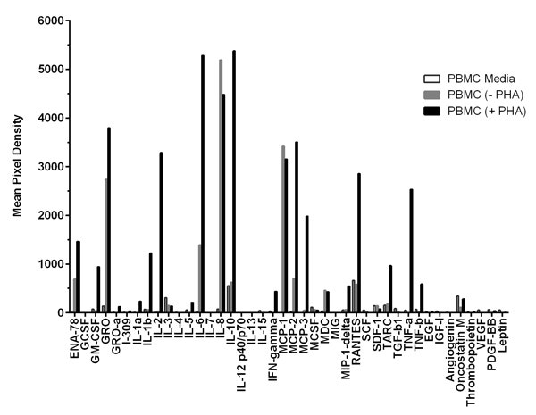
Cytokine Array - Human Cytokine Antibody Array (Membrane, 80 Targets)(AB133998)
Detect 80 human cytokines at once on a single membrane array. A cost-effective way to profile immune and inflammatory signaling from small sample volumes.
See how Danaher Life Sciences can help
FAQ's
What Sample Types Can Be Used for Multiplex Biomarker Panels?
Multiplex biomarker panels can be applied to a wide range of sample types, including serum, plasma, whole blood, tissue lysates, cell culture supernatants, cerebrospinal fluid and formalin-fixed tissue sections. The most appropriate sample depends on the biomarkers of interest, assay platform and study objectives.
How Do Multiplex Assay Results Compare to ELISA?
Multiplex assays and ELISA often show good correlation for the same biomarker, but absolute values are not always interchangeable. Differences in antibodies, calibrators, assay formats and sample dilutions can shift reported concentrations. For consistent comparisons, use the same platform throughout a study and, when needed, confirm key biomarkers with an established singleplex assay such as ELISA.
Can Biomarkers with Different Concentration Ranges Be Measured in the Same Panel?
Yes, but this can be challenging. Multiplex assays often contain biomarkers that vary widely in abundance. Successful panel development requires careful optimization to ensure highly abundant analytes do not interfere with the detection of lower-concentration targets.
What Causes Unexpected Results in Multiplex Biomarker Assays?
Common approaches include classical machine learning, deep learning and hybrid methods. These techniques are used for tasks such as image segmentation, cell classification, feature extraction and spatial analysis.Unexpected results may arise from cross-reactivity, matrix effects, sample quality issues, reagent interference, instrument variability or biomarker concentrations that fall outside the validated assay range. Quality control review can help identify potential technical causes before biological conclusions are drawn.
How Should Results Below or Above the Quantification Range Be Interpreted?
Results below the lower limit of quantification or above the upper limit of quantification should be interpreted with caution, as measurement accuracy may be reduced outside the validated range. Depending on the assay, sample dilution, repeat testing or alternative analytical approaches may be required.
What Is Fit-for-Purpose Validation?
Fit-for-purpose validation matches the extent of assay validation to how the data will be used. Exploratory discovery studies may need only evidence of acceptable precision, sensitivity and specificity. In contrast, assays supporting clinical studies or decision-making require more rigorous evaluation of accuracy, precision, quantification range, stability and reproducibility against predefined acceptance criteria.
References
- Japp NC, Souchek JJ, Sasson AR, Hollingsworth MA, Batra SK, Junker WM. Tumor Biomarker In-Solution Quantification, Standard Production, and Multiplex Detection. J Immunol Res. 2021;2021(1):9942605.
- Otoo JA, Schlappi TS. REASSURED multiplex diagnostics: a critical review and forecast. Biosensors (Basel). 2022;12(2):124.
- Jet T, Gines G, Rondelez Y, Taly V. Advances in multiplexed techniques for the detection and quantification of microRNAs. Chem Soc Rev. 2021;50(6):4141-4161.
- Wang Y, Gao Y, Yin Y, Pan Y, Wang Y, Song Y. Nanomaterial-assisted microfluidics for multiplex assays. Microchim Acta. 2022;189(4):139.
- Liu K, Liu F, Li G, Zheng Y. Multiplex imaging analysis of the tumor immune microenvironment for guiding precision immunotherapy. Front Immunol. 2025;16:1617906.
- Donniacuo A, Mauro A, Cardamone C, Basile A, Manzo P, Dimitrov J, et al. Comprehensive profiling of cytokines and growth factors: pathogenic roles and clinical applications in autoimmune diseases. Int J Mol Sci. 2025;26(18):8921.
- Jiang C, Fu Y, Liu G, Shu B, Davis J, Tofaris GK. Multiplexed profiling of extracellular vesicles for biomarker development. Nano-Micro Lett. 2022;14(1):3.
- Ni W, Han EX, Cyr M, Mackay S, Zhou J. Applying single-cell highly multiplexed secretome proteomics to characterize immunotherapeutic products and predict clinical responses. Proteomics. 2023;23(13-14):2200242.
- Yoo KH, Szymura S, Dong Z, Kandel A, Cha S-c, Kwak LW. Predictive markers for the efficacy of CAR T-cell therapy: the interplay between CAR T-cell fitness and systemic immunity. Blood Adv. 2025;9(24):6432-6442.
- Icoz K, Tas Z, Azizieh F. Transforming cytokine diagnostics: AI, multiplexing, and point-of-care biosensing technologies. Microchim Acta. 2025;192(11):784.
- Islam MA, Masson J-F. Plasmonic biosensors for health monitoring: inflammation biomarker detection. ACS Sens. 2025;10(2):577-601.
- Sheng W, Zhang C, Mohiuddin T, Al-Rawe M, Zeppernick F, Falcone FH, et al. Multiplex immunofluorescence: a powerful tool in cancer immunotherapy. Int J Mol Sci. 2023;24(4):3086.
- Paulo JA, Schweppe DK. Advances in quantitative high-throughput phosphoproteomics with sample multiplexing. Proteomics. 2021;21(9):2000140.
- Kreitmann L, Miglietta L, Xu K, Malpartida-Cardenas K, D'Souza G, Kaforou M, et al. Next-generation molecular diagnostics: Leveraging digital technologies to enhance multiplexing in real-time PCR. TrAC Trends Anal Chem. 2023;160:116963.
- Chen Q, Yi J, Liu Y, Yang C, Sun Y, Du J, et al. Clinical diagnostic value of targeted next-generation sequencing for infectious diseases. Mol Med Rep. 2024;30(3):153.
- Duan H, Cheng T, Cheng H. Spatially resolved transcriptomics: advances and applications. Blood Sci. 2023;5(1):1-14.
- Andreou C, Weissleder R, Kircher MF. Multiplexed imaging in oncology. Nat Biomed Eng. 2022;6(5):527-540.
- Harms PW, Frankel TL, Moutafi M, Rao A, Rimm DL, Taube JM, et al. Multiplex immunohistochemistry and immunofluorescence: a practical update for pathologists. Mod Pathol. 2023;36(7):100197.
- Li J-R, Pan X, Lin Y, Zhao Y, Liu Y, Li Y, et al. Spatial proximity of immune cell pairs to cancer cells in the tumor microenvironment as biomarkers for patient stratification. Cancers (Basel). 2025;17(14):2335.
- Panitz V, Končarević S, Sadik A, Friedel D, Bausbacher T, Trump S, et al. Tryptophan metabolism is inversely regulated in the tumor and blood of patients with glioblastoma. Theranostics. 2021;11(19):9217.
- Ly R, Ly N, Sasaki K, Suzuki M, Kami K, Ohashi Y, et al. Nontargeted serum lipid profiling of nonalcoholic steatohepatitis by multisegment injection-nonaqueous capillary electrophoresis-mass spectrometry: a multiplexed separation platform for resolving ionic lipids. J Proteome Res. 2022;21(3):768-777.
- Wu Y, Li Y, Jin K, Zhang L, Li J, Liu Y, et al. CRISPR-dCas12a-mediated genetic circuit cascades for multiplexed pathway optimization. Nat Chem Biol. 2023;19(3):367-377.
- Anh NK, Thu NQ, Tien NTN, Long NP, Nguyen HT. Advancements in mass spectrometry-based targeted metabolomics and lipidomics: implications for clinical research. Molecules. 2024;29(24):5934.
- Dunbar SA. Nucleic acid sample preparation techniques for bead-based suspension arrays. Methods. 2023;219:22-29.
- Zhang X, Wei X, Wu C-X, Men X, Wang J, Bai J-J, et al. Multiplex profiling of biomarker and drug uptake in single cells using microfluidic flow cytometry and mass spectrometry. ACS Nano. 2024;18(8):6612.
- Nguyen GT, Bennett JL, Liu S, Hancock SE, Winter DL, Glover DJ, et al. Multiplexed screening of thousands of natural products for protein-ligand binding in native mass spectrometry. J Am Chem Soc. 2021;143(50):21379-21387.
- de Korne-Elenbaas J, Caduff L, Lison A, McLeod R, Pitton M, Gan C, et al. Design, validation, and implementation of multiplex digital PCR assays for simultaneous quantification of multiple targets. Lett Appl Microbiol. 2025;78(1):ovae137.
- Tanak AS, Muthukumar S, Krishnan S, Schully KL, Clark DV, Prasad S. Multiplexed cytokine detection using electrochemical point-of-care sensing device towards rapid sepsis endotyping. Biosens Bioelectron. 2021;171:112726.
- Dinc R, Ardic N. Artificial intelligence for exosomal biomarker discovery for cardiovascular diseases: multi-omics integration, reproducibility, and translational prospects. Cells. 2026;15(3):304.