Cell DIVE Multiplex Imaging Solution
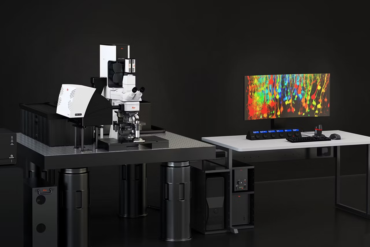

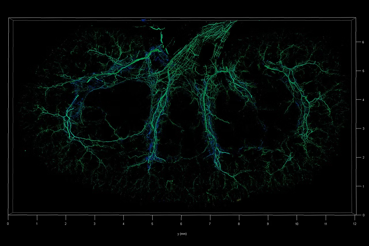
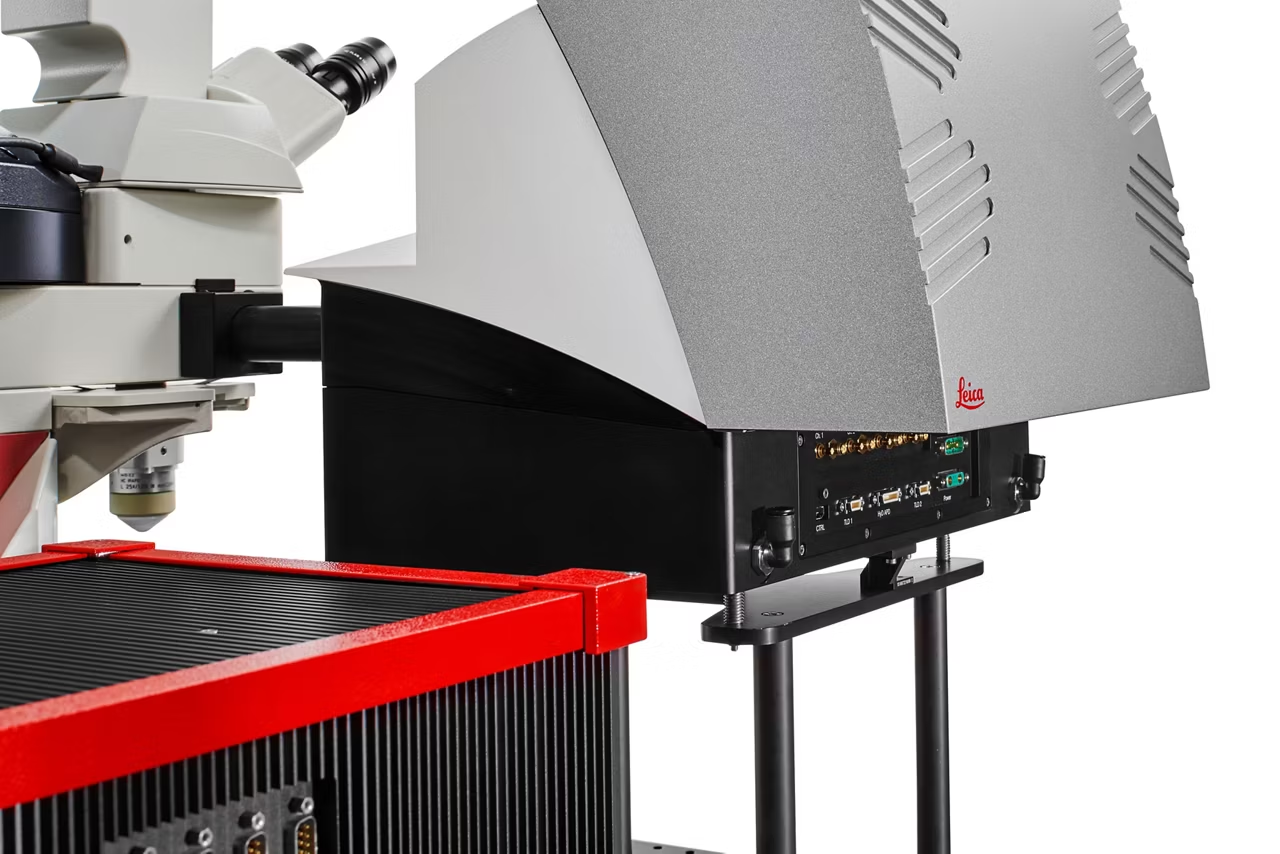
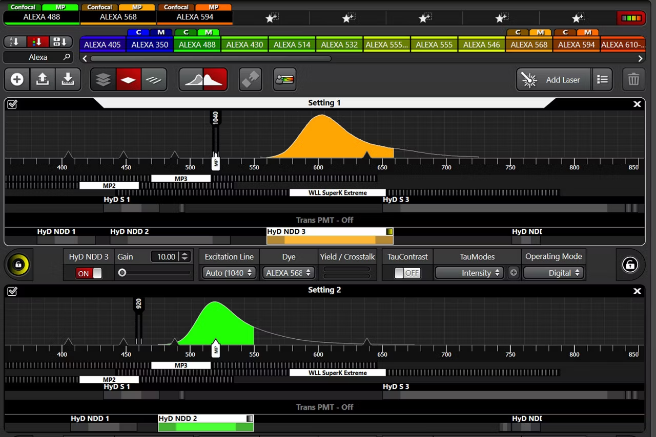
What if every scientist could map normal and diseased tissue by cell type, biomarker profile, and specific features?
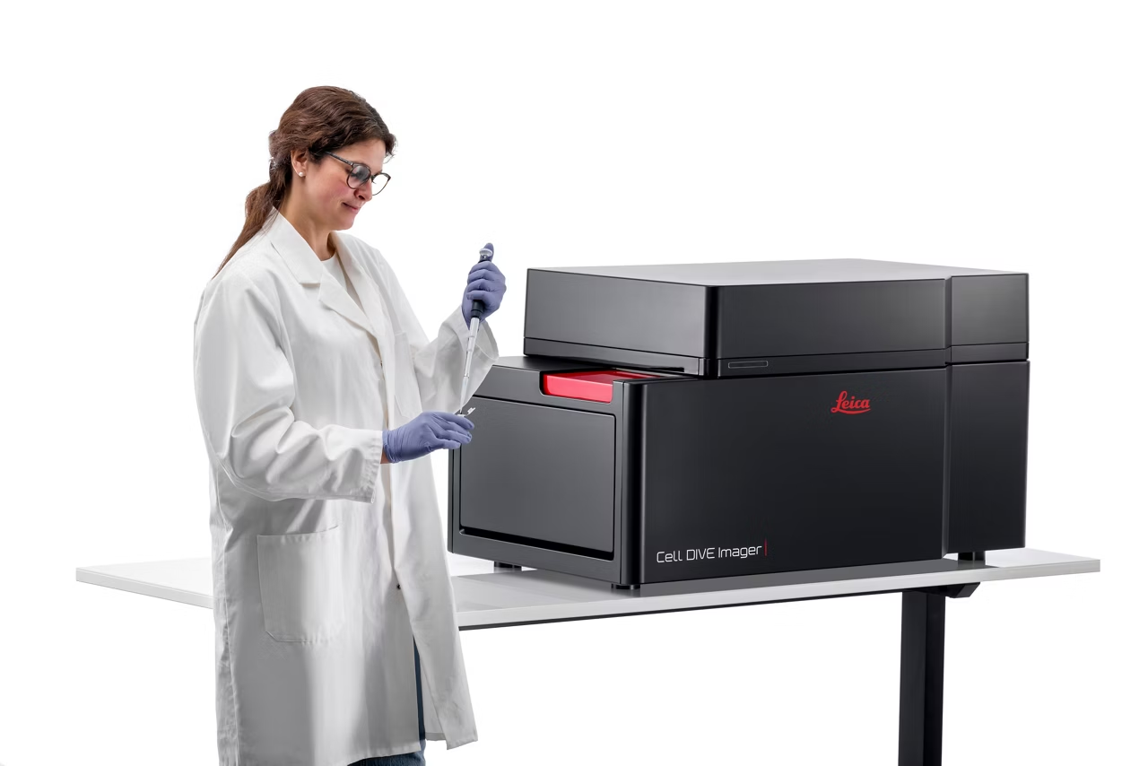
Cell DIVE is a precise, open multiplexing solution that lets your research dictate the level of automation required, which antibodies to use, how to build your antibody panel and more.
Cell DIVE keeps evolving to support your ever-changing research needs. With new technical innovations in software and automation, you can get more out of every experiment.
Cell DIVE multiplexed imaging solution
Explore how Cell DIVE multiplexed imaging can uncover unique cell phenotypes and their distribution within the tissue microenvironment.
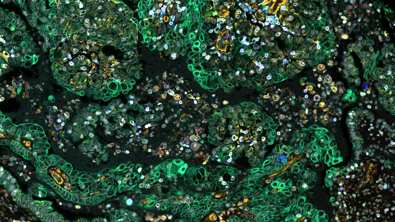
Capture crystal-clear 2D images of 60+ protein biomarkers, now with the option to add z-stack imaging in any channel.
Crystal Clear Whole Tissue Imaging

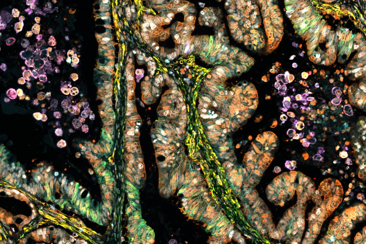
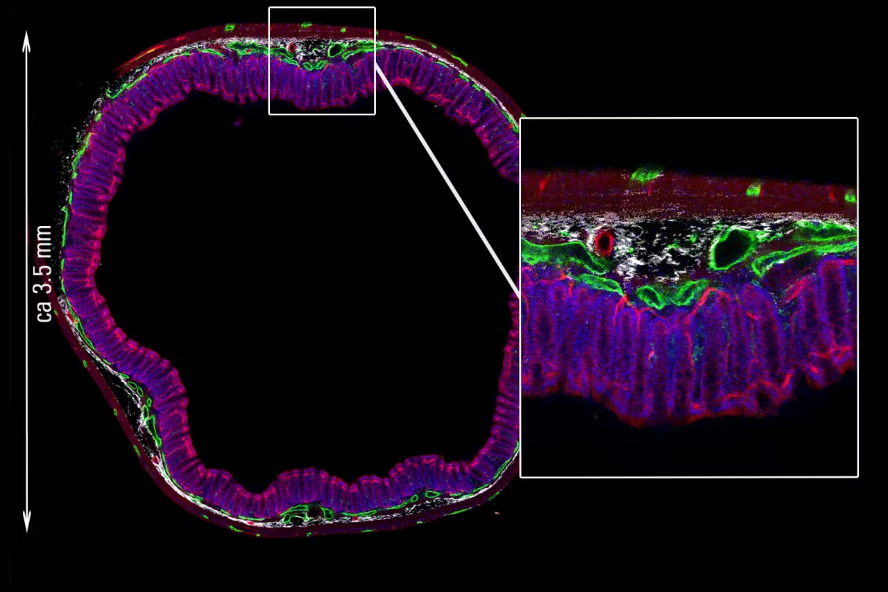
Cell DIVE helps researchers deepen their understanding of the tissue microenvironment by offering outstanding spatial mapping of single cells within context. Unveil whole tissue imaging down to the single cell level, automatically calibrated and corrected to enable quality analysis downstream.
See for yourself and explore
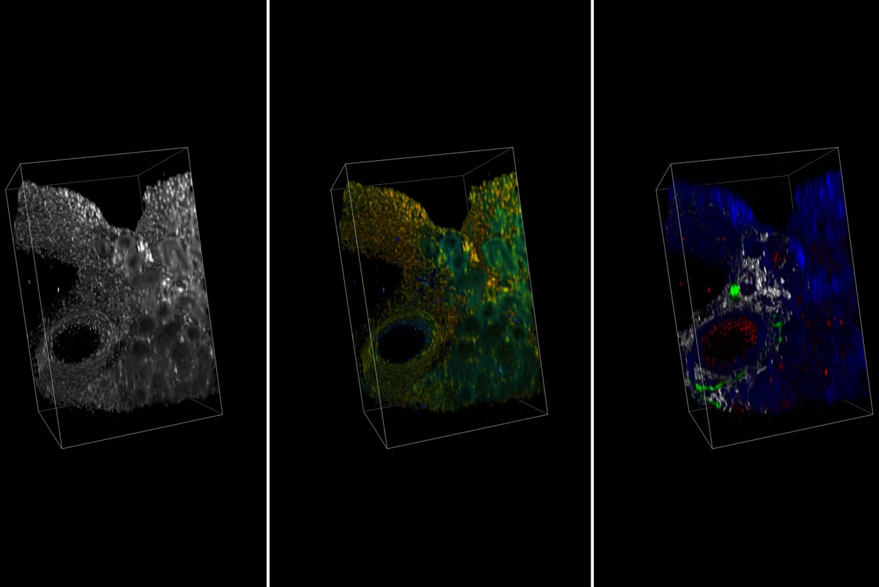
Explore a pancreatic adenocarcinoma data set with over 25 biomarkers to see how Cell DIVE image quality can advance your tissue research.
Built by scientists, for scientists, from a decade of discovery and development

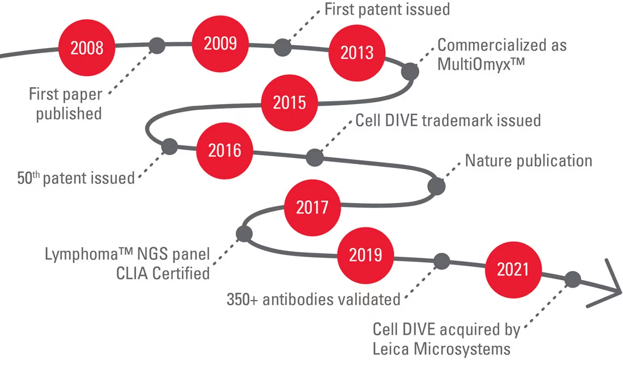
Cell DIVE multiplexed imaging is backed by 10+ years of rigorous research, testing, and validation. Deployed in the United States and evaluated through international collaboration, our science, algorithms, and methodology are proven, so you can trust your results. Have peace of mind from a proven workflow: benefit from a proven, patent protected, end to end iterative staining process.
Be free to choose your study's antibodies

An adaptable and antibody agnostic system, Cell DIVE offers you the freedom to design your study as you wish, now or in the future.
Cell DIVE does not dictate where you source antibodies, offering cost savings and the flexibility to design your study as you wish. Design your own completely custom panel, use a multiplex reagent kit, or save time on study design and reagent preparation with Cell DIVE validated antibodies available from our partners at Cell Signaling Technology (CST). Leverage the expertise of CST, a trusted name in antibody manufacturing, and discover over 100 antibodies ready for use with Cell DIVE.
You can also respond to changing research needs in real time or revisit your study in the future, with Cell DIVE's adaptable workflow and tissue preserving capability.
Scale your research as it suits you
Cell DIVE with ClickWell enables easily scalable multiplexing by giving you options for automating workflows to fit your needs. Choose the level of automation that is right for your research today and adapt as your needs evolve.
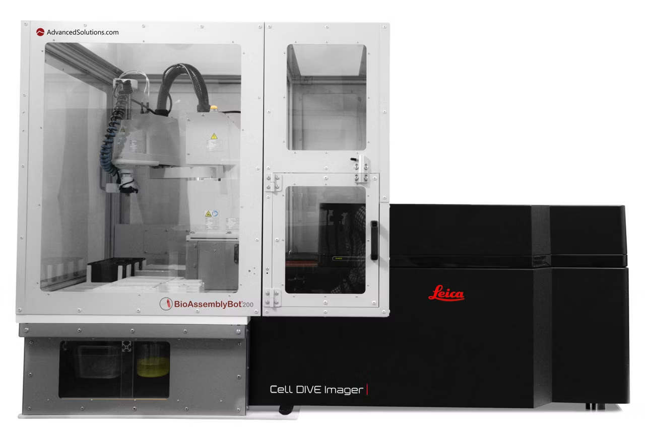
Streamline your work by pairing your Cell DIVE multiplexed imaging solution with BioAssemblyBot 200 from Advanced Solutions Life Sciences. BAB 200 is a customized multi-tasking robot that seamlessly integrates with the Cell DIVE imager to automate staining and imaging for up to 15 slides at a time.
Support RNA & multi-omics workflows, the BAB 200 now features an integrated slide heater for consistent thermal control across transcriptomics workflows.
Supercharge your research with Cell DIVE Connect
Increase your lab’s multiplexing output with Cell DIVE Connect, a new feature that allows you to add capacity without complexity. Cell DIVE Connect is a network-based software deployment that links multiple Cell DIVE Imagers to enable protocol sharing, optimize imager utilization, and efficiently manage multiple slides and studies.
Designed for running large complex studies, Cell DIVE Connect gives you sub-cellular registration in final data from images acquired on any Cell DIVE Imager on the network.
Transforming tissue research with open multiplexing.
Data you can trust
Crystal clear whole tissue imaging down to the single cell level.
Choose your antibodies

350+ validated antibodies so you can design studies your own way.
Scale your research

Multiplex imaging that's scalable and efficient.

Aivia AI Image Analysis Software
Explore AI Image Analysis Software transforms image processing with up to 74% faster analysis time. Achieve high-quality, reproducible results effortlessly.