










Complete Flow Cytometry Guide: Mechanism, Principles, Types, Protocol & Applications
Key Takeaways
- Flow cytometry enables single-cell, multiparameter analysis using light scattering and fluorescence
- It supports >40 parameters simultaneously, enabling deep cellular characterization
- Core systems include fluidics, optics, and electronics, each driving data acquisition
- Applications span drug discovery, immunology, and clinical diagnostics
- Advanced formats such as spectral and mass cytometry expand analytical depth
- Key challenges include sample prep sensitivity, cost, and reproducibility limitations
What is Flow Cytometry?
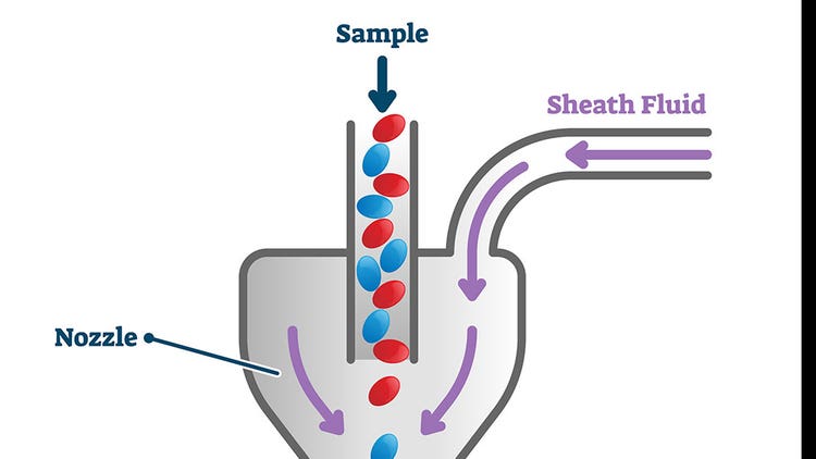
Flow cytometry is a laser-based analytical technique used to measure and analyze multiple phenotypic characteristics of single cells as they flow through a laser beam in a fluid stream.
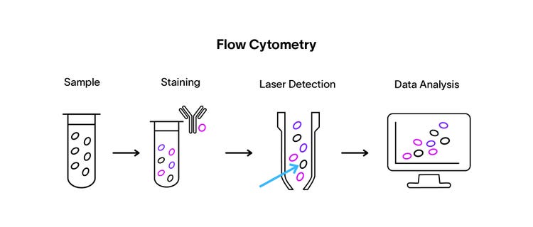
During the assay, cell components, such as surface proteins, are labeled with fluorescent tags, usually attached to antibodies. When fluorescently tagged cells pass through the laser beam, they emit light at different wavelengths, which is captured by the detector near the beam. The fluorescence and light-scattering profiles of individual cells can be used to study their identity.
Characteristics or properties of cells commonly analyzed using flow cytometry include cell size, cell volume, cell viability and cell cycle.
In 1950, the technique was first employed to measure the volume of cells in a sample. However, it is now widely used for a myriad of purposes in biotechnology and pharma labs, such as cell sorting, assessing the purity of a cell population, verifying the monoclonality of a cell line, and analyzing protein abundance.
In this article, you will learn further about the flow cytometry principles, instrumentation, applications and advancements in technology.
See how Danaher Life Sciences can help
Principles of Flow Cytometry
Flow cytometry is a powerful assay for the qualitative and quantitative analysis of cell properties. Flow cytometry runs on the principles of light scattering, excitation and emission. Fluorescently tagged cell components get excited when they pass through a laser beam, producing light of different wavelengths. The fluorescence is used to analyze cellular properties.
The following parameters analyze cells:
Visible light scatter
The forward scatter helps measure the size in the forward direction. The side scatter at 90 degrees measures the granularity and complexity of a cell.
Fluorescence
Cells can emit fluorescence from dyes, such as propidium iodide, reporter genes fused to fluorescent proteins like GFP, or fluorescently conjugated antibodies.
What are the steps of Flow Cytometry?
Flow cytometry follows a stepwise analytical workflow designed for precise, high-throughput cell analysis:
Step-by-step process
-
Sample labeling
- Cells are labeled with fluorescent antibodies targeting specific markers
- Enables detection of proteins or cellular states
-
Hydrodynamic focusing
- Cells are aligned into a single-file stream using sheath fluid
-
Laser excitation
- Cells pass through a laser beam
-
Fluorophores become excited and emit light signals
-
Signal detection
-
Detectors capture:
- Forward scatter (FSC) → cell size
- Side scatter (SSC) → complexity/granularity
- Fluorescence signals → marker expression
-
-
Data analysis
- Signals are converted into digital data for visualization and gating analysis
Flow Cytometry Instruments
Flow Cytometry vs Related Techniques
Feature | Flow Cytometry | FACS (Cell Sorting) | Microscopy |
|---|---|---|---|
Core function | Multiparametric analysis | Cell analysis + sorting | Visual imaging |
Throughput | High (thousands/sec) | Moderate–high | Low |
Data output | Quantitative | Quantitative + physical separation | Qualitative |
Cell isolation | No | Yes | No |
Multiplexing capability | High (>40 parameters) | High | Limited |
Single-cell resolution | Yes | Yes | Yes |
Types of Flow Cytometers
Traditional Flow Cytometers
These cytometers channel the cells through a stream of sheath fluid where they traverse beams from lasers that provide light scattering or fluorescence spectrum information.
Acoustic Focusing Cytometers
These cytometers use ultrasonic waves to direct a stream of cells to the laser beam for further analysis. It minimizes sample clogging that’s observed with some other cytometers.
Cell Sorters
specialized flow cytometers that sort and collect individual cells based on desired parameters, such as the presence or absence of specific phenotypic markers. Cell samples are deposited into wells on a plate or collected into tubes. Cell sorters can facilitate simple 2-way sorts (positive/negative) or multi-parameter sorts that allow users to combine desired parameters.
Flow Cytometry Imagers
This is a spin on traditional flow cytometers used to analyze single cells for multiple fluorescence parameters, but also includes an imaging component that takes photos of the cells as they pass through the imaging area. It can provide subcellular localization information of the fluorescent probes.
Multicolor flow cytometry
The technique is also known as polychromatic cytometry. It analyzes the subpopulation of target cells based on multiple experimental parameters. In immunology, it’s used to detect specific populations of an immune cell at the single-cell level by combining a quantitative proteomics approach.
Spectral flow cytometry
This technique captures the full emitted spectrum of a fluorescent dye. It provides greater depth of information and benefits such as reduced need for compensation, increased panel size and richer data sets.
Mass cytometry or cytometry by Time-of-Flight (CyTOF)
This is a next-generation flow cytometry protocol and involves the use of metal-tagged antibodies for the morphological and functional analysis of single cells.
Flow cytometry panel design: key principles
A panel is a set of fluorophore-conjugated antibodies designed to identify specific cell populations.
Key design considerations
Marker selection
- Choose biologically relevant surface or intracellular markers
Fluorophore matching
- Bright dyes - low-abundance targets
- Dim dyes - high-abundance targets
Spectral overlap
- Overlapping emission profiles require compensation
Instrument compatibility
- Panel must align with laser configuration and filters
Understanding Gating Strategies
Gating is the process of defining cell populations by drawing boundaries on scatter or fluorescence plots.
Key gating workflow
FSC/SSC gating
- Removes debris and isolates the main cell population
Singlet gating
- Eliminates doublets (cell clumps)
Live/dead discrimination
- Excludes non-viable cells
Hierarchical gating
- Sequential filtering improves accuracy
Featured Flow Cytometer Product
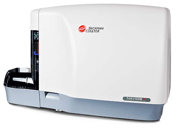
Navios EX Flow Cytometers
Powerful, Dependable Flow Cytometer
Strong and stable cytometer performance is fundamental when analyzing important clinical samples as every event matters. Designed specifically for the clinical laboratory the NAVIOS EX 10-color flow cytometer delivers proven reliability and stability performance with improved sample traceability and reporting functions.
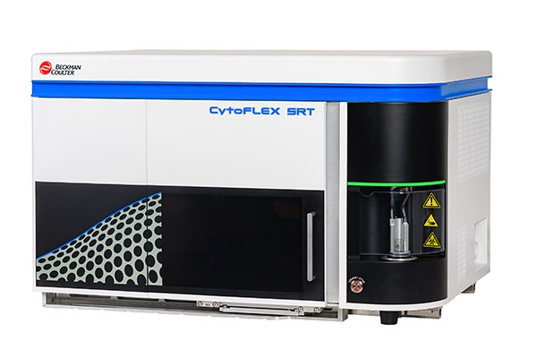
CytoFLEX SRT Benchtop Cell Sorters
CytoFLEX SRT Benchtop Cell Sorter Overview
CytoFLEX SRT Cell Sorter is a benchtop sorter. It is capable of meeting requirements for a wide range of sorting needs. And like the CytoFLEX Platform, it includes innovative technologies that simplify the setup and operation, empowering investigators to focus on the research questions. The Violet-Blue-Yellow Green-Red (V-B-Y-R) Series has 15 fluorescent detectors when fully activated.
See how Danaher Life Sciences can help
Flow Cytometry Applications
Here are a few well-known applications of flow cytometry:
Proteomics in early discovery
Flow cytometry is a great tool for proteomics in early discovery. Assays can simultaneously assess dozens of cell-surface or intracellular proteins. The choice of antibodies can provide information on protein isoforms. Use of an imaging flow cytometer will elucidate subcellular location or trafficking.
Immunophenotyping for cellular composition
In immunophenotyping, flow cytometry provides information on the composition of a cell population based on lineage markers. This helps researchers uncover cellular heterogeneity and uncover rare cell populations that may be valuable for future study.
Gene expression analysis in cell signaling pathways
Flow cytometry can highlight gene expression tied to important cell signaling pathways. Stimulated cells can be analyzed for up- or downregulation of specific genes, allowing researchers to hypothesize about regulatory mechanisms. This includes genes associated with cell cycle phases.
Disease diagnosis and monitoring in clinical labs
Disease diagnosis and monitoring can be conducted via flow cytometry in clinical labs. This is especially relevant for cancer and infectious diseases, where rapid testing is required. Flow cytometry assays can be qualified as an In Vitro Diagnostic (IVD) or validated as a Laboratory Developed Test (LDT).
Quality Control and Standardization in Flow Cytometry
Standardization and optimization of a flow cytometry assay are essential to ensure valid, reproducible results. Some of the requirements for flow cytometry labs include:
Documentation of Steps
Sample preparation and initial sample quality can affect the integrity of flow cytometry data. It is important to document all steps to increase reproducibility properly.
Calibration and Performance Checks
Calibration and performance checks of the instrument, including tracking maintenance in logbooks, are required for all labs, especially clinical labs.
Reagent QC
Maintaining rigorous reagent QC is crucial for managing the inevitable lot-to-lot variability in flow cytometric analysis. Regular titration of antibodies is required to minimize false-positive results.
Standardization Initiatives and Guidelines
These should be followed in labs. For instance, ISO 15189 accreditation is required in Europe for flow cytometry labs in accordance with the EU IVD Regulation 2017/746.
Flow cytometry troubleshooting
Assembled a troubleshooting table with problems and solutions
Problem | Likely Cause | Solution |
|---|---|---|
High background | Non-specific antibody binding | Titrate antibody to optimal concentration; add Fc receptor blocking step before staining |
Low cell count | Poor sample preparation | Optimize single-cell dissociation method; check centrifuge speed and timing to avoid cell loss. |
False positives | Dead cells bind the antibody non-specifically | Add a viability dye (e.g., propidium iodide or DAPI) to exclude dead cells during gating. |
Poor/weak signal | Mismatched fluorophore and laser line | Verify fluorophore excitation matches instrument laser; check filter configuration in cytometer settings. |
High CV (coefficient of variation) | Instrument drift or inconsistent setup | Run QC calibration beads at the start of each session; check laser delay and voltage settings. |
High autofluorescence | Cell type or fixation method | Switch to a brighter fluorophore; use the autofluorescence subtraction available in spectral cytometers. |
Doublets in data | Cells clumping during sample prep | Add doublet discrimination gating using pulse width (FSC-W vs FSC-A plot) |
Inconsistent results between runs | Lot-to-lot reagent variability | Perform antibody titration for each new lot; use internal reference controls across runs. |
Considerations and Challenges in Flow Cytometry
Flow cytometry poses several challenges, including sample preparation that can lead to low cell viability and suboptimal cell counts during specimen evaluation. Other important considerations include the cost associated with instrumentation, labor and training.
Reproducibility issues can also plague the field of flow cytometry due to variability in manual data analysis (gating) methods. Manual gating is prone to bias and can be subjective.
Employing flow cytometry for clinical testing requires labs and patients to consider reimbursement approvals and amounts.
