AI Image Analysis in Spatial Biomarker Research
Spatial biomarker research can redefine how scientists study disease by revealing not only what biological signals are present, but also where they occur within tissues and how they interact. As imaging datasets grow larger and more complex, advanced image analysis methods are needed to help researchers extract deeper insights faster, more consistently and with greater precision. This article explores how AI image analysis supports spatial biomarker discovery, from image acquisition and analysis to biomarker validation and translational research.
Key Takeaways
- AI image analysis can accelerate the interpretation of complex spatial biology datasets while improving consistency and reducing manual effort
- Spatial biomarkers provide biological context by revealing where cells, proteins and genes are located and how they interact within tissues
- AI-powered segmentation, feature extraction and spatial mapping support identifying cell populations, tissue architecture and cell-cell interactions
- Applications span cellular phenotyping, tissue microenvironment analysis, high-plex imaging and advanced 3D model systems such as organoids
- Successful implementation depends on high-quality imaging data, robust preprocessing, representative annotations and rigorous validation to ensure reliable and biologically meaningful results
What Is AI Image Analysis?
AI image analysis uses artificial intelligence and machine learning algorithms to detect, classify and measure features within digital images, helping researchers extract meaningful information from complex tissue and cellular image data.1
Traditional image analysis relies on predefined rules and thresholds set by researchers to classify cells using fixed parameters. However, these manual approaches falter when analyzing heterogeneous tissues, dynamic cellular morphologies or large multiplexed imaging datasets. This is where AI image analysis excels.1
Spatial biomarker research is an ideal example of how AI/ML approaches can advance image analysis, as spatial biology technologies generate vast amounts of high-dimensional image data. They help researchers transform these complex datasets into actionable biological insights by automating image processing, improving reproducibility and accelerating data interpretation.1
Ultimately, AI-driven analysis uncovers rare cell populations, cell-to-cell interactions and tissue architecture that traditional methods may overlook.
AI Image Analysis vs. Traditional Image Analysis
Include a compact comparison table showing how the main image-analysis approaches differ.
See how Danaher Life Sciences can help
While AI-based approaches can improve throughput and uncover complex patterns, no single method is universally optimal. The most appropriate approach depends on the biological question, imaging modality, dataset quality, available annotations and the level of validation required.1
What are spatial biomarkers?
Conventional biomarkers reveal what is present in a sample, whereas spatial biomarkers reveal both what is present and where it is located within the tissue.2
Examples of spatial biomarker measurements include:
- The distance between immune and tumor cells3
- The distribution of specific cell populations within tissue regions4
- Cell-to-cell interaction patterns5
These measurements provide context that cannot be captured by bulk tissue analysis alone. Because tissue location and cellular relationships can influence disease progression and treatment response, capturing spatial information helps researchers gain a more complete understanding of disease biology.6
Why Spatial Biomarkers Need AI-Driven Image Analysis
Advances in spatial biology have dramatically increased the complexity and scale of imaging datasets. Modern multiplexed and 3D imaging technologies can profile dozens of biomarkers across thousands to millions of cells, generating data volumes that are difficult to analyze using manual or traditional rule-based approaches alone.7
Manual and rule-based methods can specifically suffer from overlapping signals, heterogeneous tissue architecture and subtle phenotypical differences. Results may also vary between observers, particularly when identifying complex cell states or spatial relationships within the tissue microenvironment.7
AI-driven image analysis addresses these challenges by recognizing patterns that may be difficult to define using fixed rules. By automating feature detection and classification across large datasets, AI can improve the sensitivity and specificity of spatial biomarker measurements while increasing analytical throughput and consistency. As a result, researchers can extract richer biological insights from increasingly complex spatial datasets.7
AI Image Analysis Workflow for Spatial Biomarker Research
Acquire images and check quality
Capture images using microscopy, multiplexed imaging or other spatial biology platforms. Use consistent acquisition settings, quality checks and metadata capture to support reliable downstream analysis.8
Output: Quality-checked images with acquisition metadata.
Prepare and organize the data
Prepare images by denoising, normalizing, registering and removing artifacts as needed. Keep data traceable and organized for integration with molecular, clinical and other experimental datasets.9
Output: Preprocessed, traceable image datasets ready for analysis.
Identify cells and map spatial features
AI-based methods identify cells and tissue regions, then measure features such as marker expression, morphology and cellular proximity. These measurements help researchers characterize tissue organization.11
Output: Quantified cellular features and spatial relationships.
Interpret patterns and identify candidates
Use clustering, dimensionality reduction and visualization to explore cell groups, tissue neighborhoods and biological differences.13 Integrate spatial findings with molecular and clinical data, using interpretable analysis and expert review to assess candidate biomarker signatures.14
Output: Candidate spatial biomarker signatures for validation.
Validate findings for the intended use
Assess accuracy, sensitivity, specificity and reproducibility using expert annotations, independent datasets and orthogonal methods as appropriate. Link validated measurements to the intended research question, such as target validation, patient stratification research or mechanism-of-action studies.15
Output: Performance evidence supporting the intended research or translational use.
Understanding the AI methods
Classical machine learning versus deep learning
Classical machine-learning approaches typically rely on predefined image features, such as intensity, texture, shape or morphology, to classify cells and tissue regions. In contrast, deep learning methods learn these features directly from image data, making them particularly effective for analyzing complex tissue architecture, heterogeneous cell populations and high-dimensional imaging datasets.10
Supervised versus unsupervised methods
AI models can implement supervised or unsupervised approaches. Supervised methods are typically trained to identify predefined cell types, tissue features or biomarker patterns, whereas unsupervised methods can reveal previously unrecognized cellular states and spatial relationships.12
Applications in Spatial Biomarker Research
Cellular and Subcellular Phenotyping
AI image analysis can automatically identify cell types and cellular states based on marker expression, morphology and tissue location. It can also determine the subcellular localization of biomarkers, distinguishing where proteins are expressed in the cell and directly influencing biological function and signaling activity.
For example, researchers can measure the nuclear translocation of transcription factors in tumor cells to assess pathway activation and evaluate target engagement in mechanism-of-action studies.16
Tissue Microenvironment and Spatial Architecture
AI-based spatial analysis helps map the distribution of immune cells, stromal components and vascular structures within tissues. By measuring cell proximity and defining cellular neighborhoods, researchers can investigate how different cell populations interact within the tissue microenvironment, a key step in immunotherapy development. For example, the spatial proximity of cytotoxic T cells to tumor cells can be used to characterize immune infiltration patterns and assess potential response to immunotherapies.17
High-Plex and Multiplexed Assays
High-plex imaging technologies can measure dozens of biomarkers within a single tissue section, generating datasets that are difficult to interpret manually. AI image analysis can streamline this interpretation by resolving multiple cell populations, identifying subtle phenotypic differences and characterizing biomarker co-expression patterns while preserving spatial context. By combining multiplex imaging with AI-based analysis, researchers can discover distinct immune-cell neighborhoods associated with treatment response that would not be apparent from a single biomarker alone. 18
Organoids, 3D Models and Advanced Cell Systems
Advanced 3D models, such as organoids and organ-on-chip systems, generate data that aligns more closely with human physiology than 2D models do. Simultaneously, the image data obtained from them becomes more complex, requiring robust analysis tools. AI/ML image analysis can address this gap by enabling the quantification of volumetric and temporal changes in morphology, tissue organization and biomarker expression across large numbers of 3D samples.19
AI image analysis is widely used in drug discovery workflows involving tumor organoids. Researchers can monitor changes in tumor organoid growth, architecture and cell-state composition following drug treatment to evaluate compound efficacy and toxicity in a physiologically relevant model system.19
Benefits of AI Image Analysis
- Throughput: AI can process large image and cell datasets more rapidly than manual review, supporting higher-volume studies and shorter analysis timelines.20
- Consistency: Standardized analysis can reduce observer variability and improve reproducibility, provided models are appropriately trained and validated.20
- Biological context: Combining quantitative image features with spatial and molecular information can reveal subtle patterns and provide richer evidence for biomarker and translational research decisions.20
What Makes Results Reliable?
- Consistent image acquisition and quality control: Standardize illumination, staining, focus, resolution, tissue preparation and instrument settings and define image-quality thresholds and metadata requirements to reduce technical variability across datasets.21
- Documented preprocessing and batch-effect assessment: Record normalization, registration, background correction and artifact-removal steps, because inconsistent preprocessing can obscure biological differences or create artificial ones. Assess batch effects before model training or inference and use appropriate reference controls.22
- Representative annotations and training data: Use clear annotation guidelines and subject-matter expertise to limit incomplete, inconsistent or subjective labels. Include relevant tissue types, disease states and imaging conditions to support broader applicability.23
- Traceable data, metadata and model versions: Maintain the infrastructure and governance needed to trace datasets, metadata, model versions and analytical outputs throughout the workflow, supporting reproducibility, security and regulatory readiness where appropriate.23
- Independent validation and expert review: Test performance on independent datasets and compare results with expert annotations or orthogonal methods, because development-set performance may not generalize across samples, instruments or laboratories.15 Maintain human oversight and align validation with the intended research or translational use, particularly when model outputs are difficult to interpret.24
Featured Product
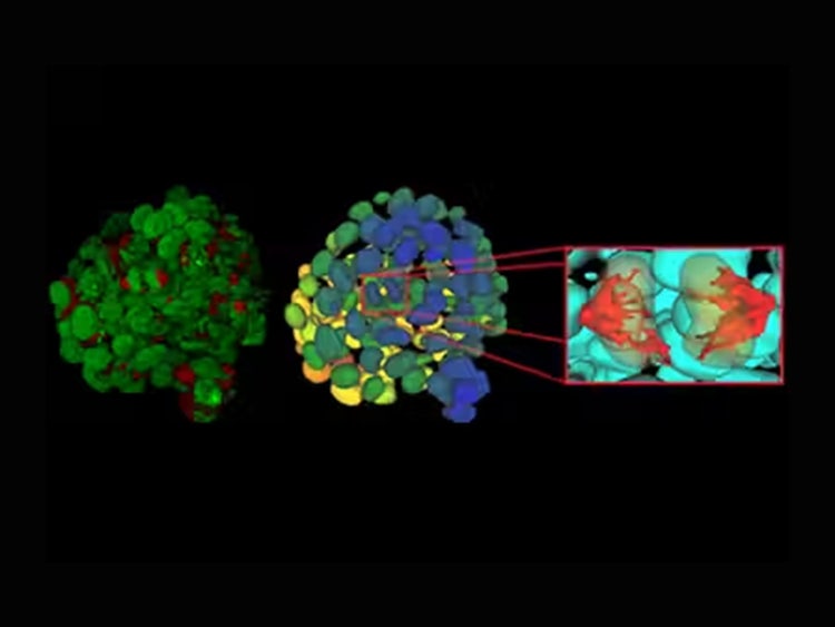
Aivia AI Image Analysis Software
AI image analysis software that runs a complete workflow, from deep-learning cell segmentation to automatic phenotyping and data exploration, for complex 3D and multiplexed images.
Cell DIVE Multiplex Imaging Solution
An open multiplex imaging solution for high-plex tissue analysis at subcellular resolution, mapping 60+ biomarkers per scan across normal and diseased tissue.

See how Danaher Life Sciences can help
FAQ's
What is the difference between AI image analysis and traditional image analysis?
Traditional image analysis relies on predefined rules and thresholds to identify and measure features within an image. AI image analysis learns patterns directly from data, allowing it to adapt to biological variability and recognize complex cellular and tissue features.
How does AI improve microscopy image analysis?
AI can automate image interpretation at scale, reducing manual review time while improving consistency. It can also detect subtle image features and complex patterns that may be difficult to identify using conventional approaches.
What is the role of AI in spatial biomarker research?
AI helps transform complex imaging datasets into biologically meaningful insights. It supports cell identification, spatial mapping, biomarker quantification and the analysis of tissue organization and cell-cell interactions.
What types of AI algorithms are used for spatial biomarker image analysis?
Common approaches include classical machine learning, deep learning and hybrid methods. These techniques are used for tasks such as image segmentation, cell classification, feature extraction and spatial analysis.
Can AI image analysis be used for both 2D tissue sections and 3D organoids or spheroids?
Yes. AI image analysis can be applied to both 2D and 3D imaging data, helping researchers assess morphology, biomarker expression, tissue architecture and treatment responses in increasingly complex model systems.
What are common image segmentation errors and how can they be reduced?
Common errors include missed cells, merged cell boundaries, incorrect classifications and segmentation artifacts caused by poor image quality. These issues can be reduced through high-quality image acquisition, representative training data, robust quality control and independent validation of model performance.
References
- Tarhini AA, El Naqa I. Artificial intelligence for multiscale spatial analysis in oncology: current applications and future implications. Int J Mol Sci. 2025;26(16):8002.
- Williams HL, Frei AL, Koessler T, Berger MD, Dawson H, Michielin O, et al. The current landscape of spatial biomarkers for prediction of response to immune checkpoint inhibition. NPJ Precis Oncol. 2024;8(1):178.
- Li J-R, Pan X, Lin Y, Zhao Y, Liu Y, Li Y, et al. Spatial proximity of immune cell pairs to cancer cells in the tumor microenvironment as biomarkers for patient stratification. Cancers (Basel). 2025;17(14):2335.
- Failmezger H, Hessel H, Kapil A, Schmidt G, Harder N. Spatial heterogeneity of cancer-associated protein expression in immunohistochemically stained images as an improved prognostic biomarker. Front Oncol. 2022;12:964716.
- Cohn DE, Forder A, Marshall EA, Vucic EA, Stewart GL, Noureddine K, et al. Delineating spatial cell-cell interactions in the solid tumour microenvironment through the lens of highly multiplexed imaging. Front Immunol. 2023;14:1275890.
- Mi H, Sivagnanam S, Ho WJ, Zhang S, Bergman D, Deshpande A, et al. Computational methods and biomarker discovery strategies for spatial proteomics: a review in immuno-oncology. Brief Bioinform. 2024;25(5):bbae421.
- Saha P, Yasmin A, Jha R, Passi A, Kaur M, Jindal S, et al. Integrative approaches in lung cancer diagnosis: bridging molecular biomarkers and AI-driven imaging. Biomarkers. 2026:1-31.
- Bhattiprolu S. AI-driven microscopy: from classical analysis to deep learning applications. Methods Microsc. 2025;(0).
- Dimililer K, Erdem BD, Kayali D, Olawale OP. Image preprocessing phase with artificial intelligence methods on medical images. In: Artificial Intelligence and Image Processing in Medical Imaging. Amsterdam: Elsevier; 2024. p. 51-82.
- Van Buren K, Li Y, Zhong F, Ding Y, Puranik A, Loomis CA, et al. Artificial intelligence and deep learning to map immune cell types in inflamed human tissue. J Immunol Methods. 2022;505:113233.
- Ali M, Benfante V, Basirinia G, Alongi P, Sperandeo A, Quattrocchi A, et al. Applications of artificial intelligence, deep learning, and machine learning to support the analysis of microscopic images of cells and tissues. J Imaging. 2025;11(2):59.
- Pantanowitz L, Pearce T, Abukhiran I, Hanna M, Wheeler S, Soong TR, et al. Nongenerative artificial intelligence in medicine: advancements and applications in supervised and unsupervised machine learning. Mod Pathol. 2025;38(3):100680.
- Torro R, Díaz-Bello B, El Arawi D, Dervanova K, Ammer L, Dupuy F, et al. Celldetective, an AI-enhanced image analysis tool for unraveling dynamic cell interactions. eLife. 2026;14:RP105302.
- Liu F, Beck S, Yang L, Luo H, Zhang K. Advancing AI for multi-omics and clinical data integration in basic and translational cancer research. Nat Rev Cancer. 2026:1-16.
- Armstrong AJ, Liu VY, Selvaraju RR, Chen E, Simko JP, DeVries S, et al. Development and validation of an artificial intelligence digital pathology biomarker to predict benefit of long-term hormonal therapy and radiotherapy in men with high-risk prostate cancer across multiple phase III trials. J Clin Oncol. 2025;43(32):3494-3504.
- Abel J, Jain S, Rajan D, Padigela H, Leidal K, Prakash A, et al. AI-powered quantification of nuclear morphology in cancers enables prediction of genome instability and prognosis. NPJ Precis Oncol. 2024;8(1):134.v
- Liang H, Zhu H, Wang L, Yu J, Feng R, Yan X, et al. AI-driven spatial analysis of tumor-infiltrating lymphocytes predicts chemo-immunotherapy response in triple-negative breast cancer. Front Immunol. 2026;17:1797753.
- Cheung AM, Han W, Zhou S, Wang D, Ramanathan V, Qu O, et al. Spatial analysis of immune cells in breast cancer using k-nearest neighbor graphs and Louvain-community clustering of immunofluorescent protein multiplexing images. In: 17th Int Workshop Breast Imaging (IWBI 2024). 2024:1317414.
- Huang K, Li M, Li Q, Chen Z, Zhang Y, Gu Z. Image-based profiling and deep learning reveal morphological heterogeneity of colorectal cancer organoids. Comput Biol Med. 2024;173:108322.v
- Alum EU. AI-driven biomarker discovery: enhancing precision in cancer diagnosis and prognosis. Discov Oncol. 2025;16(1):313.v
- Krichen M, Aoun NB. AI-driven medical image analysis: challenges and limitations. In: AI for Medical Image Analysis: Reconciling Innovation and Ethical Considerations. Cham: Springer; 2025. p. 213-232.
- Dehbozorgi P, Ryabchykov O, Bocklitz T. A systematic investigation of image pre-processing on image classification. IEEE Access. 2024;12:64913-64926.
- Kumbhar S, Bhinge S, Bhatia M. Analytical and computational challenges in AI-driven biomarker assays for neurodegenerative diseases: current limitations, validation strategies, and future perspectives. Adv Biomark Sci Technol. 2026.
- Rundo L, Militello C. Image biomarkers and explainable AI: handcrafted features versus deep learned features. Eur Radiol Exp. 2024;8(1):130.