Spatial Biology
What is Spatial Biology?
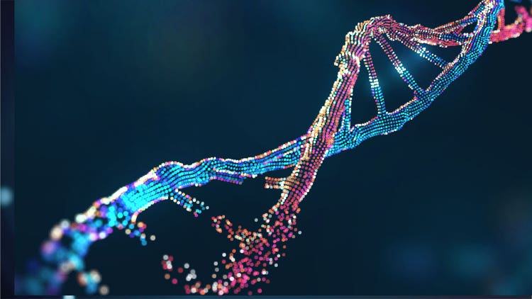
The body's building blocks dynamically mediate essential biological processes by interacting with one another as they move within their native microenvironments. Spatial biology studies the organization and movement of cells and molecules to elucidate mechanisms in health and disease.
This field provides critical insights into tumor microenvironments, proliferation, metastasis and abnormalities in tissue architecture. Unlike single-cell approaches, spatial biology focuses on positional information, enabling precise gene expression mapping, protein localization, morphology, clustering and migration, impacting disease progression.¹
Spatial biology encompasses several subfields collectively known as spatial omics. These include:
- spatial transcriptomics, which maps transcriptional activity across tissues²
- spatial proteomics, which localizes proteins, antibodies and biomarkers³
- spatial genomics, which identifies genomic architecture in the nucleus and variations throughout tissues⁴
- spatial metabolomics, which profiles the distribution of metabolites such as sugars and lipids⁵
Key Takeaways
- Spatial biology maps genes, proteins, metabolites and cells within preserved tissue architecture, retaining positional context often lost during tissue dissociation for single-cell analysis.
- Major spatial-omics subfields include spatial transcriptomics, proteomics, genomics and metabolomics, each emphasizing different molecular layers.
- Spatial profiling measures the distribution of molecular targets, while spatial phenotyping classifies cells and analyzes their organization and interactions within tissue.
- Cancer research is a leading application area: mapping tumor microenvironments supports biomarker discovery, investigation of therapeutic response and precision-oncology research.
Core Concepts in Spatial Biology
Spatial profiling and phenotyping
Spatial biology aims to profile biomolecules directly in situ, retaining their positional context within tissues. This process, called spatial profiling, captures the distribution of genes, transcriptional machinery, proteins and metabolites across cell or tissue samples while maintaining tissue integrity. ⁶
Spatial phenotyping, which identifies and classifies cell types based on their organization and interactions with their microenvironments, is closely linked to spatial profiling. ⁷
These approaches collectively provide an integrated view of tissue organization and cellular diversity.
Spatial atlas and spatial context
Advancements in high-throughput spatial biology have given rise to spatial atlases, catalogs of molecular and cellular landscapes in healthy and diseased tissues. These atlases provide frameworks for comparing tissue and cellular organization across individuals, conditions or experimental models. By integrating large-scale spatial datasets, researchers can uncover cellular communication patterns and spatial heterogeneity underlying disease profiles.⁸
Spatial resolution and organization
Spatially resolved data is a critical component of spatial biology. Spatial resolution indicates the level of organizational and dynamic information that can be collected from the sample. Spatially resolved data differ from single-cell data in the positional context of the molecules they harbor. Thus, it provides a richer understanding of how cellular states and interactions are embedded within tissue architecture.⁹
Spatial biology vs. single-cell analysis
Conventional single-cell methods dissociate tissue before analysis, capturing detailed molecular information while losing each cell’s original position relative to its neighbors. Spatial biology preserves tissue context, linking transcripts, proteins or metabolites to their location within a sample. This added context helps researchers investigate cellular neighborhoods, tissue architecture and microenvironmental effects that are difficult to resolve from dissociated single-cell data alone. Together, single-cell and spatial approaches can provide a more complete view of cellular identity and tissue organization.
Molecular Targets and Biomolecules
Each subfield of spatial biology focuses on distinct classes of molecular targets and applies specific technologies to map them in situ.
Gene expression and transcriptomics
Spatial transcriptomics captures the positional context of transcriptional machinery, including the localization of RNA molecules and transcripts within intact tissues. It is based on in situ hybridization, in which RNA is localized by tagging it with complementary DNA or modified nucleic acids.⁹ Researchers can map gene expression patterns by combining this labeling method with next-generation sequencing or fluorescent imaging while preserving the native tissue architecture. Thus, they can uncover region-specific transcriptional programs and cell–cell interactions.²
Protein localization and proteomics
Spatial proteomics uses approaches such as antibody-based imaging and mass spectrometry to determine the distribution of proteins across tissue sections and how this distribution influences their function. These methods can reveal cell–type–specific protein expression and highlight spatial variations in signaling networks and functional domains within tissues. Spatial proteomics is commonly used to characterize the tissue-level spatial variations in cancer, autoimmune diseases, cardiovascular diseases and neurological disorders.³
Metabolite mapping and biomarkers
Spatial metabolomics profiles metabolites, lipids and other small molecules across tissues. Mapping these biomolecules using mass spectrometry, segmentation and clustering analyses provides insight into metabolic pathways, microenvironmental conditions and biochemical heterogeneity. Similarly, spatial mapping of clinically relevant biomarkers supports the study of disease mechanisms and therapeutic responses as a function of native tissue organization.⁵
Featured Product
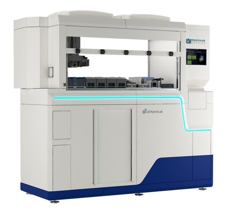
CellXpress.ai Automated Cell Culture System
The future of cell culture backed by machine learning and data-driven science.
Techniques and Technologies in Spatial Biology
Imaging and cytometry
Imaging and quantification are fundamental in spatial biology. Techniques such as imaging mass cytometry, multiplex imaging, immunofluorescence and fluorescence imaging allow simultaneous detection of multiple proteins, RNA molecules, metabolic byproducts and other biomolecules within tissue sections.⁹ Advanced microscopy technologies, mainly super-resolution microscopy, further enhance the spatial resolution beyond the diffraction limit, providing insights into cellular organization and molecular interactions on a nanometer scale.¹⁰
Advanced analysis platforms
The advancements in automated imaging systems and integrated computational biology support have enabled high-plex spatial biology workflows. These platforms streamline the workflow from sample preparation to image acquisition and data analysis, enabling simultaneous measurement and localization of multiple targets across thousands of cells. Each high-plex solution has distinct advantages in resolution, throughput and molecular coverage, giving researchers flexibility when selecting the appropriate method for their biological question.¹¹
Innovations in spatial biology
Innovations in spatial biology aim to enhance throughput and drive multiplex imaging, which allows the detection of hundreds of targets in a single sample. AI/ML-driven approaches, such as deep learning, strengthen the analysis of high-throughput workflows by facilitating cell type classification, spatial pattern recognition and predictive modeling. These advances are expected to improve the speed and accuracy of comprehensive, high-resolution mapping of tissue microenvironments in disease research.¹²
Applications of Spatial Biology
Cancer research
Cancer cells are in constant contact with components in the tumor microenvironment, such as the extracellular matrix, immune cells, growth factors and cytokines. Therefore, spatial biology has become a critical tool in cancer research, providing detailed insights into the heterogeneity of the tumor microenvironment.¹³ By mapping the spatial distribution of immune, stromal and tumor cells, researchers can better understand cell–cell interactions that influence tumor growth and metastasis. Furthermore, spatial profiling of target molecules in the tumor microenvironment empowers more accurate prediction of therapeutic responses, guiding the design of targeted treatments in precision oncology.¹,⁹
Oncology is a leading application area for spatial biology, with tumor microenvironment profiling increasingly used to identify candidate spatial biomarkers associated with immunotherapy response, adding tissue-context information that bulk or dissociated single-cell sequencing alone cannot provide.
Developmental biology and tissue mapping
Spatial profiling is crucial for developmental biology when mapping tissue architecture and identifying patterns that govern organogenesis.¹⁴ Researchers can develop comprehensive benchmarks for understanding normal tissue formation and cellular differentiation processes by generating spatial atlases of organs at different developmental stages. These atlases serve as foundational resources for studying normal development and developmental disorders.⁸
Disease research
Alongside cancer, spatial biology studies various diseases by tracking changes in tissue organization and cellular interactions. Investigating spatial alterations in tissue microenvironments enables researchers to characterize disease progression, identify early pathological events and study the interplay between different cell types in health and disease. This spatially resolved perspective is invaluable for uncovering mechanisms overlooked in single-cell analyses.¹⁵⁻¹⁷
See how Danaher Life Sciences can help
FAQs
What is spatial biology and why is it important?
Spatial biology maps molecules and cells within preserved tissue architecture. Retaining positional context helps researchers understand how tissue organization, cellular interactions and local microenvironments influence biological processes, disease progression and therapeutic response.
What are the main branches of spatial omics?
Main branches include spatial transcriptomics (maps RNA), spatial proteomics (maps proteins), spatial genomics and epigenomics (study DNA and chromatin) and spatial metabolomics (maps metabolites and lipids). These approaches are integrated to study multiple molecular layers within tissue.
Spatial transcriptomics vs spatial proteomics?
Spatial transcriptomics maps RNA to show gene location in tissue, while spatial proteomics maps proteins to reveal their localization, abundance and functions. Since RNA and protein levels often don't match, these approaches offer complementary views of tissue biology.
What techniques and technologies are used in spatial biology?
Spatial biology employs sequencing-based methods such as multiplexed RNA imaging, immunofluorescence, imaging mass cytometry and mass spectrometry imaging. Computational analysis, machine learning and data integration help identify cells, classify types and analyze spatial patterns.
How is spatial biology used in oncology?
In oncology research, spatial biology maps tumor, immune and stromal cells within the tumor microenvironment. Researchers use these data to study tumor heterogeneity, cellular neighborhoods, disease progression, metastasis and treatment response, as well as to identify candidate spatial biomarkers for precision-oncology research.
How does spatial biology differ from single-cell analysis?
Conventional single-cell methods analyze dissociated cells to obtain detailed molecular data but lose tissue location, whereas spatial biology preserves tissue context to examine cellular neighborhoods. Together, these approaches offer complementary insights into cell identity and tissue structure.
References
- Seferbekova Z, Lomakin A, Yates LR, Gerstung M. Spatial biology of cancer evolution. Nat Rev Genet 2023;24(5):295-313.
- Williams CG, Lee HJ, Asatsuma T, Vento-Tormo R, Haque A. An introduction to spatial transcriptomics for biomedical research. Genome Med 2022;14(1):68.
- Wu M, Tao H, Xu T, Zheng X, Wen C, Wang G, et al. Spatial proteomics: unveiling the multidimensional landscape of protein localization in human diseases. Proteome Sci 2024;22(1):7.
- Bouwman BA, Crosetto N, Bienko M. The era of 3D and spatial genomics. Trends Genet 2022;38(10):1062-1075.
- Planque M, Igelmann S, Campos AMF, Fendt S-M. Spatial metabolomics principles and application to cancer research. Curr Opin Chem Biol 2023;76:102362.
- Moffitt JR, Lundberg E, Heyn H. The emerging landscape of spatial profiling technologies. Nat Rev Genet 2022;23(12):741-759.
- Gulati GS, D’Silva JP, Liu Y, Wang L, Newman AM. Profiling cell identity and tissue architecture with single-cell and spatial transcriptomics. Nat Rev Mol Cell Biol 2025;26(1):11-31.
- Haniffa M, Taylor D, Linnarsson S, Aronow BJ, Bader GD, Barker RA, et al. A roadmap for the human developmental cell atlas. Nature 2021;597(7875):196-205.
- Lewis SM, Asselin-Labat M-L, Nguyen Q, Berthelet J, Tan X, Wimmer VC, et al. Spatial omics and multiplexed imaging to explore cancer biology. Nat Methods 2021;18(9):997-1012.
- Yang T, Luo Y, Ji W, Yang G. Advancing biological super-resolution microscopy through deep learning: a brief review. Biophys Rep 2021;7(4):253.
- Aung TN, Bates KM, Rimm DL. High-plex assessment of biomarkers in tumors. Mod Pathol 2024;37(3):100425.
- Xu H, Cong F, Hwang TH. Machine learning and artificial intelligence–driven spatial analysis of the tumor immune microenvironment in pathology slides. EU Focus 2021;7(4):706-709.
- Nicolini A, Ferrari P. Involvement of tumor immune microenvironment metabolic reprogramming in colorectal cancer progression, immune escape, and response to immunotherapy. Front Immunol 2024;15:1353787.
- Choe K, Pak U, Pang Y, Hao W, Yang X. Advances and challenges in spatial transcriptomics for developmental biology. Biomolecules 2023;13(1):156.
- Miyamoto AT, Shimagami H, Kumanogoh A, Nishide M. Spatial transcriptomics in autoimmune rheumatic disease: potential clinical applications and perspectives. Inflamm Regen 2025;45(1):6.
- Kiessling P, Kuppe C. Spatial multi-omics: novel tools to study the complexity of cardiovascular diseases. Genome Med 2024;16(1):14.
- Piwecka M, Rajewsky N, Rybak-Wolf A. Single-cell and spatial transcriptomics: deciphering brain complexity in health and disease. Nat Rev Neurol 2023;19(6):346-362.