Visoria P Polarization Microscope
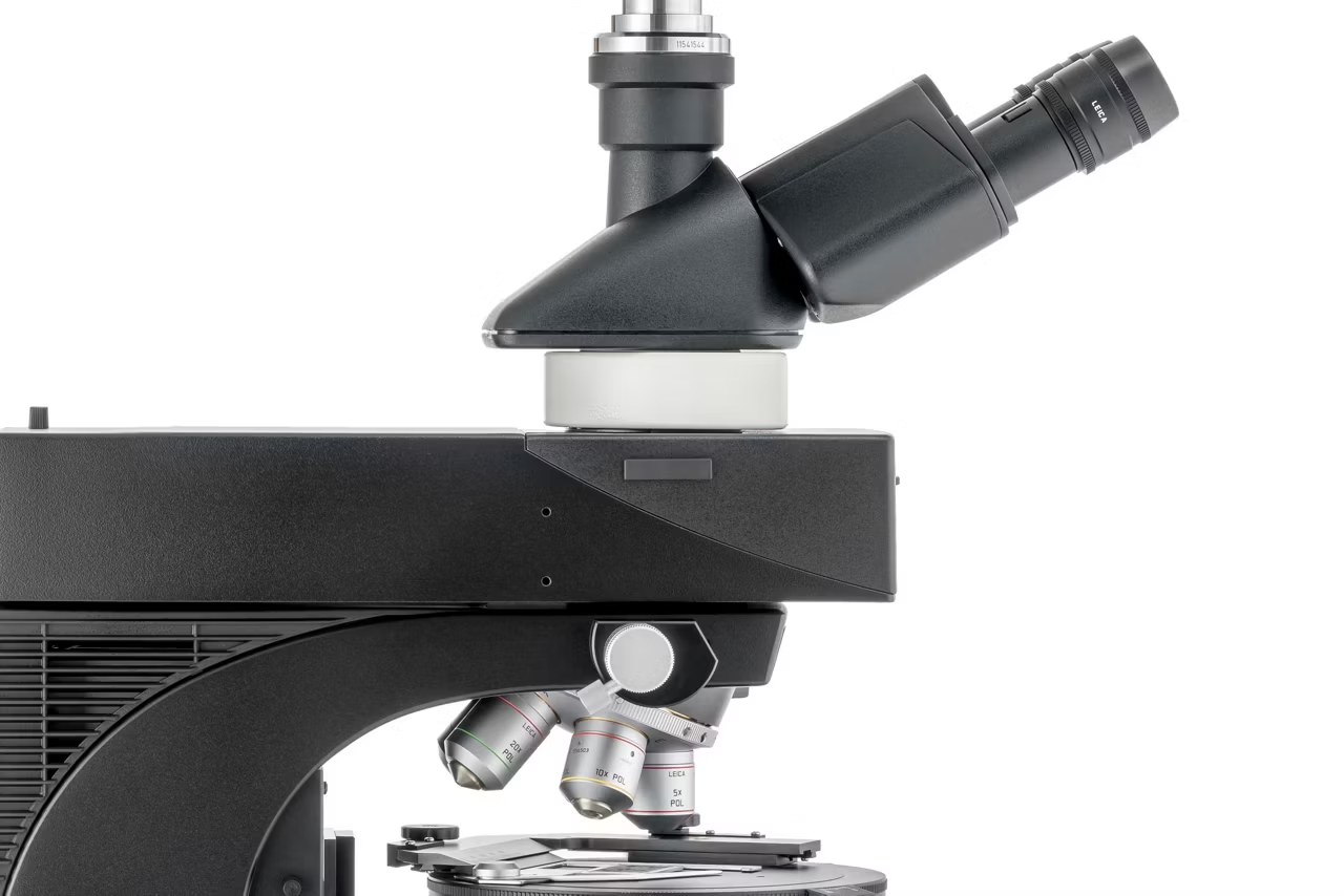
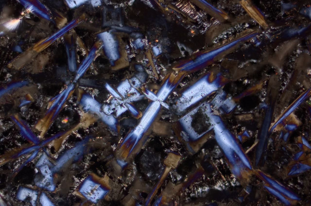
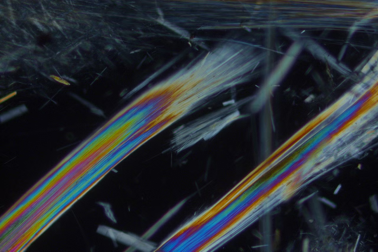
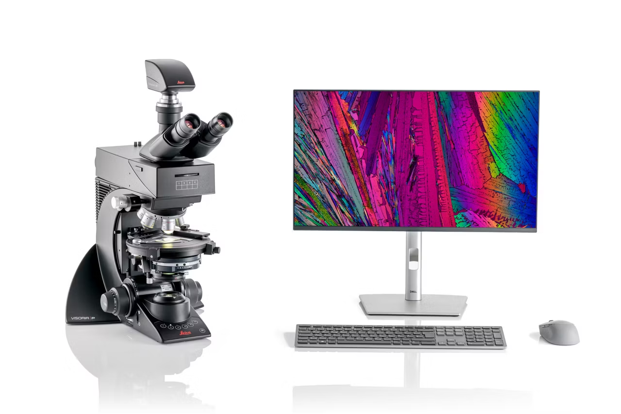
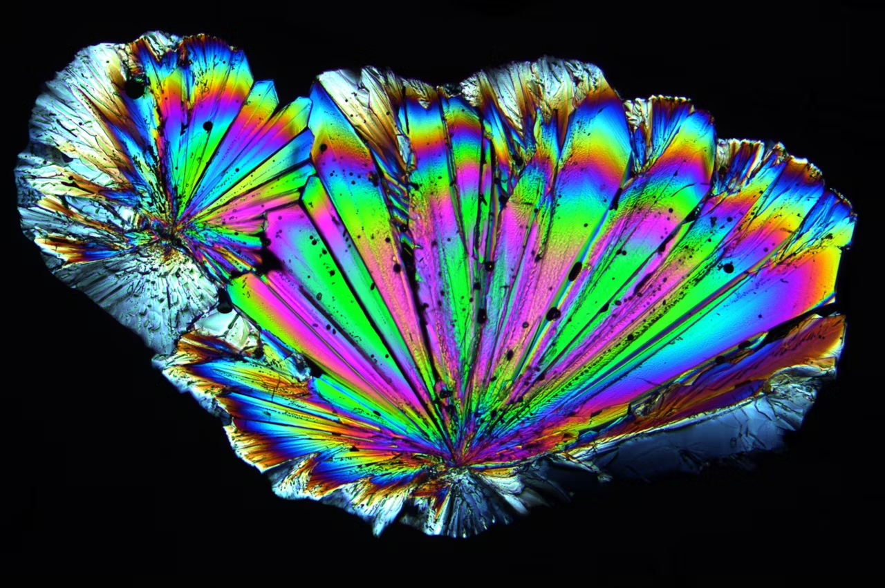
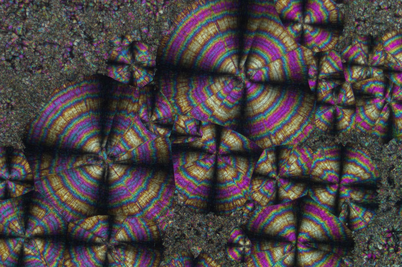

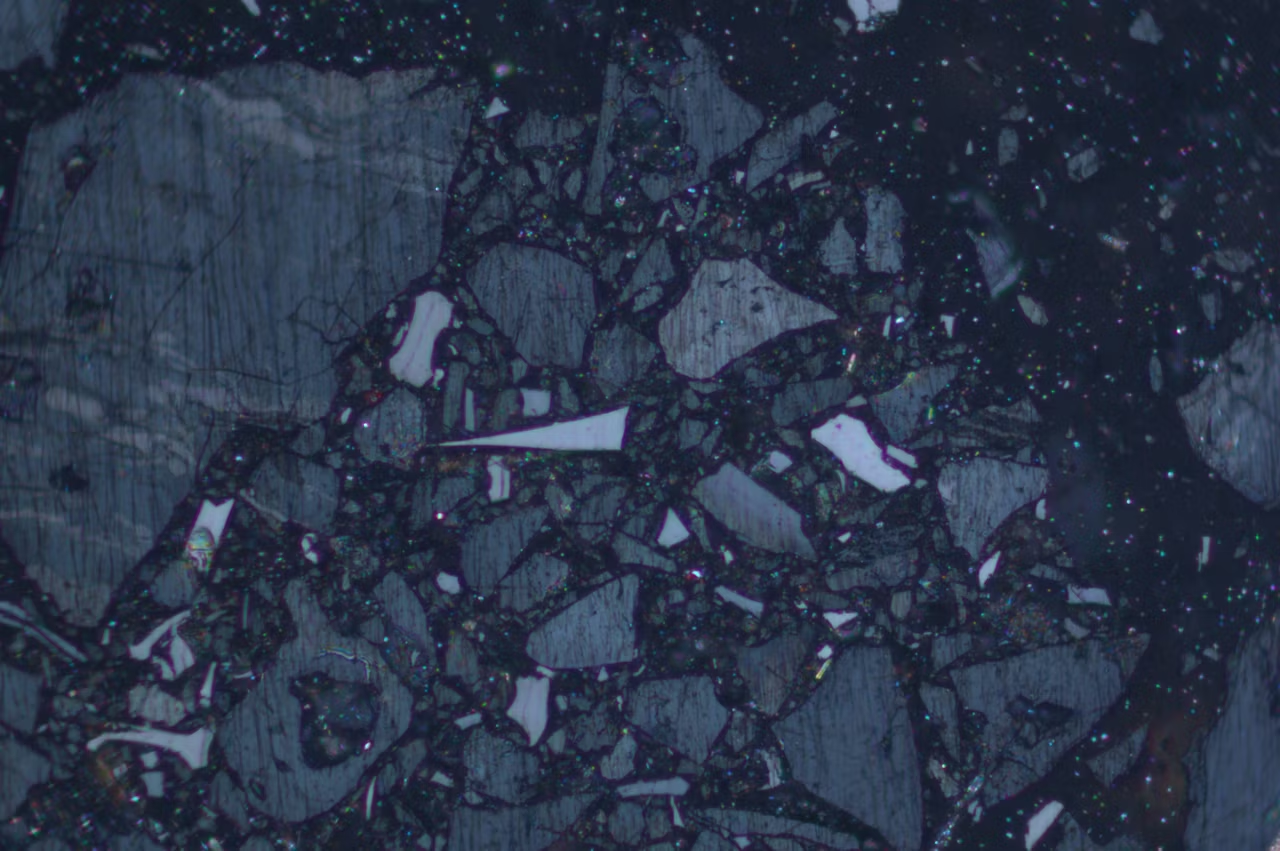


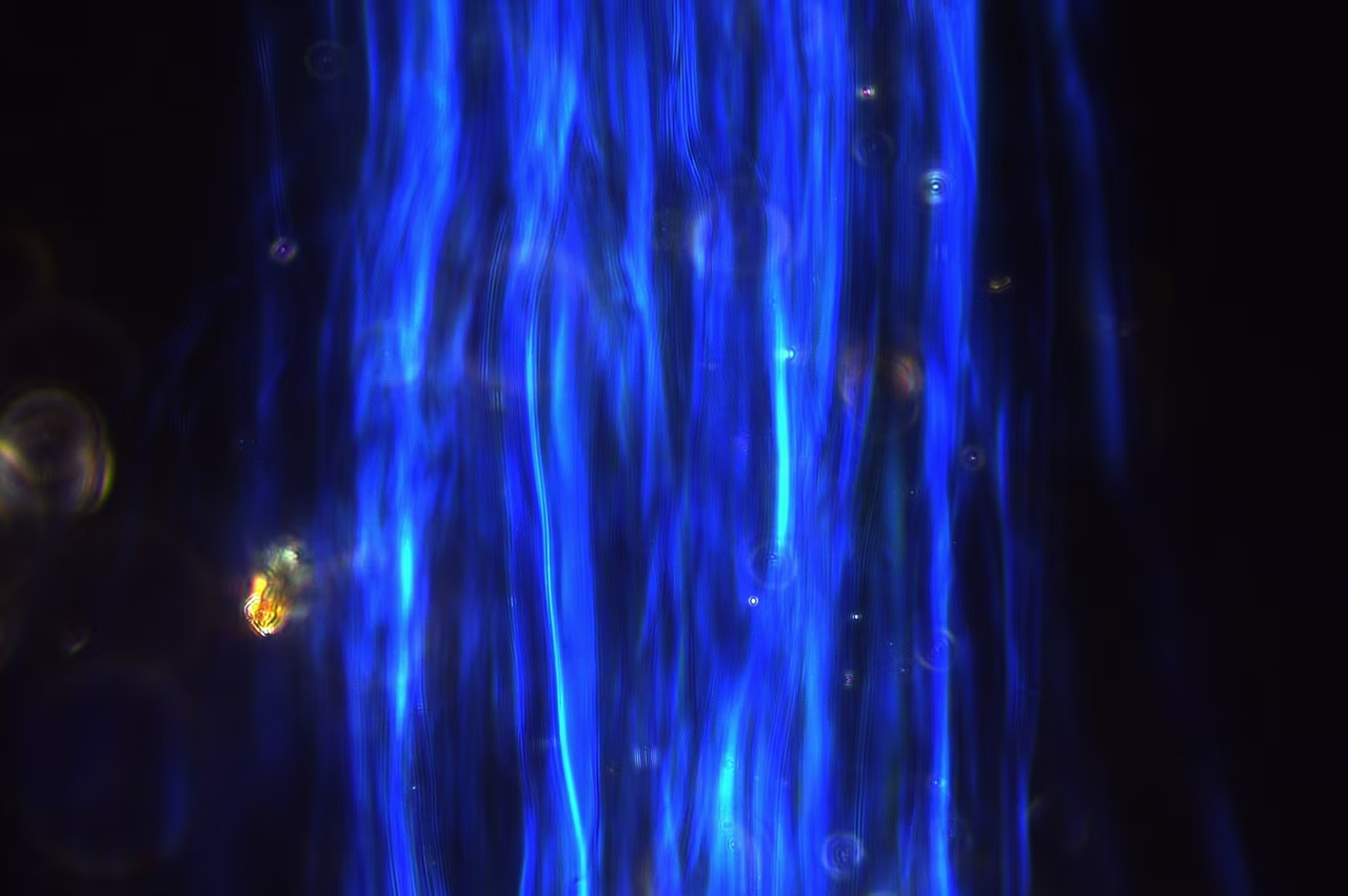
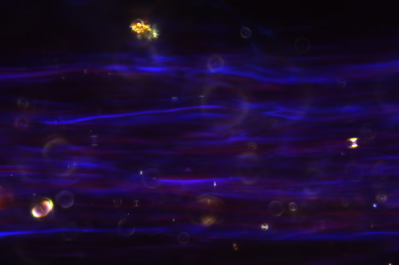
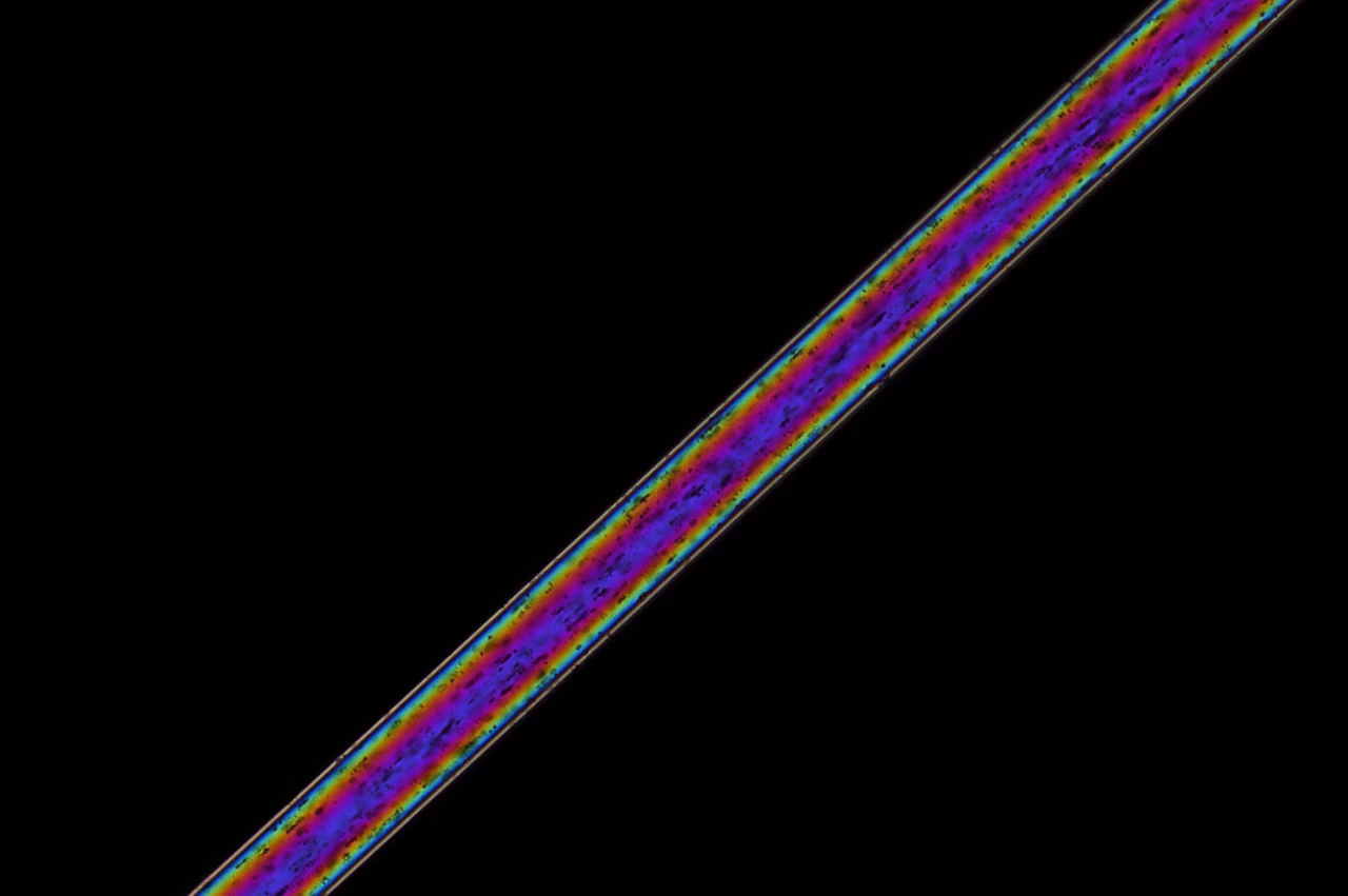
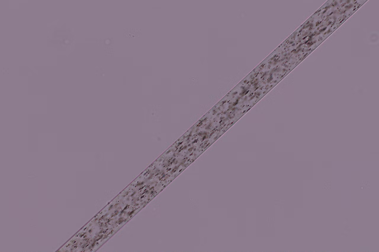
Experience enhanced efficiency and comfort in your daily microscopy routine. The Visoria P polarization microscope uses polarized light to study the optical properties of materials and geological samples.
Streamline your workflows with encoded functions, optimized light settings, and other microscope features. You can also be more comfortable and minimize strain thanks to the microscope’s ergonomic design.
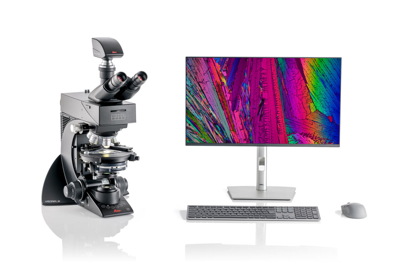
Visoria P Digital Polarization Microscope for Industrial Applications (Transparent samples only)
Experience enhanced efficiency and comfort in your daily microscopy routine. The Visoria P polarization microscope uses polarized light to study the optical properties of materials and geological samples. Streamline your workflows with encoded functions, optimized light settings, and other microscope features. You can also be more comfortable and minimize strain thanks to the microscope’s ergonomic design. This configuration includes the Visoria P digital polarization and is suited for industrial applications (transparent samples only).
Visoria P Polarization Microscope for Geosciences Including Conoscopy with Flexacam c5
Experience enhanced efficiency and comfort in your daily microscopy routine. The Visoria P polarization microscope uses polarized light to study the optical properties of materials and geological samples. Streamline your workflows with encoded functions, optimized light settings, and other microscope features. You can also be more comfortable and minimize strain thanks to the microscope's ergonomic design. This configuration includes the Visoria P polarization microscope as well as Flexacam c5 microscope camera and is suited for geosciences applications (incl. Conoscopy)
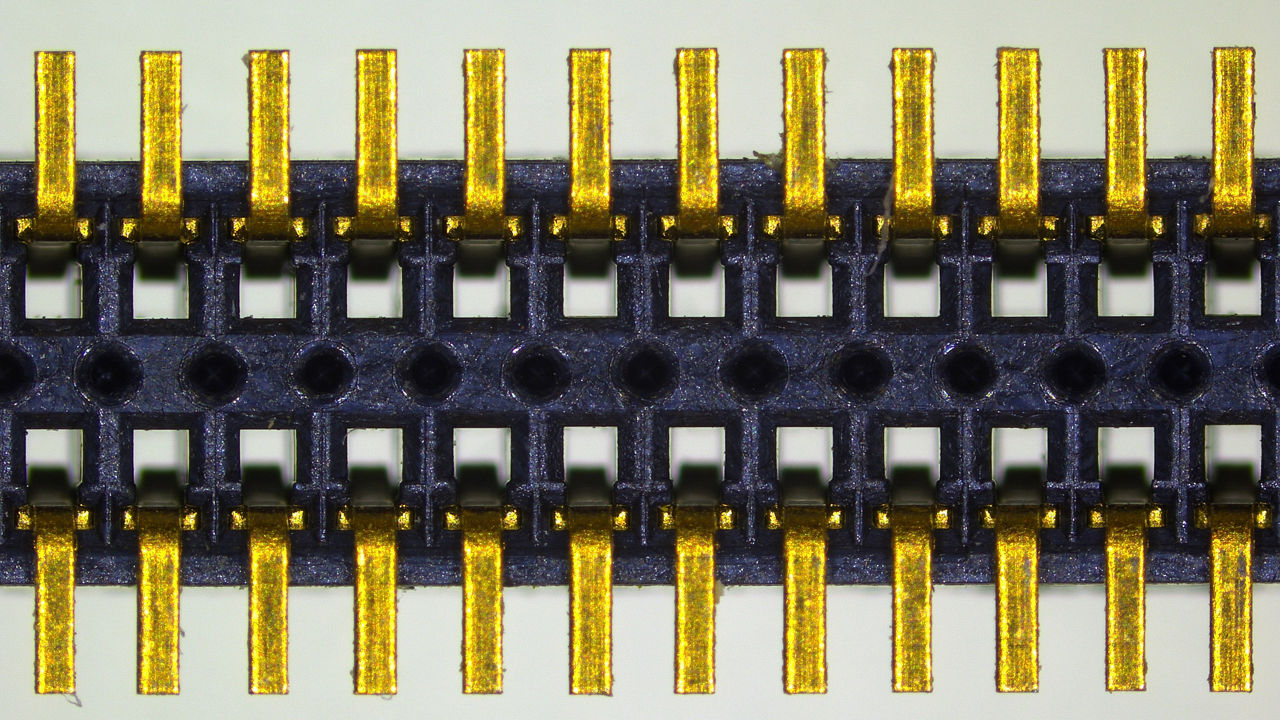
Visoria P Polarization Microscope for Industrial Applications with Flexacam c5 (transparent samples only)
Experience enhanced efficiency and comfort in your daily microscopy routine. The Visoria P polarization microscope uses polarized light to study the optical properties of materials and geological samples. Streamline your workflows with encoded functions, optimized light settings, and other microscope features. You can also be more comfortable and minimize strain thanks to the microscope's ergonomic design. This configuration includes the Visoria P polarization microscope as well as Flexacam c5 microscope camera and is suited for industrial applications (transparent samples only)

Visoria P Polarization Microscope for Industrial Applications (transparent samples only)
Meet Visoria P
For polarization microscopy
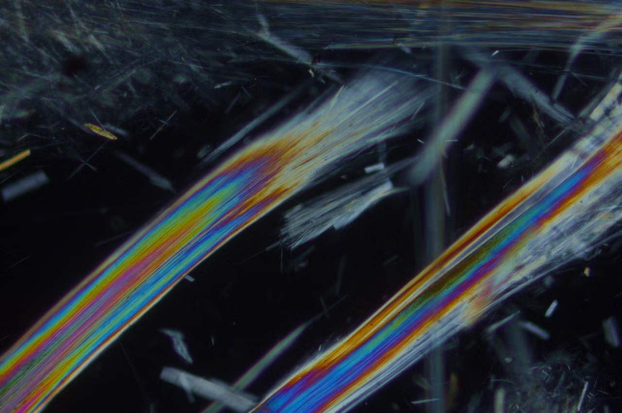
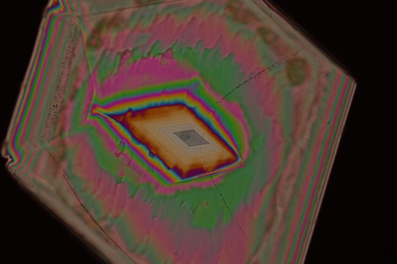
The Visoria P polarization microscope delivers key results when investigating birefringent (optically anisotropic) materials like minerals, rocks, coal, plastics, polymers, liquid crystals, glass,and concrete.
Save time with optimized light settings
Spend more time viewing and examining samples with Visoria P.
If you change the microscope’s magnification or contrast method, there is no need to manually adjust the brightness thanks to the light management function. The illumination settings are automatically applied thanks to the microscope’s encoding.
Simplify your documentation

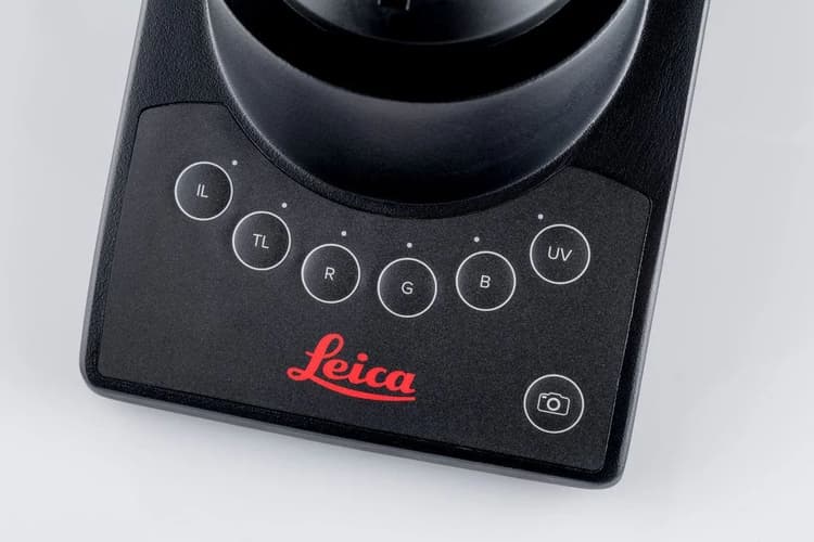
You can quickly capture sample details with a press of a button while keeping your eyes on the image. The button for image acquisition is easily accessible on the Visoria P microscope stand.
When you store an image for documentation, selected system settings are automatically saved along with the meta data of the image.
The scale bar is automatically adjusted and added to the image which increases efficiency and saves you valuable time.
Operate your microscope with ease

Perform daily routines rapidly and reliably thanks to the intuitive operation of Visoria P.
- Easily find the appropriate aperture for each objective with color coding.
- Protect your specimens and objectives from accidental damage with the built-in focus stop.
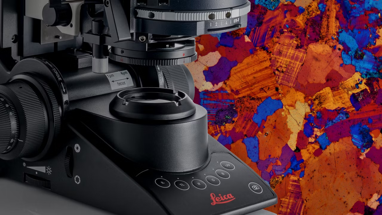
- For finer focus at higher magnification, use the three-gear focusing system - coarse, medium, and fine.
Work without eyepieces by going digital

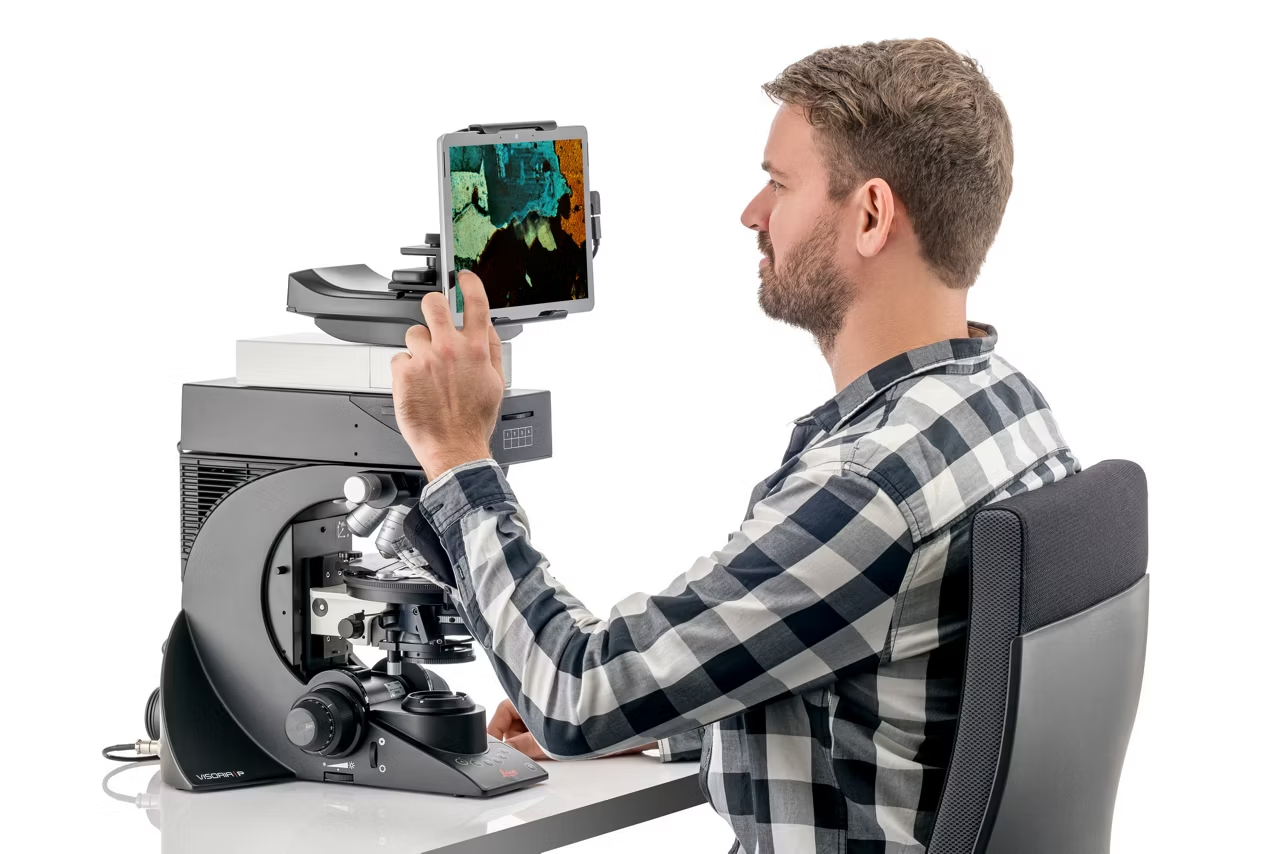
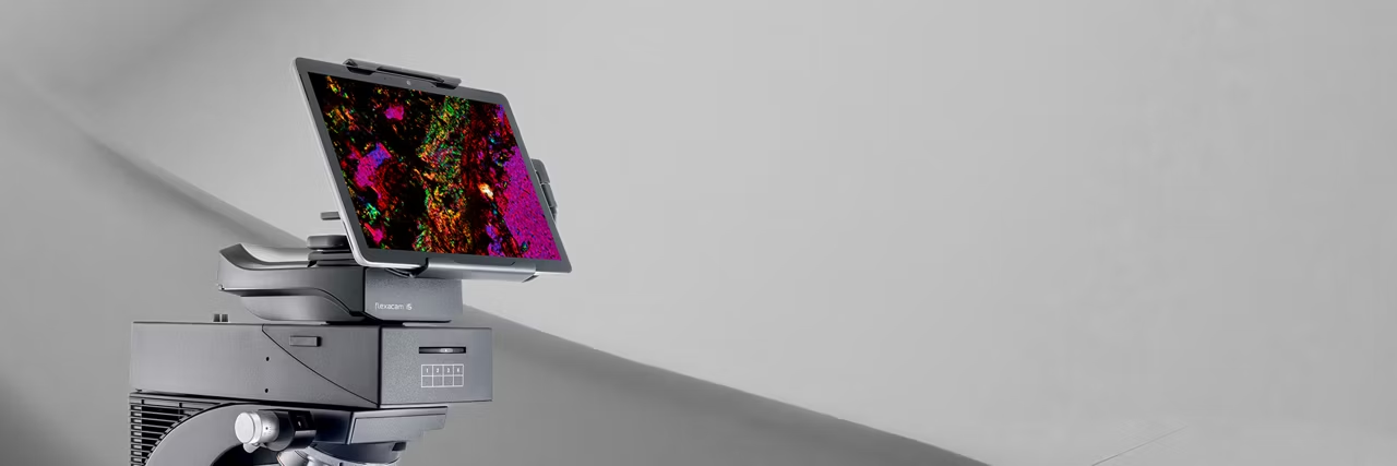
The Visoria P digital polarization microscope without eyepieces offers a number of practical benefits:
- Work in a comfortable and relaxed position by viewing images directly on a tablet.
- Visualize and document your work steps quickly and discuss image results easily with your colleagues.
- Save space on your workbench without the need for a computer.
Stay comfortable while working

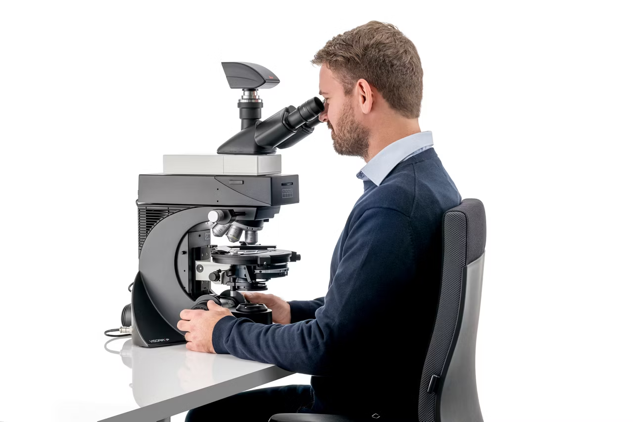
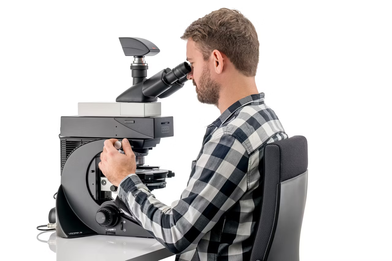
Visoria P adapts to your needs, allowing a proper posture and reducing the risk of neck and back strain during long hours at the microscope.
Work comfortably with aligned shoulders and optimal hand and arm positioning thanks to the symmetrical layout and height adjustment of the focus control knobs.
Adapt your microscope with ergo accessories

You can maintain an upright posture thanks to the adaptability of Visoria P. Choose from a range of ergonomic accessories to suit your needs.
- Ergonomic modules: Insert ErgoModules below the tube to adjust the eyepiece height for a comfortable sitting posture.
- Ergonomic lift: The optional ErgoLift enables easy height adjustments of the microscope.
Determine optical properties of materials with conoscopy

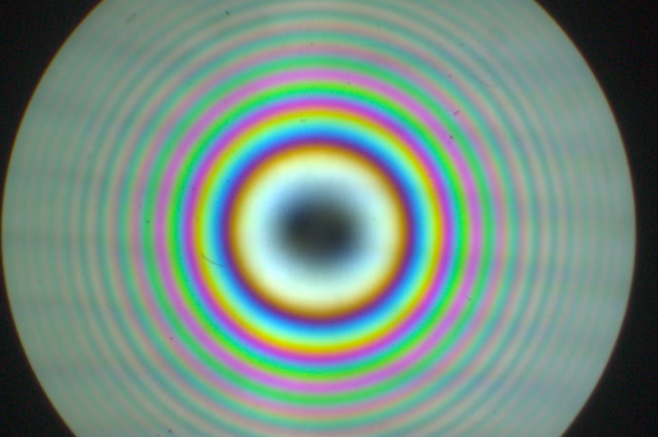
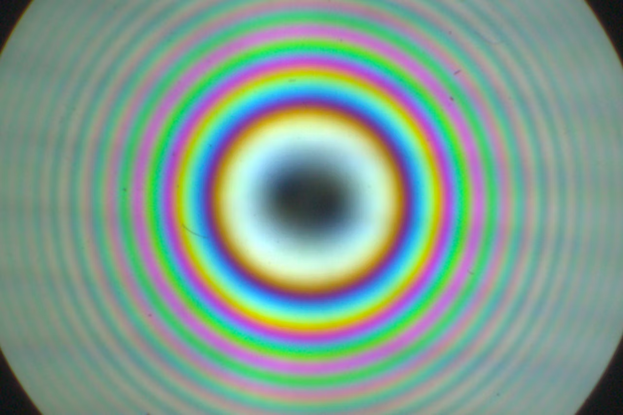
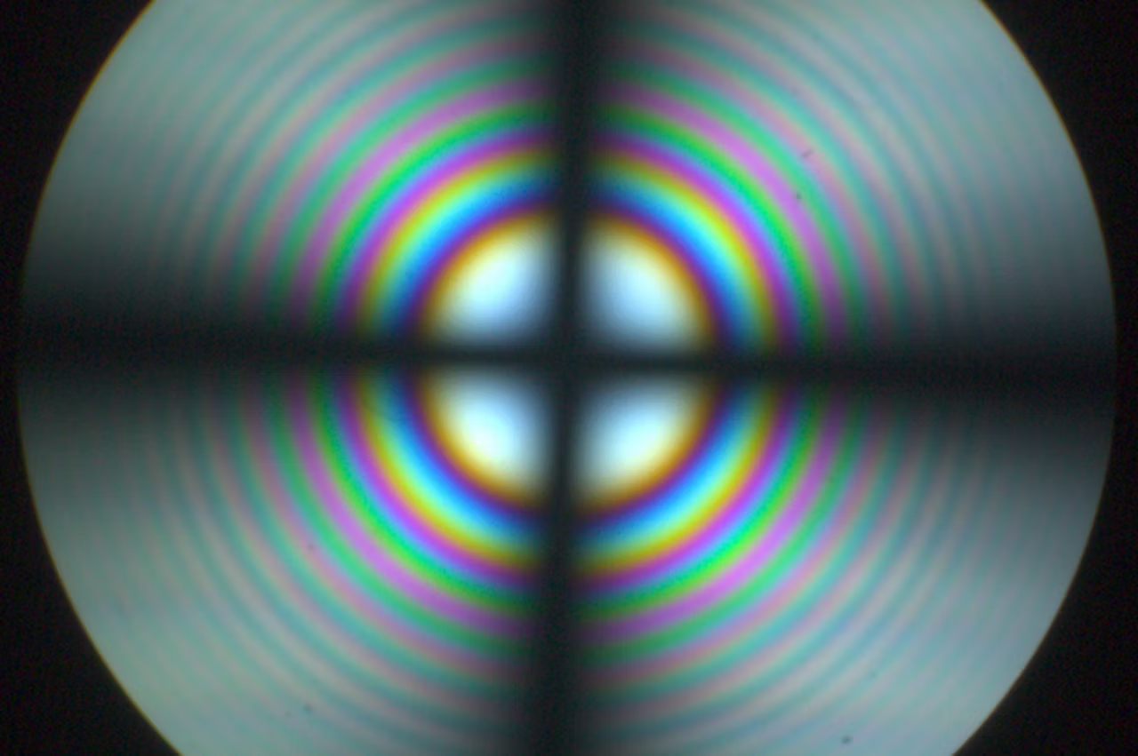
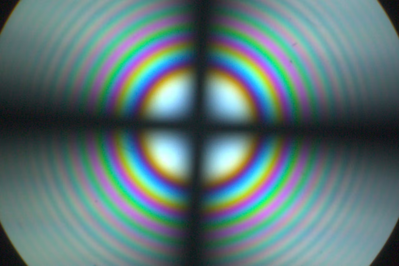
Conoscopy is useful for evaluating the optical properties of anisotropic materials.
Choose from three conoscopy modules
- Bertrand lens cube
- Bertrand lens module (A/B module)
- Advanced conoscopy module.
Specialized objectives for coal and asbestos analysis

For the analysis of coal, Visoria P helps you to identify different sample components using specialized oil-immersion objectives.
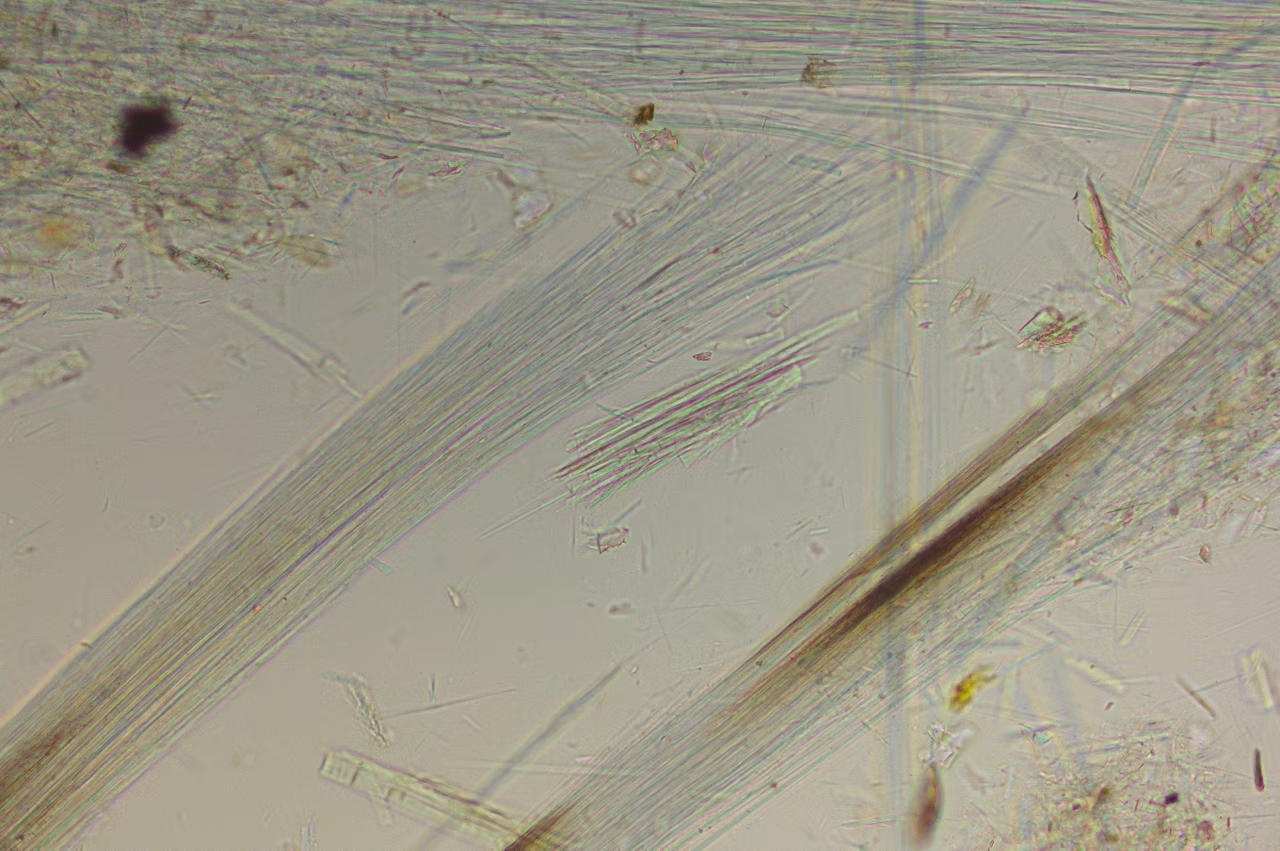
To determine the presence of asbestos in a sample, the dispersion color method is typically used. For this analysis, you can take advantage of the dispersion staining objectives.
Powered by the Enersight software platform
Simplify and streamline your workflow with the Visoria P polarization microscope and Enersight software platform. It helps you compare, measure, and share data seamlessly with a single intuitive interface.
Key advantages
- Optimize images by automatic correction of shading due to uneven illumination.
- Get high-quality images with optimal illumination and camera parameters by using the Quick Brightness function.
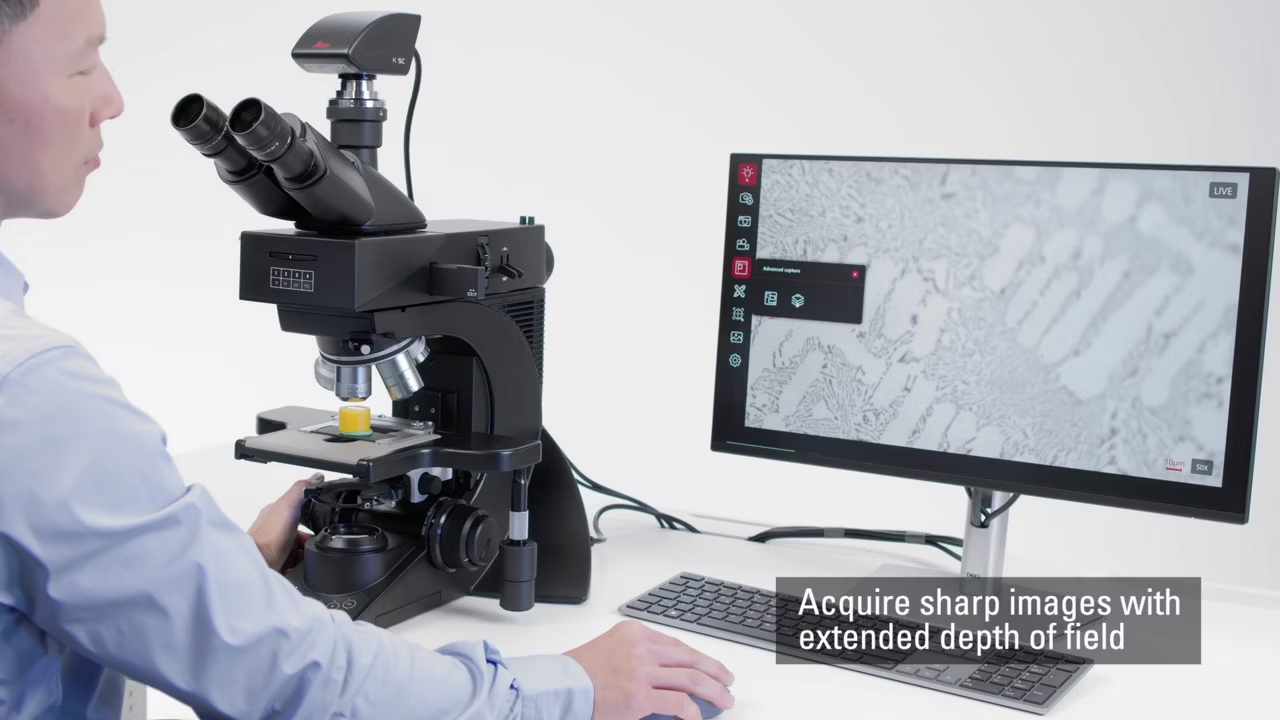
- Acquire sharp images with extended depth of field (EDOF).
- Observe specimens with a larger field of view and higher resolution using the XY Stitching with Manual Stage function.
- Achieve insightful overlays by combining images from up to four fluorescence channels.
- Gain a better understanding of specimens by merging multiple images from different contrast methods, such as brightfield and fluorescence.
Power up routines with efficiency and comfort
Efficiency through encoded functions
Increase the efficiency of your routine microscopy tasks. Save valuable time with simplified documentation and encoded microscope functionality, allowing you to focus on your sample.
More comfort with ergonomics

Work with a comfortable posture by taking advantage of the symmetrical positioning of controls and ergonomic accessories. These features help reduce neck and back strain, even during extended hours at the microscope.
Powered by Enersight

Simplify and streamline your workflow with the Visoria P polarization microscope and Enersight software platform. It helps you compare, measure, and share seamlessly with a single intuitive interface.
| Nosepiece | Encoded 5x (M25), centerable |
| Eyepieces (FOV) | 20 / 22 / 25 mm |
| Tubes | Phototubes 50 / 50 with fixed port, 100 / 50 / 0 with fixed port, 100 / 50 / 0 with variable port |
| Digital version | Digital version with 10” screen / tablet |
| Ergonomic accessories | Wide range of ergonomic accessories available (ErgoTubes, ErgoLift, ErgoModules) |
| Encoded illumination management | IL and TL: High-power white LED, encoded 4-color fluorescence illumination, further external light sources on request (non-encoded) |
| Incident light axis | Manual encoded, 4-fold filter turret, color-coded diaphragm assistant;field and aperture diaphragm, slots for analyzer / polarizer, two filter positions |
| Fluorescence light axis | Optional |
| Incident light (IL) | Methods: Brightfield (with BF cube or Smith reflector), DIC, fluorescence, oblique illumination, qualitative and quantitative polarization |
| Transmitted light axis | Manual, fixed and flip-top condenser operation with color-coded diaphragm assistant |
| Transmitted light (TL) | Methods: Brightfield, darkfield, phase contrast, DIC, qualitative and quantitative polarization |
| Operation | 360° rotatable polarization stage with verniers and brake, 360° rotatable polarization stage with verniers, 45° clickstop position and brake. Stages are exchangeable and height adjustable. Further stages on request. |
| Stage | Scanning stage 75 x 50 mm, additional manual xy stages available, stage bracket for adaptation of 3rd party stages, bject clamps, sample holder without point counting, sample holder with point counting |
| Focus drive | Height-adjustable focus knobs, 19 mm travel range, maximum 28 mm total stage stroke depending on stage and condenser type, 2-gear focus drive (coarse / fine) with 1 mm scale, 3-gear focus drive (coarse / medium / fine) with 140, 4 and 1 µm scale, torque adjustment, and adjustable upper focus stop |
| Conoscopy | Bertrand lens cube, Bertrand lens module (A/B module), advanced conoscopy module (focusable) |
| Analyzer | Fixed, 180°, 360° |
| Polarizer | Fixed, 0 / 45 / 90°, 90° with rotatable lambda plate, 360°, fixed with lambda plate |
| Compensators | Tilting compensators up to 5 or 30 orders, quartz wedge, lambda and 1/4 lambda plate |
| Supply voltage | 100–240 V AC, 50 / 60 Hz, power consumption max. 15 W |
| Ambient conditions | 15–35°C, relative humidity max. 80% up to 30°C (non-condensing) |
| Size and weight | Length: 410 mm, width: 331 mm, height: 505 mm, approx. 18 kg (depending on configuration) |
| Stand | Illumination toggle buttons with status indicator, image capture button, antimicrobial surface with AgTreat (ISO 22196) |
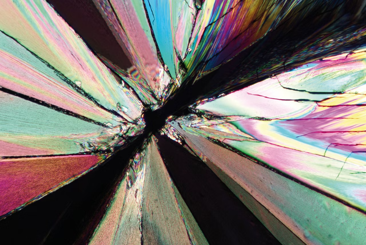
DM4 P, DM750 P & Visoria P Polarization Microscopes
Polarization microscopes with strain free optics, LED illumination, rotatable stage and Bertrand lens for minerals, polymers and pharmaceuticals. Get a quote.
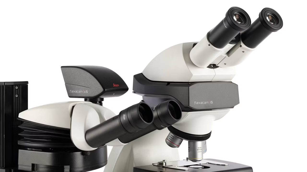
Flexacam c5 & i5 Microscope Cameras
Flexacam c5 and i5 microscope cameras deliver high resolution images with precise colors. Powered by Enersight software for easy use. Request a quote today.
Enersight Microscope Software Platform
Enersight simplifies image acquisition and documentation across industry, life sciences and clinical pathology with one consistent interface. Get a quote!
K3C & K3M The Microscope Camera K3 series
Microscope Camera K3 series images challenging samples with precision. k3-m. Request a quote!