Visoria P Polarization Microscope for Geosciences Including Conoscopy with Flexacam c5
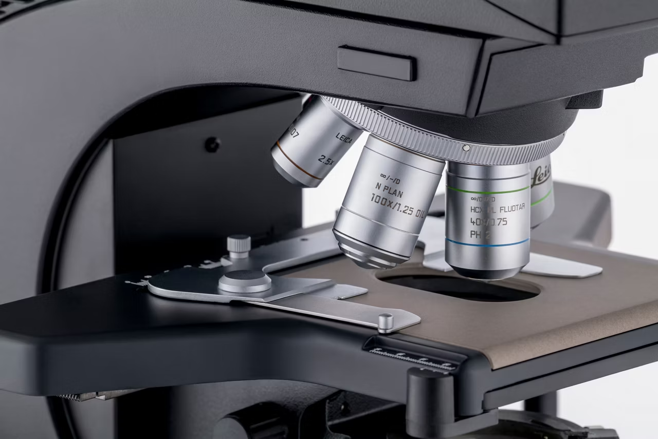
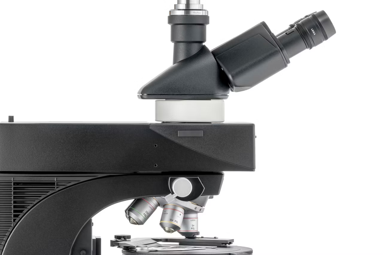
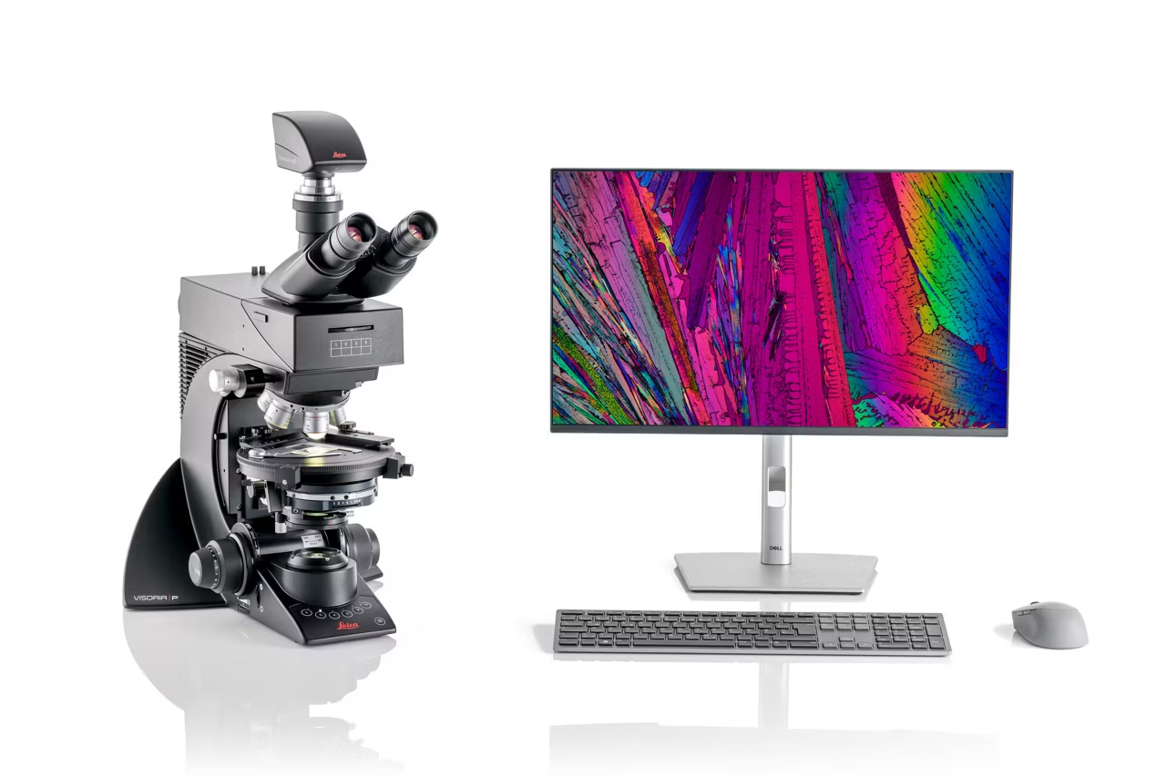
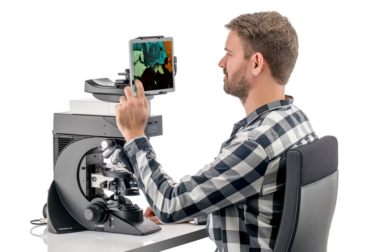
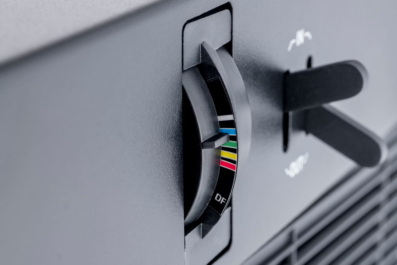
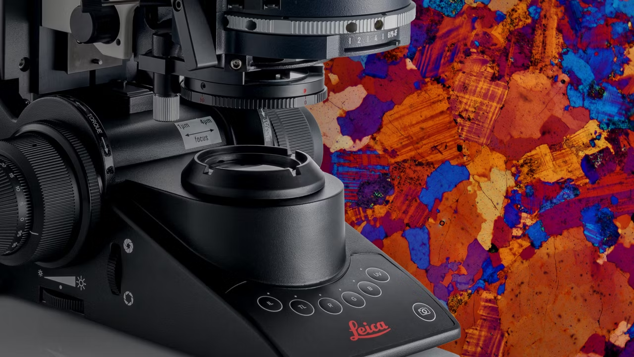
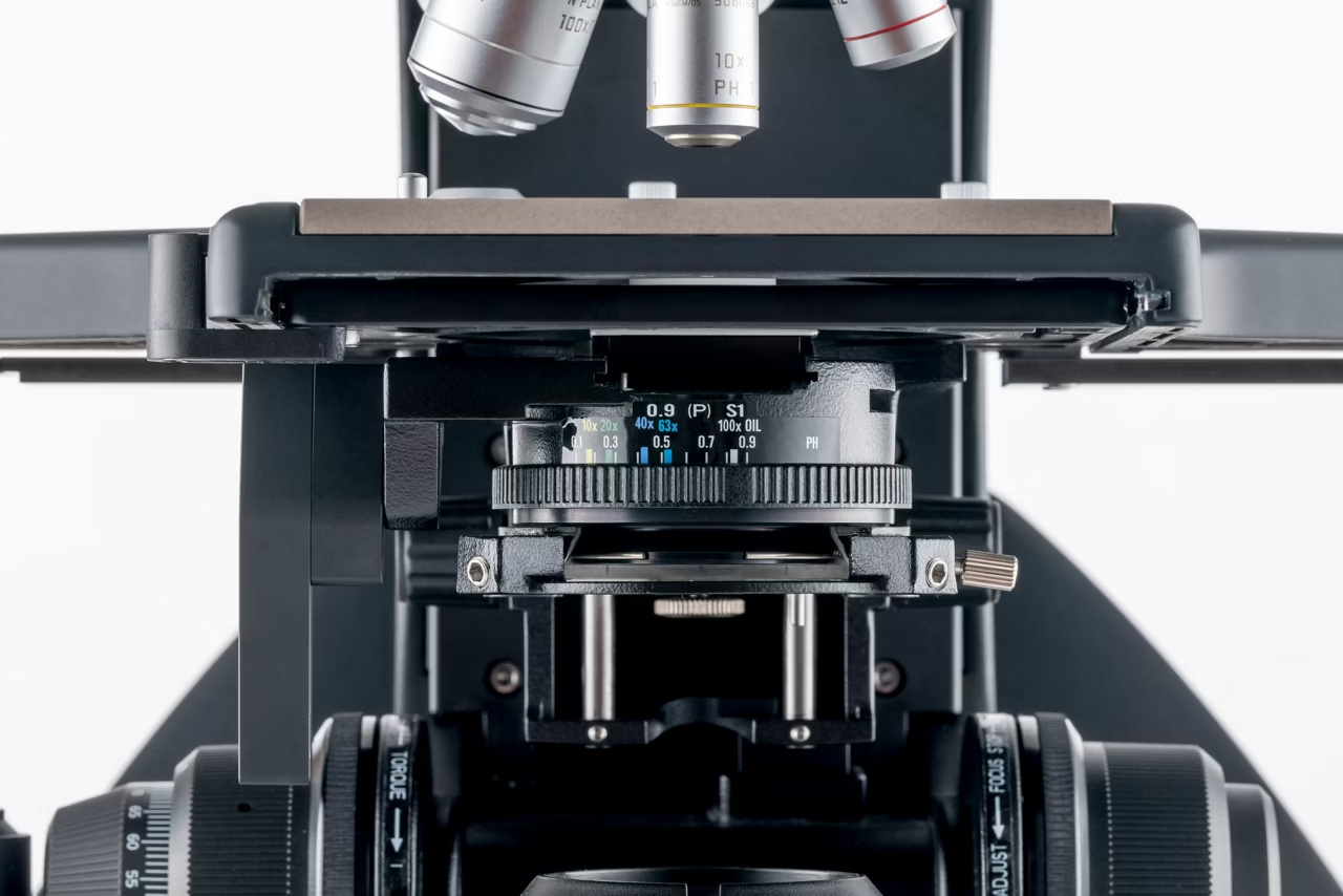
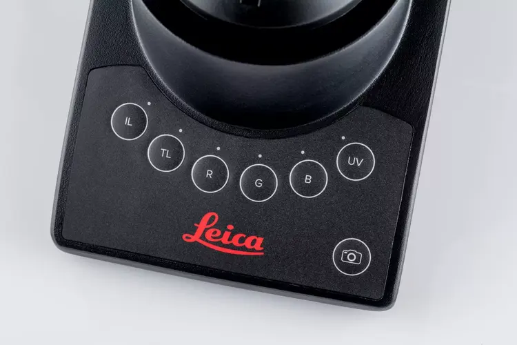
Experience enhanced efficiency and comfort in your daily microscopy routine. The Visoria P polarization microscope uses polarized light to study the optical properties of materials and geological samples.
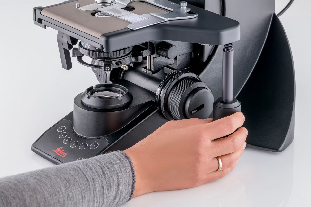
Streamline your workflows with encoded functions, optimized light settings, and other microscope features. You can also be more comfortable and minimize strain thanks to the microscope’s ergonomic design.
This configuration includes the Visoria P polarization microscope as well as Flexacam c5 microscope camera and is suited for geosciences applications (incl. Conoscopy)
- Visoria P Polarization microscope with illumination management
- Binocular phototube
- Two eyepieces (10x), field of view 22 mm, thereof one with cross hair reticule
- Flexacam c5 microscope camera, incl. c-mount 0.55x
- High-power white LED
- Contrast methods (transmitted light): Quantitative polarization, Brightfield
- Encoded nosepiece, 5x
- Objectives (N PLAN POL): 5x, 10x, 20x, 40x, 63x
- 360° rotatable polarization stage with verniers, 45° clickstop position and brake Object guide without point counting
- 360° analyzer, Polarizer, Whole-wave plate f. compensator slot
- Bertrand lens cube
- 3-step focus drive with height-adjustable focus knobs
- Enersight microscope software
- Dust protection cover
- Quick start guide and user manual
| Contrasting Method(s) | Brightfield, Qualitative Polarization |
| Long Working Distance | Optional |
| Microscope Stage Type(s) | Manual |
| Microscope Camera Option(s) | Single Camera |
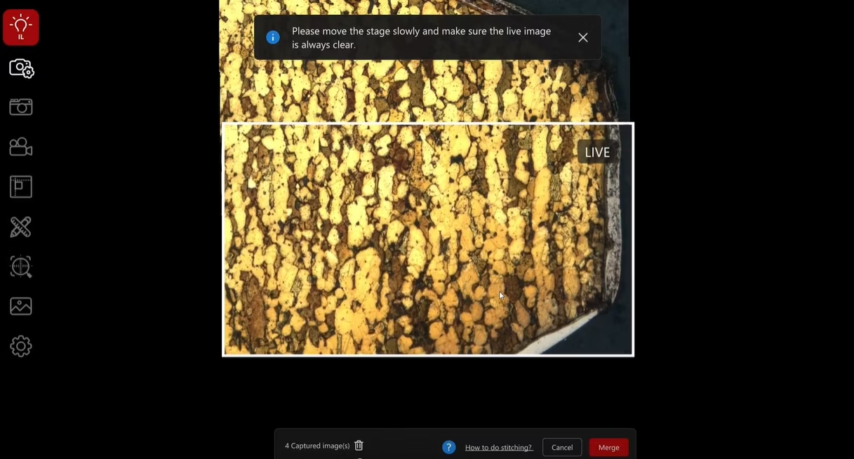
| Microscopy Software Available | 3D Visualization, Documentation, Measuring, Tile Stitching |
| Has Eyepiece | Yes |
| Microscope Type(s) | Upright Microscope, Light Microscope |
| Maximum Optical Magnification (x times) | 630 |

Bertrand lens cube, fixed
11555083
1
QTY
C-Mount HC 0.55x
11541544
1
QTY
Flexacam c5
12730535
1
QTY
Visoria P
11561615
1
QTY
Focus drive 3-step
11561605
1
QTY
Ground plate
11561609
1
QTY
Lamp housing LH113 LED
11504199
1
QTY
Focus knobs for Visoria
11561608
1
QTY
Tube HC L1TP 4/5/7, binoc. phototube
11551506
1
QTY
Eyepiece HC PLAN s 10x/22 Br.M +reticule
11557807
1
QTY
Eyepiece HC PLAN s 10x/22 Br. M
11507820
1
QTY
Pol rotating stage 45° with clamp
11551079
1
QTY
Object Guide without Point Counting
13613661
1
QTY
Analyzer360°, 30x5 with grey filter
11555080
1
QTY
Condenser achr.-apl. A 0.9 (P), CC
11501183
1
QTY
Polarizer L ICT/P
11555034
1
QTY
Whole-wave plate f. compensator slot
11553388
1
QTY
Dust cover DML/Visoria/3000 Fluo/Photo
11501074
1
QTY

Microscope Objective N PLAN 63x/0,80 POL
11556056
1
QTY

Microscope Objective N PLAN EPI 5x/0,12 POL
11556075
1
QTY

Microscope Objective N PLAN EPI 10x/0,25 POL
11556517
1
QTY

Microscope Objective N PLAN 20x/0,40 POL
11556083
1
QTY

Microscope Objective N PLAN 40x/0,65 POL
11556084
1
QTY

Visoria B Laboratory Microscope for Histopathology or Cytopathology
Visoria B laboratory microscope with encoded functions for efficient histopathology and cytopathology applications in clinical labs. Order now!
Visoria B Laboratory Microscope for Hematopathology or Clinical Microbiology
Visoria B laboratory microscope with brightfield darkfield phase contrast and encoded nosepiece for clinical microbiology. Order now!
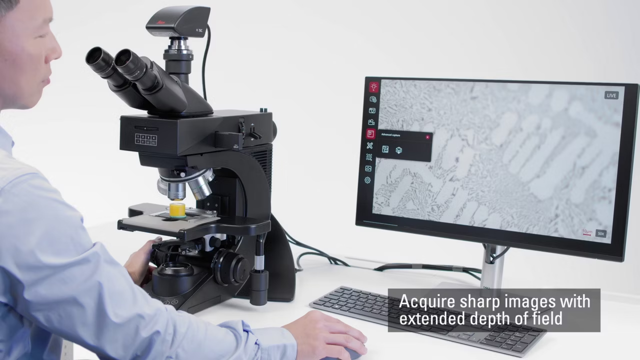
Visoria M Digital Materials Microscope for Inspection and Measurement of Fine Details and Surface Structures
Visoria M digital microscope for inspection & measurement of fine details and surface structures in metals electronics and polymers. Request a quote.
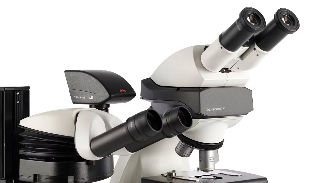
Visoria M Materials Microscope with Flexacam c5 for Inspection and Measurement of Fine Details and Surface Structures
Visoria M materials microscope with Flexacam c5 for high-resolution inspection of fine details and surface structures in industrial QA. Buy now!
Enersight Extended Depth of Field (EDOF)
Acquire sharp images of your sample with extended depth of field (EDOF). It involves the vertical Z movement of the sample to capture in-focus image information
Enersight Layer Thickness
Acquire sharp images of your sample with extended depth of field (EDOF). It involves the vertical Z movement of the sample to capture in-focus image information