Introduction to Multiplex Imaging
Definition and Scope
Multiplex imaging refers to the simultaneous detection and visualization of multiple molecular targets within a single biological sample. It broadens the scope of information that can be collected from a biological sample at once.¹ Related concepts include:
- High-plex imaging: The ability to detect dozens or hundreds of markers in one assay, providing a comprehensive molecular profile²
- Multi-channel imaging: Acquisition of multiple optical channels (fluorescence, brightfield or other modalities) with multiple detectors or filters to visualize distinct markers simultaneously³
- Multi-omics imaging: Integration of different molecular layers, such as transcriptomics, proteomics and metabolomics, within the spatial context of tissues⁴
Technological Evolution
The complex and heterogeneous composition of tissue and tumor microenvironments required a progression from single-marker assays, which detect one protein or molecule at a time, to multi-target detection platforms capable of analyzing numerous molecules simultaneously. Simultaneously, advances in spatial biology, immunofluorescence, and imaging methods have empowered detailed localization of biomolecules within intact cellular and tissue architectures. These innovations demonstrate significant potential for clinical diagnostics and translational research by accelerating biomarker discovery, patient stratification and fostering a deeper understanding of complex biological systems.¹
Types and Modalities of Multiplex Imaging
Optical and Fluorescence-Based Imaging
Optical multiplex imaging captures multiple signals from different molecular markers using light-based detection methods with high spatial resolution. It is frequently used in routine histology workflows.⁵ On the other hand, fluorescence imaging visualizes multiple markers using several fluorophores with different excitation/emission spectra. Fluorophores with overlapping spectra are handled with spectral unmixing techniques to separate signals, improving accuracy and sensitivity.⁶
Varieties of optical and fluorescence-based multiplex imaging include:
- Confocal multiplex imaging, which enhances resolution and reduces background by optically sectioning thick samples with a pinhole-based detection system⁷
- 3D multiplex imaging, which extends confocal or light-sheet microscopy into spatial analysis, allowing precise mapping of markers within intact specimens⁸
- Whole-slide multiplex immunofluorescence (mIF) empowers high-throughput analysis of entire tissue slides, facilitating biomarker quantification and spatial pattern analysis in clinical and research applications⁹
Label-Based Multiplexing
Label-based multiplexing involves tagging techniques for the simultaneous detection of multiple markers. Types of label-based multiplex imaging are:
- Antibody-based multiplex imaging with tagged antibodies to detect multiple markers ¹⁰
- Multiplex immunofluorescence (mIF) using multiple fluorophores for simultaneous detection ¹¹
- Multiplex immunohistochemistry (mIHC) using chromogens that produce color upon a physical or chemical alteration ¹²
- Cyclic immunofluorescence and iterative staining multiplexing use repeated staining, bleaching and imaging cycles to streamline the analysis of diverse tissue samples¹³
Mass Spectrometry Multiplex Imaging
Combining mass spectrometry with multiplex imaging enables the spatial mapping of multiple biomarkers in tissue microenvironments. Imaging mass cytometry is one such integrative method that applies metal-tagged antibodies to detect high numbers of biomarkers. Using metal-tagged antibodies instead of fluorophores eliminates the risk of interference due to spectral overlaps or autofluorescence. Mass spectrometry imaging (MSI) employs molecular mass-based detection to analyze the spatial distribution of various molecular species. These methods allow quantification of multiple proteins and their visualization, revealing significant structural details, such as post-translational modifications.⁴
Featured Product
Multiplex Imaging and Analysis

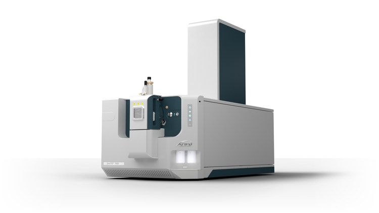
ZenoTOF 7600 System for Mass Spectrometry
A high-resolution mass spectrometry solution that combines powerful MS/MS sensitivity, fragmentation technology and a step-change in data independent acquisition
Omics-Integrated and High-Plex Imaging
Multiplex imaging can integrate transcriptomics, proteomics and metabolomics data to map RNA, proteins and metabolites with spatial context. Omics-integrated high-plex imaging facilitates the mapping of thousands of markers in tissue sections, providing insight into tissue microenvironments.¹⁴
Supporting Technologies and Key Concepts
Several technologies form the backbone of multiplex imaging.
Many multiplex imaging modalities rely on highly specific antibodies to target biomarkers through conjugation to detectable tags, such as chromogens, fluorophores and metals.¹⁰ Furthermore, high-throughput proteomics can quantify and localize proteins across tissues at single-cell resolution.¹⁵ Pipelines combining proteomics and multiplex imaging streamline the several steps, starting with image acquisition and preprocessing, segmentation and feature extraction, followed by statistical or machine learning–based analyses. These pipelines should emphasize robustness and reproducibility, especially for clinical diagnostics applications, but must be flexible enough to accommodate high-throughput for large-scale research and clinical applications.¹⁶
Complexities and Challenges in Image Analysis
Multiplex imaging is more complicated and challenging than singular biomarker detection in many ways.
High-Dimensional Data Interpretation
Multiplex imaging generates vast amounts of high-dimensional data from tissue sections, with dozens or even hundreds of markers per sample. This complexity requires specialized computational approaches to extract meaningful biological insights while accounting for signal overlap, tissue heterogeneity and sample variability.¹⁷
Modern imaging data classification tools, such as clustering algorithms and phenotyping, can overcome the challenges of data size and complexity. These tools assist in categorizing cells into subtypes based on multiplex data. Furthermore, quantitative imaging tools generate additional insight by measuring marker intensity, co-localization and spatial relationships.¹⁸
Spatial and Computational Analysis
Advancements in imaging technologies have enhanced spatial resolution, but analyzing subcellular details across entire tissues poses computational and storage challenges. A key challenge is standardizing multiplex image analysis, as differences in staining protocols, imaging hardware and analysis software can impact reproducibility and cross-study comparisons.¹⁹
Spatial analysis workflows can be difficult to work with, as they comprise rigorous image registration, segmentation and quantification. Although AI/ML-driven spatial analysis can be adopted to automate these steps, extensive training datasets and careful validation are required to avoid bias.¹⁹
Image Processing and Workflow Optimization
Multiplex imaging faces imaging-related challenges, ranging from autofluorescence and uneven illumination to tissue distortion. Furthermore, meticulous staining optimization is required to minimize signal bleed-through. On the other hand, multiplex assay development demands iterative refinement of antibody panels, fluorophore combinations and imaging modalities to maximize data quality.¹⁹
Workflow optimization in multiplex imaging faces several key challenges during several steps, from sample preparation to image analysis and segmentation, demanding end-to-end workflow solutions for standardization.²⁰
Advanced Computational Technologies
AI/ML-driven technologies can be leveraged to automate multiplex imaging workflows and improve data interpretation.¹⁸ Deep learning and convolutional neural networks (CNNs) can analyze complex tissue structures, enabling automated segmentation and pattern recognition.²¹ AI/ML methods help interpret high-dimensional multiplex data, classify cell types and uncover phenotypic patterns, essential for patient stratification. In addition, combining AI/ML with quantitative imaging improves reproducibility and cross-study comparability.¹⁸ Collectively, AI-driven analysis of multiplex imaging data cultivates model robustness, reduces variability and facilitates translation of the findings into clinical settings.
Applications of Multiplex Imaging
Cellular Analysis and Phenotyping
Multiplex imaging can improve biomarker research and target discovery by generating detailed cell phenotyping and classification. This helps researchers distinguish cell types and states within complex tissues. Multiplexed protein marker analysis and quantification can reveal comprehensive information about tissue microenvironments, advancing our understanding of cell–cell interactions and tissue heterogeneity.¹
Oncology and Tumor Microenvironment
Multiplex imaging is particularly valuable for oncology, as tumor development relies heavily on the interplay between tumor cells and other components in distinctly heterogeneous tumor microenvironments (TME).¹⁷ Multiplex analysis can reveal immune response patterns, as the biomarkers on different immune cells indicate whether they exhibit activating (i.e., cytotoxic) or immunosuppressive phenotypes. These insights guide researchers when designing and optimizing immunotherapy strategies.²²
Clinical Diagnostics and Pathology
In clinical settings, multiplex imaging improves the accuracy and level of detail from tissue biomarker and section analysis, which is essential for precision diagnostics. Simultaneously, several markers can be discovered and analyzed, helping clinicians conduct therapeutic response profiling and patient stratification.²³
Conclusion
Multiplex imaging harbors tremendous research and clinical benefits. Detecting multiple biomarkers within a single tissue sample supports a more holistic outlook on disease mechanisms and patient-specific tissue models, unlike the reductionist single-marker assays. Thus, it improves diagnostic accuracy and predictive power when monitoring patient responses to targeted therapies and immunotherapies.
Pipelines integrating multiplex imaging can close the gap between research and clinic, streamlining the translation of biomarker discovery into diagnostics and therapeutic evaluation. These workflows show great promise for advancing precision medicine.
See how Danaher Life Sciences can help
FAQs
What is multiplex imaging, and how does it work?
It is a technique that detects and visualizes multiple molecular targets within a single tissue sample, preserving spatial context. It uses labeled antibodies or probes in optical, fluorescence or mass-based detection to simultaneously capture signals from several markers.
What is multiplex immunofluorescence (mIF)?
mIF employs fluorophore-conjugated antibodies and advanced imaging systems to detect multiple proteins in one assay.
What are the main types of multiplex imaging?
Common types include optical/fluorescence-based imaging, mass spectrometry imaging and cyclic or iterative staining platforms.
What is high-plex imaging?
High-plex imaging involves simultaneously profiling dozens or hundreds of biomarkers for comprehensive tissue characterization.
How is mass spectrometry used in multiplex imaging?
Techniques like imaging mass cytometry detect metal-tagged antibodies, allowing high-plex, label-based analysis without spectral overlap.
References
- Semba T, Ishimoto T. Spatial analysis by current multiplexed imaging technologies for the molecular characterisation of cancer tissues. Br J Cancer 2024;131(11):1737-1747.
- Aung TN, Bates KM, Rimm DL. High-plex assessment of biomarkers in tumors. Mod Pathol 2024;37(3):100425.
- Arridge SR, Ehrhardt MJ, Thielemans K. (An overview of) Synergistic reconstruction for multimodality/multichannel imaging methods. Philos Trans R Soc A 2021;379(2200):20200205.
- Zhang H, Lu KH, Ebbini M, Huang P, Lu H, Li L. Mass spectrometry imaging for spatially resolved multi-omics molecular mapping. npj Imaging 2024;2(1):20.
- Ugolini F, Pasqualini E, Simi S, Baroni G, Massi D. Bright-field multiplex immunohistochemistry assay for tumor microenvironment evaluation in melanoma tissues. Cancers (Basel) 2022;14(15):3682.
- Chen K, Yan R, Xiang L, Xu K. Excitation spectral microscopy for highly multiplexed fluorescence imaging and quantitative biosensing. LSA 2021;10(1):97.
- Lochhead JJ, Williams EI, Reddell ES, Dorn E, Ronaldson PT, Davis TP. High resolution multiplex confocal imaging of the neurovascular unit in health and experimental ischemic stroke. Cells 2023;12(4):645.
- Cho W, Kim S, Park Y-G. Towards multiplexed immunofluorescence of 3D tissues. Mol Brain 2023;16(1):37.
- Doyle J, Green BF, Eminizer M, Jimenez-Sanchez D, Lu S, Engle EL, et al. Whole-Slide Imaging, Mutual Information Registration for Multiplex Immunohistochemistry and Immunofluorescence. Lab Invest 2023;103(8):100175.
- Hickey JW, Neumann EK, Radtke AJ, Camarillo JM, Beuschel RT, Albanese A, et al. Spatial mapping of protein composition and tissue organization: a primer for multiplexed antibody-based imaging. Nat Methods 2022;19(3):284-295.
- Sheng W, Zhang C, Mohiuddin T, Al-Rawe M, Zeppernick F, Falcone FH, et al. Multiplex immunofluorescence: a powerful tool in cancer immunotherapy. Int J Mol Sci 2023;24(4):3086.
- Harms PW, Frankel TL, Moutafi M, Rao A, Rimm DL, Taube JM, et al. Multiplex immunohistochemistry and immunofluorescence: a practical update for pathologists. Mod Pathol 2023;36(7):100197.
- Eng J, Bucher E, Hu Z, Zheng T, Gibbs SL, Chin K, et al. A framework for multiplex imaging optimization and reproducible analysis. Commun Biol 2022;5(1):438.
- Carstens JL, Krishnan SN, Rao A, Sorace AG, Seeley EH, Ferri-Borgogno S, et al. Spatial multiplexing and omics. Nat Rev Methods Primers 2024;4(1):54.
- Mund A, Brunner A-D, Mann M. Unbiased spatial proteomics with single-cell resolution in tissues. Mol Cell 2022;82(12):2335-2349.
- Zahumensky J, Malinsky J. Live cell fluorescence microscopy—an end-to-end workflow for high-throughput image and data analysis. Biol Methods Protoc 2024;9(1):bpae075.
- Liu K, Liu F, Li G, Zheng Y. Multiplex imaging analysis of the tumor immune microenvironment for guiding precision immunotherapy. Front Immunol 2025;16:1617906.
- Parra ER, Ilié M, Wistuba II, Hofman P. Quantitative multiplexed imaging technologies for single-cell analysis to assess predictive markers for immunotherapy in thoracic immuno-oncology: promises and challenges. Br J Cancer 2023;129(9):1417-1431.
- Wilson CM, Ospina OE, Townsend MK, Nguyen J, Moran Segura C, Schildkraut JM, et al. Challenges and opportunities in the statistical analysis of multiplex immunofluorescence data. Cancers (Basel) 2021;13(12):3031.
- Windhager J, Zanotelli VRT, Schulz D, Meyer L, Daniel M, Bodenmiller B, et al. An end-to-end workflow for multiplexed image processing and analysis. Nat Protoc 2023;18(11):3565-3613.
- Zidane M, Makky A, Bruhns M, Rochwarger A, Babaei S, Claassen M, et al. A review on deep learning applications in highly multiplexed tissue imaging data analysis. Front Bioinform 2023;3:1159381.
- Zhao C, Germain RN. Multiplex imaging in immuno-oncology. J ImmunoTher Cancer 2023;11(10):e006923.
- Bollhagen A, Bodenmiller B. Highly multiplexed tissue imaging in precision oncology and translational cancer research. Cancer Discov 2024;14(11):2071-2088.