Mica Microhub
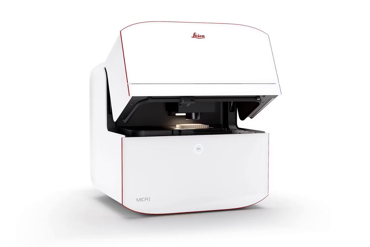
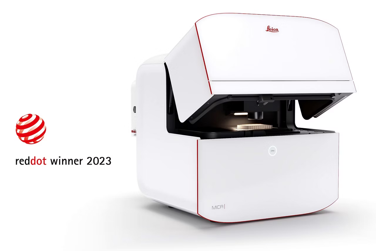
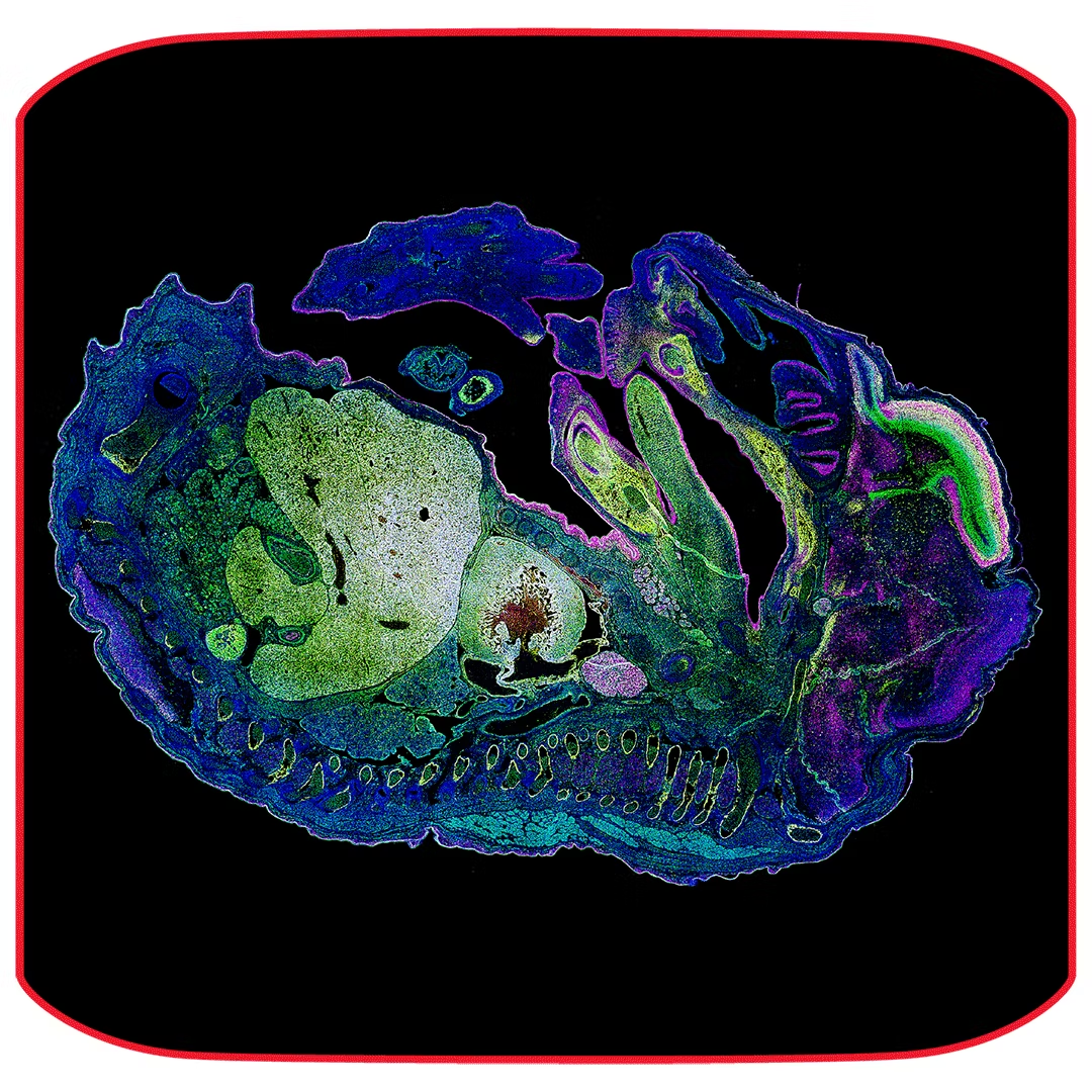
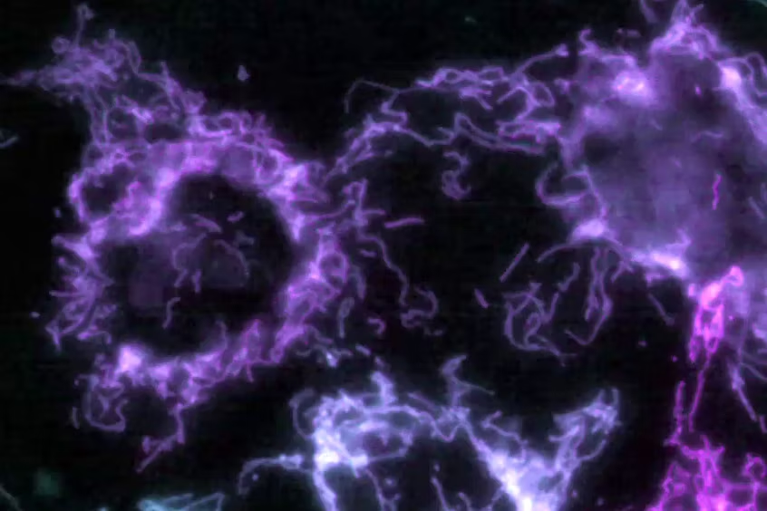
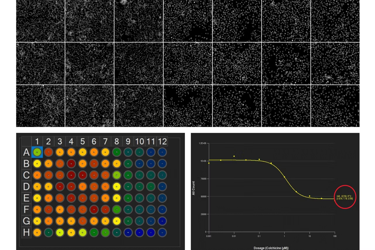
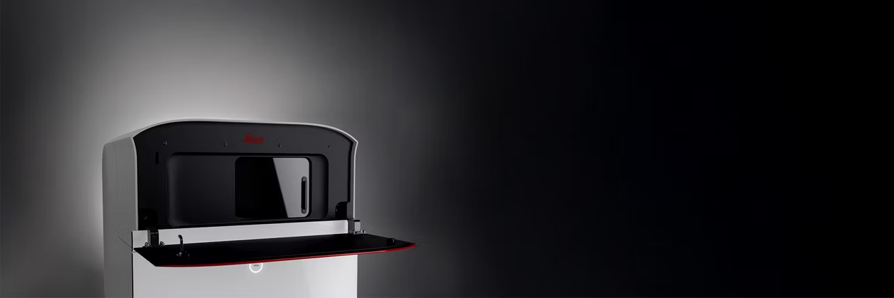
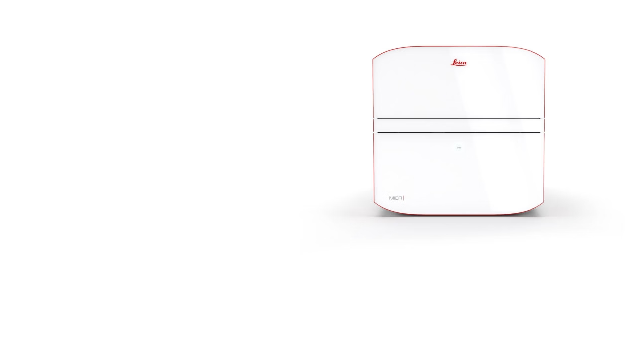
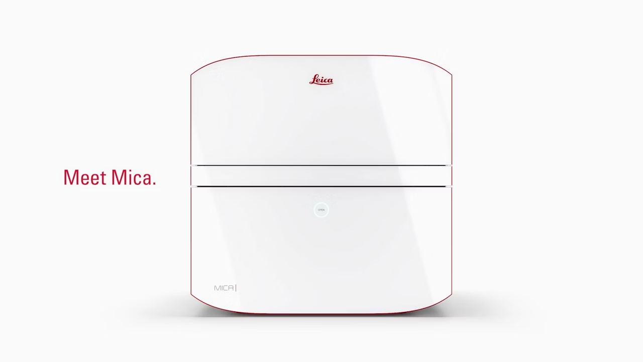
The world’s first Microhub. More than a highly automated microscope, Mica unites widefield and confocal imaging in a sample protecting, incubating environment. With the simple push of a button, you have everything you need - all in one place - to supercharge fluorescence imaging workflows and get meaningful scientific results faster.
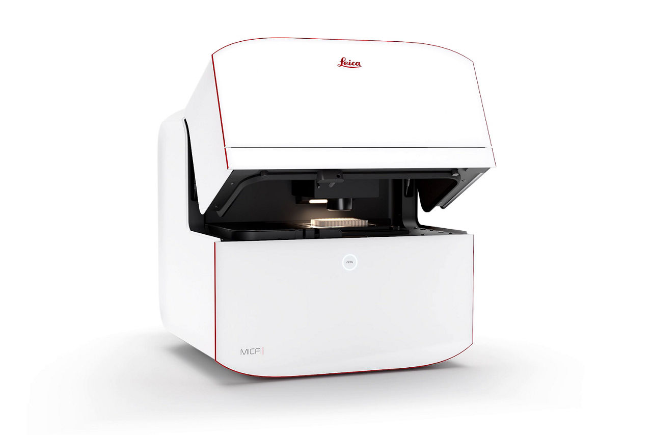
Mica Widefield Microhub Automated Microscope
Simplify advanced fluorescence imaging through extensive intelligent automation, AI‑supported analysis, and an easy-to-use interface. Now all lab members can get expert microscopy results, regardless of experience. Ideal for exploration and high-throughput fluorescence imaging of fixed samples. MICA BENEFITS: ACCESS FOR ALL: Mica requires minimal technical input and gives consistent results every time, so all lab members can leverage microscopy to make more discoveries. NO CONSTRAINTS: Get true simultaneous capture of up to four labels for fast acquisitions. RADICALLY SIMPLIFIED WORKFLOWS: Intelligent automation and AI-supported analysis of diverse samples, such as tissues on slides or well plates, enable greater efficiency and a faster track to publication.
Mica WideFocal Microhub Automated Microscope
Combine widefield and confocal imaging in an easy-to-use platform and simplify advanced fluorescence imaging. By streamlining workflows and reducing complexity through intelligent automation, AI-supported analysis, you can get expert microscopy results without all the training. Ideal for high-resolution imaging of thick or complex samples. MICA BENEFITS: ACCESS FOR ALL: Mica requires minimal technical input and gives consistent results every time, so all lab members can leverage microscopy to make more discoveries. NO CONSTRAINTS: Visualize in widefield and switch effortlessly to confocal without ever moving your sample. Get true simultaneous capture of up to four labels for fast acquisitions. RADICALLY SIMPLIFIED WORKFLOWS: Intelligent automation and AI-supported analysis of diverse samples, such as tissues on slides or well plates, enable greater efficiency and a faster track to publication.
Mica Widefield Live Cell Microhub Automated Microscope
Simplify advanced live cell and fluorescence imaging. Get reliable, expert level microscopy results faster with extensive intelligent automation, AI-supported analysis, and an interface designed for ease of use. Perform long-term live cell experiments up to 4x faster than traditional widefield systems, capturing events that may previously have been missed. Easily get quality images as Mica automatically corrects the focus during experiments. Minimise phototoxicity and maintain optimal cell health with full environmental control. Ideal for long-term live-cell studies at near physiological conditions. MICA BENEFITS: ACCESS FOR ALL: Mica requires minimal technical input and gives consistent results every time, so all lab members can leverage microscopy to make more discoveries. NO CONSTRAINTS: Get true simultaneous capture of up to four labels without spatiotemporal mismatch; at near-physiological conditions for live cells, or with fixed samples. RADICALLY SIMPLIFIED WORKFLOWS: Intelligent automation and AI-supported analysis of diverse samples, such as tissues on slides or well plates, enable greater efficiency and a faster track to publication.
Mica WideFocal Live Cell Microhub Automated Microscope
Combine widefield and confocal imaging in an incubating environment and simplify advanced live cell and fluorescence imaging. Extensive intelligent automation, AI-supported analysis, and an easy-to-use interface help you achieve expert microscopy results without all the training. Perform long-term live cell experiments up to 4x faster than traditional widefield systems, capturing events that may previously have been missed. Easily get quality images as Mica automatically corrects the focus during experiments. Minimise phototoxicity and maintain optimal cell health with full environmental control. Ideal for long-term and dynamic live cell studies of organoids, spheroids, and 3D cultures. MICA BENEFITS: ACCESS FOR ALL: Mica requires minimal technical input and gives consistent results every time, so all lab members can leverage microscopy to make more discoveries. NO CONSTRAINTS: Visualize in widefield and switch effortlessly to confocal without ever moving your sample. Get true simultaneous capture of up to four labels without spatiotemporal mismatch – at near-physiological conditions for live cells, or with fixed samples. RADICALLY SIMPLIFIED WORKFLOWS: Intelligent automation and AI-supported analysis of diverse samples, such as tissues on slides or well plates, enable greater efficiency and a faster track to publication.
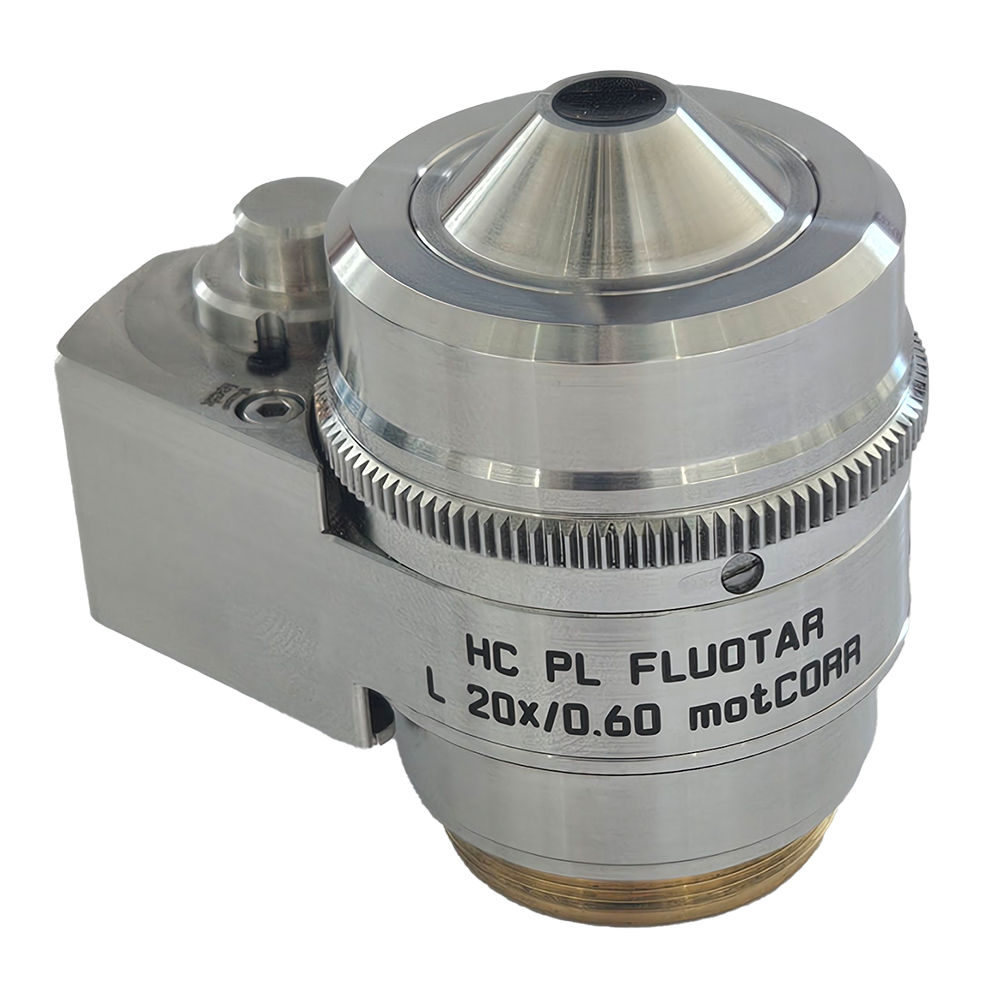
Mica 20x LWD Objective with Service Installation
20x long working distance objective for imaging on glass and plastic bottom carriers To penetrate deeper into samples using everyday standard plastic-bottom multi well plates, this Long Working Distance (LWD) objective has been optimized for use with Mica. • No need to sacrifice image quality • No need for costly glass-bottom alternatives • Avoid the frustration of switching carriers and re-optimizing your sample preparation protocols Why is long working distance important? • Focus deeper, capturing clearer images even with thicker or non-adherent samples such as organoids, spheroids and zebrafish larvae • Get increased compatibility with high-skirt well plates • Improve access to outer wells of multi well plates Please note: This objective requires service installation to ensure correct installation and calibration. A half-day service visit by our expert team is added to ensure optimal performance and system compatibility.
No constraints - 4x more data with 100% correlation

The Microhub enables you to simultaneously capture all 4 labels of different structures in a single acquisition for widefield or confocal, without ever moving your sample. This overcomes the spatiotemporal mismatch between labels of moving objects during sequential acquisition. All are powered by our patented FluoSync technology, a fast and gentle method for multicolor fluorescence imaging.
No constraints - Select the right modality in real time
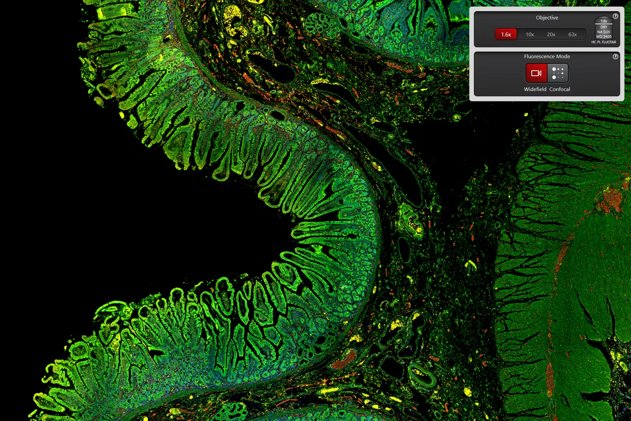
Mica unifies transmitted and fluorescence light imaging modalities. You can select from multiple imaging modalities all within one Microhub, including widefield, confocal, THUNDER imaging, LIGHTNING, Z-stacks, time-lapse and more.
This enables you to
- generate fast overviews with widefield at low magnification
- gradually zoom in on the regions of interest
- switch to confocal when and where needed without ever moving the sample to a different system
No constraints - Achieve physiological-like conditions thoughout your experiments
Live cell experiments require the cells to be in optimal shape. Typically, 2D and 3D cells in media require the temperature and the pH (via CO2) in the environment to be controlled. Stable nutrition and ion concentrations require the evaporation to be minimal. Some experiments even demand the O2 to be mimicked closer to physiological levels. Mica can provide the right conditions in the live cell configuration.
- Mica is an incubator: The entire encapsulated inner sample space can be climate controlled (temperature, CO2 and humidity regulation) and offers ideal conditions for short and long-term live cell observation.
- From dark to light: Mica also enables you to enjoy a brightly lit lab—freeing you from the constraints of sitting in a dark room for hours monitoring your experiment.
Radically simplified workflows

Intelligent automation and AI-supported analysis enable greater efficiency and a faster track to publication.
- Reduce over 60% of process steps through system intelligence
- Reduce time and effort from sample to insight by simplifying your entire workflow
- Enable 100% reproducibility and repeatability throughout your experiment
"We work with pancreatic cancer organoids so, whenever we find interesting structures and want to zoom in and have higher resolution, then, with basically one click, we can switch from the epifluorescence to confocal mode."
Dr. Anna Pastucha, Light Microscopy Facility Manager, Center for Functional Protein Assemblies, Technical University of Munich, Germany
See Mica in action
Now you can focus on your science, not figuring out your microscope.
Access for all

Everyone can now leverage microscopy to make more discoveries.
Mica provides a clear sample overview and allows you to easily change observation conditions with just a few clicks.
- 85% fewer steps to the first image
- 33% less time to the first image
- 50% of the training time
This changes everything.
Radically simplified workflows

Intelligent automation and AI-supported analysis enables greater efficiency and a faster track to publication
Access for all
Mica provides a clear sample overview and allows to easily change observation conditions with just a few clicks
No constraints

4x more data with 100% correlation. Select the right modality in real time. Achieve physiological-like conditions thoughout your experiments
| Contrasting Method(s) | Brightfield, Fluorescence, Integrated Modulation Contrast |
| Long Working Distance | No |
| Microscopy Technique | Confocal Microscopy |
| Microscope Stage Type(s) | Motorized |
| Microscope Camera Option(s) | Integrated |
| Microscopy Software Available | 2D Anaylsis, 3D Acquisition (Z-Stacking), 3D Analysis, Deconvolution, Documentation, Live Cell Imaging, Multichannel Acquisition, Multiposition Acquisition, Super Resolution, Tile Stitching, Time Lapse Imaging |
| Has Eyepiece | No |
| Microscope Type(s) | Inverted Microscope, Inverted Microscope, Digital Microscope |
| Has Resonance Scanner | No |
| Microscope Laser Type | Fixed Wave Length |
| Microscope Environmental Control | Box |
| Microscope Group | Light Microscope |
| Application(s) | Cell Biology;Cancer Research;Live Cell Imaging;Organoids and 3D Cell Culture;Model Organisms in Research;Neuroscience |
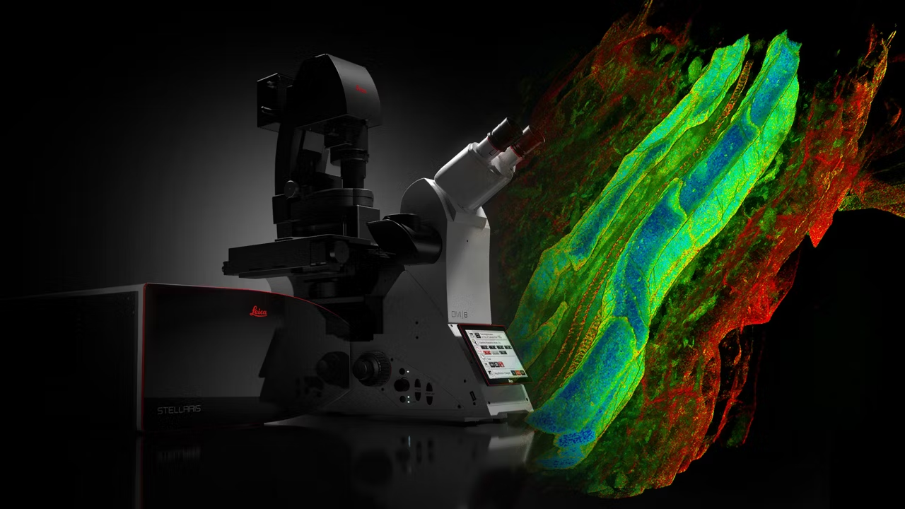
STELLARIS Confocal Microscope Platform
STELLARIS delivers confocal imaging with TauSense WLL & AI-powered Aivia analysis for deeper insights in less time. Request a quote now.

Aivia AI Image Analysis Software
Explore AI Image Analysis Software transforms image processing with up to 74% faster analysis time. Achieve high-quality, reproducible results effortlessly.

Warranty Extension+
Full microscope coverage with annual maintenance, 3 business day on-site response, unlimited parts, and all labor included. Request a quote today!
RemoteCare Smart Monitoring Option
Real time remote monitoring tracks key operating conditions, detects issues early and resolves over 30% of cases remotely. Request a quote today.