THUNDER Imager Tissue
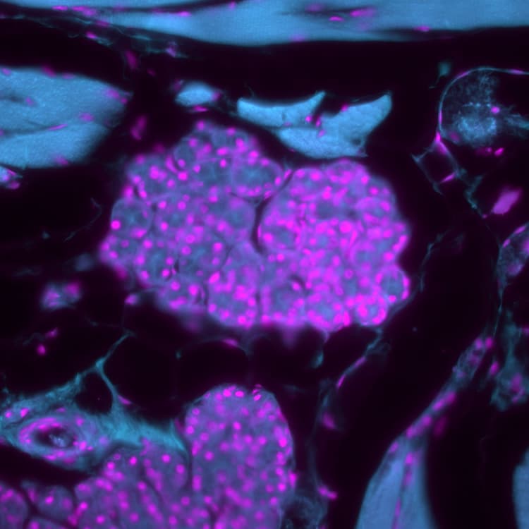

Decode 3D biology in real time*
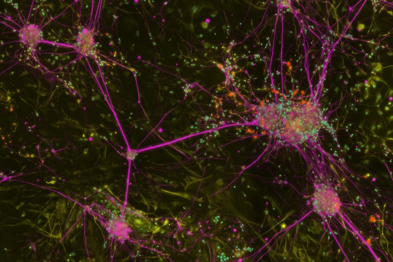
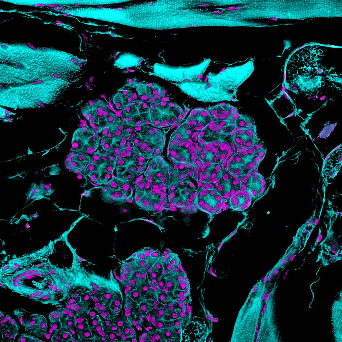
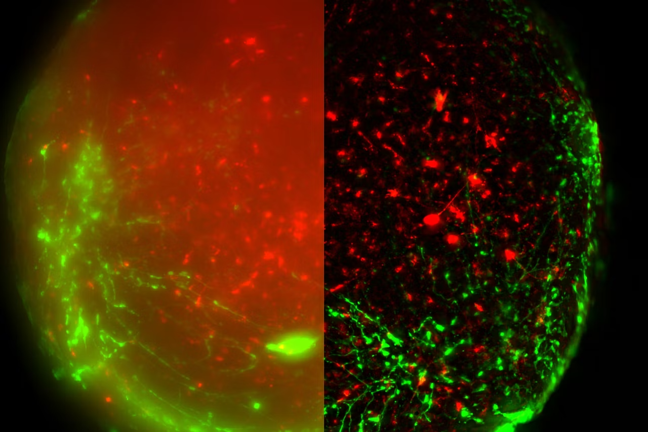
The THUNDER Imager Tissue allows real-time fluorescence imaging of 3D tissue sections typically used in neuroscience and histology research. Acquire rich, detailed images of thick tissues, free from the haze ofout-of-focus blur.
With Computational Clearing, an innovative Leica technology, fine structures deep in tissues can be resolved thanks to Computational Clearing, an innovative Leica technology. Image detailed morphological structures like axons and dendrites of neurons in a brain slice. The high image quality, even with thick tissue sections, is combined with the well-known speed, fluorescence efficiency, and ease of use of a widefield microscope.
Gain these advantages using a THUNDER Imager Tissue for your research:
- Get computationally cleared images directly in your live preview with THUNDER Live
- Rapidly acquire blur-free images showing finest details of the morphology, even deep within thick specimens
- Fast sample overviews with full electronic synchronization of the hardware
- Image and analyze challenging tissue sections with an intuitive and integrated workflow
*in accordance with ISO/IEC 2382:2015
Begin every experiment with confidence

Perform high resolution tissue imaging on thick samples without struggling to find your region of interest.
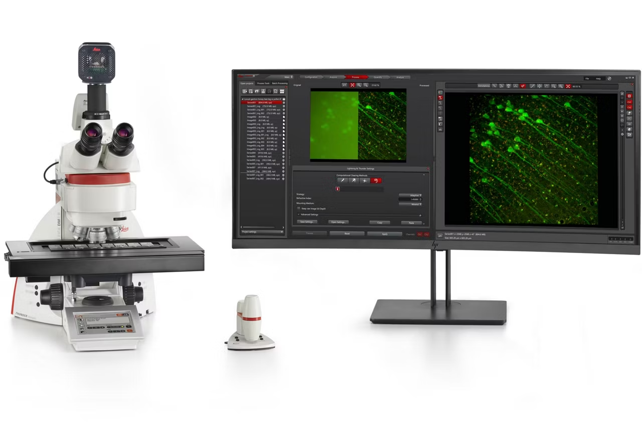
The new THUNDER Live add-on visualizes a computationally cleared image instantly in the live view and allows you to optimize ICC parameters by using live image feedback.
Your benefits with THUNDER Live:
- Computationally cleared images directly in your live preview.
- Reduced time to optimal results with immediate visual feedback.
- Intuitively and quickly find the best THUNDER ICC parameters on the fly while still in live view.
- Easy selection of important regions of interest even with thick samples.
Faster imaging
Increase your slide scanning experiment speed and perform fast and efficient multicolor acquisiton. You'll be able to assess more fluorescent markers with less photobleaching.
The advantages with the accelerated tissue imaging
- Scan your samples, such as slides, faster with fully synchronized hardware control
- Multicolor acquisitions are more than two times faster when using the Synapse controller
- Discover contextual information unbelievably fast with multicolor, whole slide scanning experiments
- Evaluate more markers, with less photobleaching, through precise control of multiline LED light sources
Resolve fine details of challenging specimens
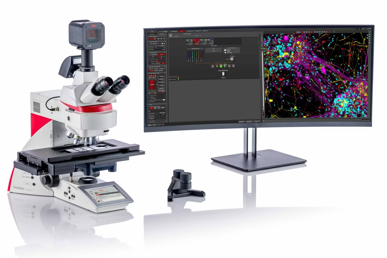
THUNDER Imager Tissue delivers instantly cleared fluorescent images with fine details of your multi-color tissue sections. Simply turn on the THUNDER Imager and start imaging! The unique Computational Clearing method will use optimal parameters to produce expert results. It does this automatically with no need for calibration or user interaction.
Achieve optimal fluorescence and contrast settings instantly via the patented Leica fluorescence intensity (FIM) and contrast managers. Choose from a range of objectives, optimized for specific applications, to ensure outstanding results even with challenging specimens.
"THUNDER Imager Tissue configurations"

Choose the configuration which best meets your requirements:
- With the Computational Clearing functionality, you can have a fully automated tissue imaging system for recording multi-color images. Including THUNDER Live for easy previews*.
- Our adaptive deconvolution can be combined with Computational Clearing to achieve brilliant imaging with 3D samples. Achieve high contrast, 3-dimensional images with maximized resolution and clarity.
The Ultimate Combination: Laser Microdissection and THUNDER Imaging
THUNDER can be combined with additional Laser Microdissection (LMD) capabilities on the same system. Since both systems are based on our upright microscopy stand, the LMD and THUNDER Imager Tissue can be combined to offer:
- Space savings with one system for multiple tasks
- Brilliant fluorescence imaging with THUNDER using LAS X
- Laser Microdissection using the unique LMD Software to visualize and mark regions of interest for subsequent dissection and collection via gravity into standard vials ready for downstream processing such as RNAseq, NGS, MS, qPCR, microarray etc.
| Microscope Platform | DM6 B |
| Display/Touch screen | SmartTouch controls the microscope conveniently and intuitively |
| Focus | Motorized and Mechanical |
| Stage | Mechanical, motorized and scanning stages |
| LAS X Navigator Software | Yes (optional) |
| Transmitted Light Illumination | LED |
| Transmitted Light Contrast Methods | BF, optional PH, DF, POL, DIC |
| Fluorescence Axis | Fully automated, with optional Internal Filter Wheel (IFW) and Excitation Manager |
| Fluorescence Illumination | LED, EL6000 |
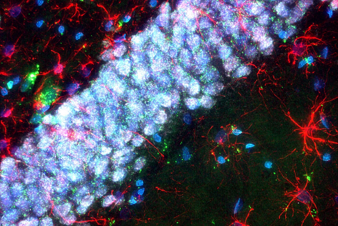
K8 Scientific CMOS Camera
Discover the K8 Scientific CMOS Camera for life science imaging. Achieve 95% quantum efficiency, low read noise and reduced phototoxicity. Request a quote!
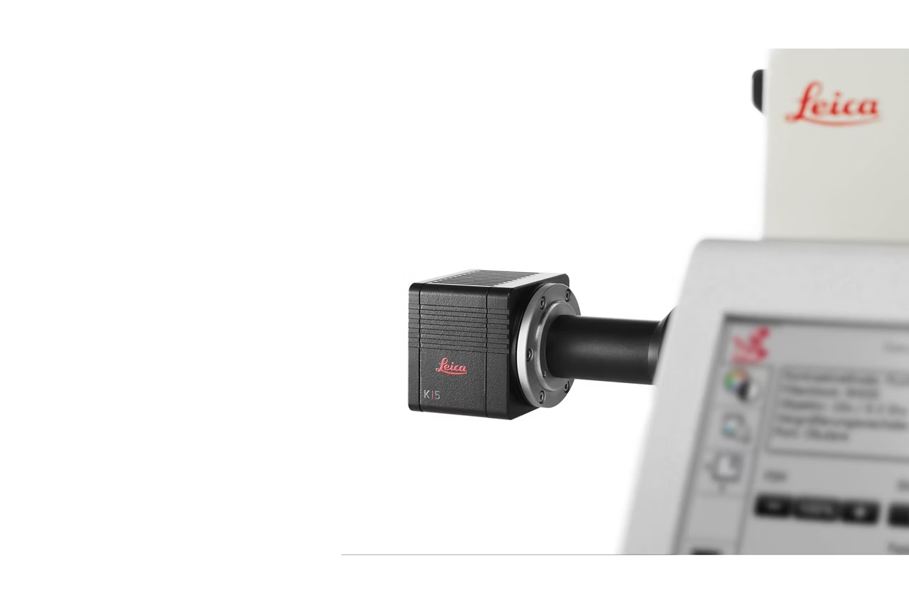
K5 Scientific CMOS Microscope Camera
Discover the K5 sCMOS Microscope Camera: 4.2MP resolution, 80% quantum efficiency. Ideal for fluorescence imaging, immunostaining, and 3D cell cultures.
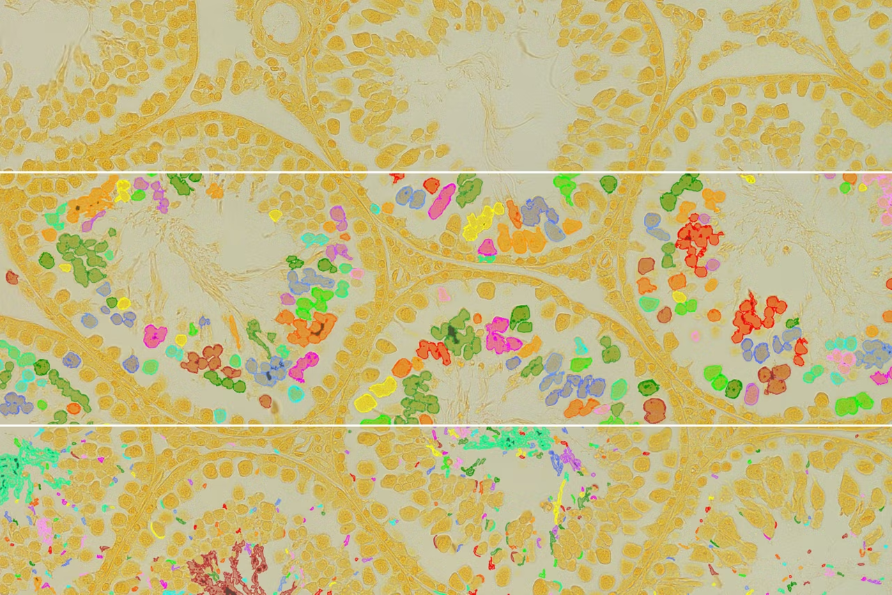
LMD Systems Laser Microdissection Microscopes
Achieve perfectly cut, contamination-free, analysis-ready dissectates with LMD7 Microscope. Well-suited for live cell culture, for cloning & re-cultivation & more.

Aivia AI Image Analysis Software
Explore AI Image Analysis Software transforms image processing with up to 74% faster analysis time. Achieve high-quality, reproducible results effortlessly.
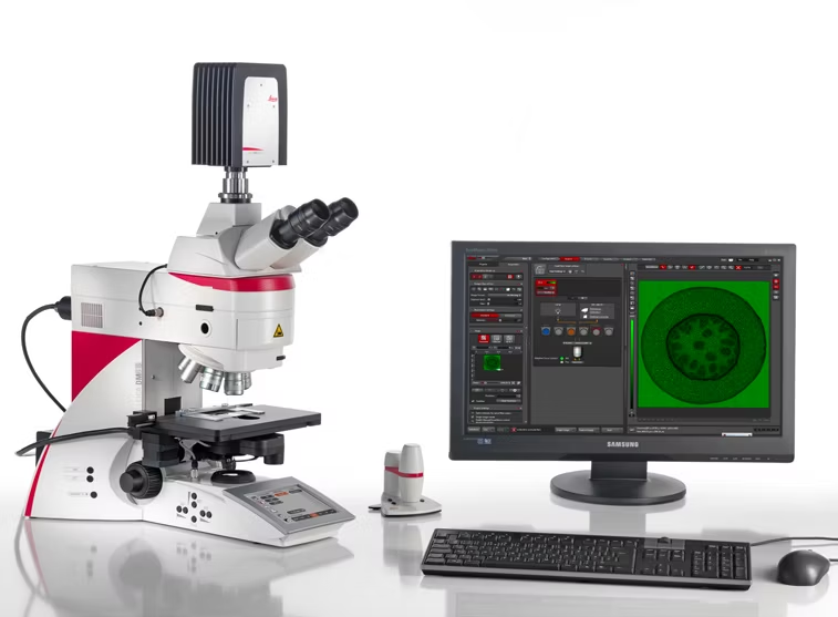
DM4 B & DM6 B Upright Microscopes
Explore the Leica DM6 B microscope to enhance workflows with LAS X Navigator sCMOS imaging & intelligent automation for flexible applications. Order now!
THUNDER Imager Model Organism
THUNDER Imager simplifies screening and imaging of live model organisms like C. elegans and Drosophila with high-speed, blur-free z-stack capability.

Warranty Extension+
Full microscope coverage with annual maintenance, 3 business day on-site response, unlimited parts, and all labor included. Request a quote today!