K5 Scientific CMOS Microscope Camera
For routine fluorescence imaging
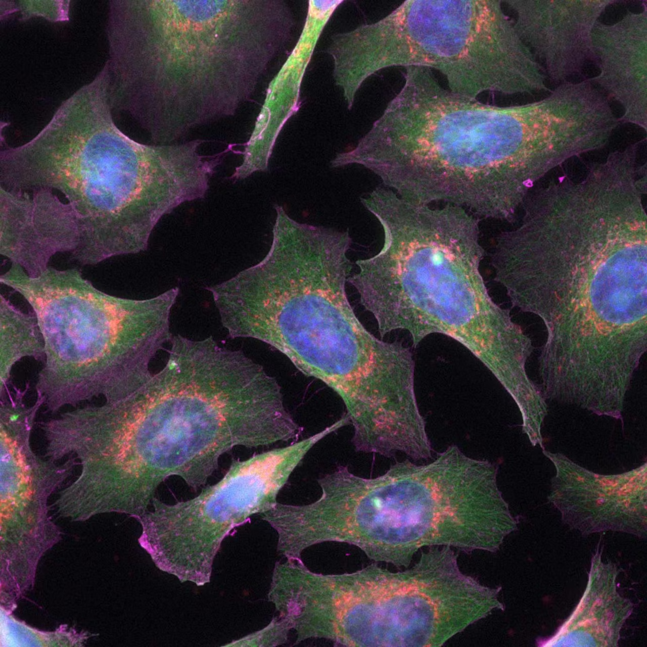
The K5 is ideal for routine fluorescence imaging applications such as immunostaining assays and 3D Cell Culture imaging.
Image a wide variety of sample types with the flexibility and power of high 4.2 megapixel resolution and fast 40 fps imaging brought to you by the K5.
Your benefits
- Bring the flexibility and power of CMOS technology into your lab and image the details of your experiment
- Efficiently segment crucial real time events
- Improve signal-to-background of your THUNDER Imager
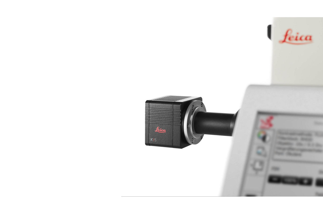
K5 Microscope Camera
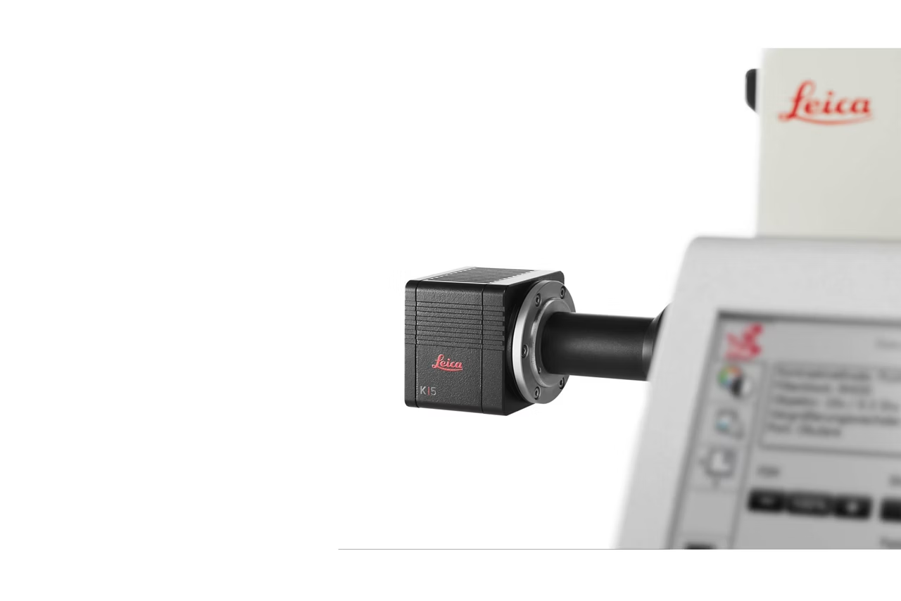
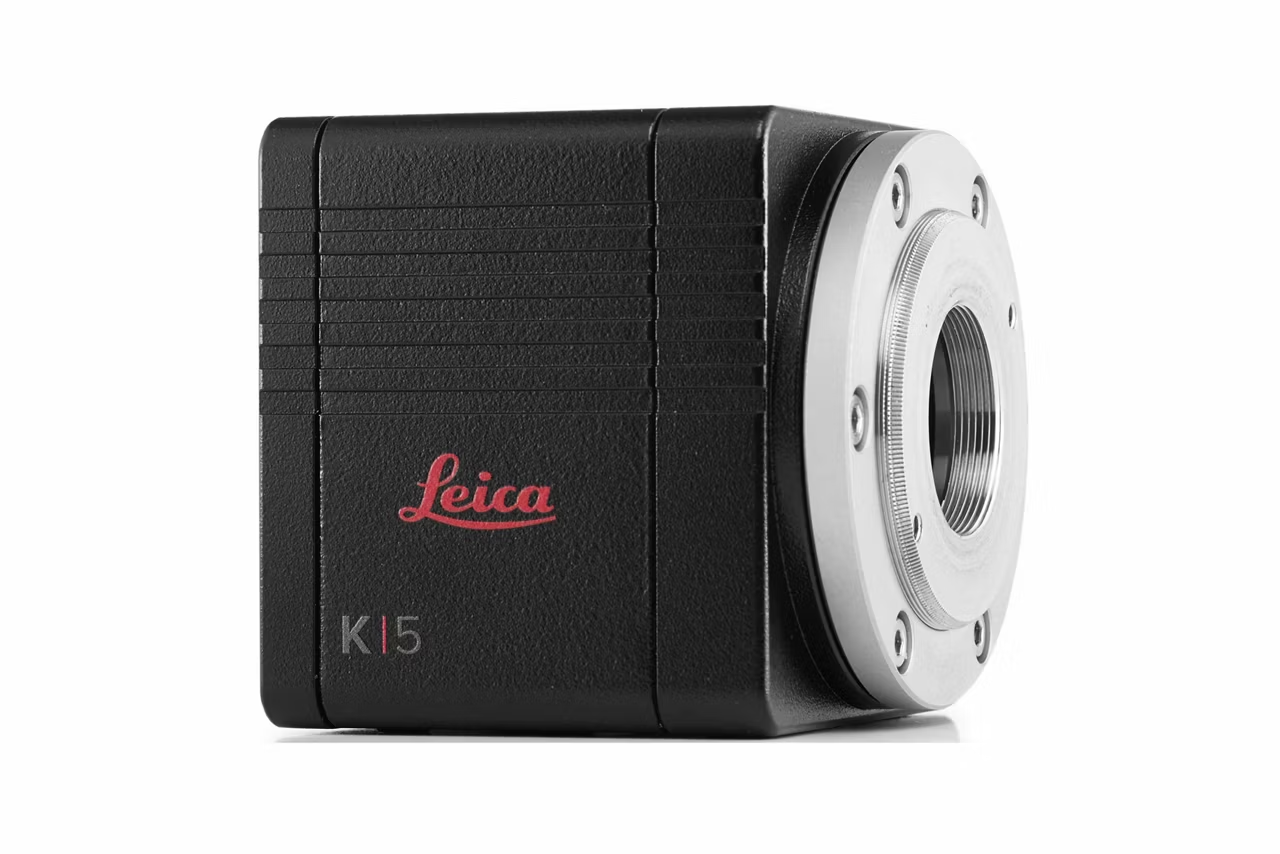
The Leica K5 - small but effective: Despite small-size dimensions of roughly 65 x 65 x 65 mm³, the new Leica k5 passive cooled sCMOS camera system provides high quantum efficiency with low readout noise making it suitable for countless applications. Trigger cable included. Main Features: - Resolution: 2048 x 2048 pixels - Pixel size: 6.5µm x 6.5µm - Superior quantum efficiency up to 80% - 21 400 : 1 dynamic range
Flexibility of imaging applications

To achieve the best imaging conditions, you can adjust the K5 camera flexible parameters to meet your sample needs:
- Image dynamic cellular events with up to 40 fps imaging
- Resolve detailed structures thanks to high 4.2 MP resolution
- Reduce exposure times and phototoxic effects with high 80% quantum efficiency.
Efficiently segment real time events
Get more biologically-relevant information with time lapse imaging thanks to the increased throughput powered by up to 40 fps observations.
Video:
Imaging cellular motility and protein dynamics in live cells requires high QE and fast frame rates. The K5‘s features make it an incredible camera for routine live cell imaging.
Here, U2OS cells expressing LifeAct-TagGFP2 were imaged for 150ms every 500ms for 5 minutes. Samples courtesy of ibidi GmbH, Gräfelfing, Germany.
Improve your images with better signal-to-background

Combine the K5‘s flexibility with the power of Computational Clearing.
Remove the haze from conventional widefield images accurately thanks to validated K5 and THUNDER Imager workflows.
Comparison:
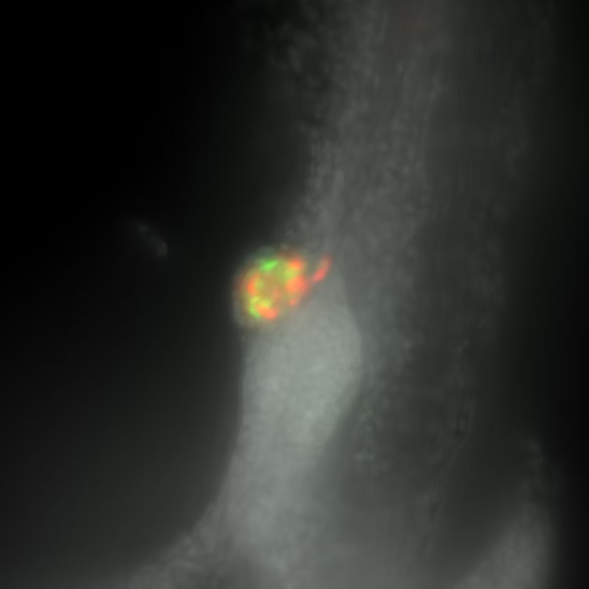
The K5 and THUNDER Imager 3D Assay enables clear identification of Alpha (GFP-green) and Beta (mCardinal-red) cells within a developing zebrafish pancreas. Imaged in the blue (Hoechst), green (GFP), and red (mCardinal) channels, this 150-image z-stack was completed with all channels within a minute.
Images courtesy of Radhan Ramadass and Yu Hsuan at the Max Planck Institute for Heart and Lung Research, Bad Nauheim, Germany.
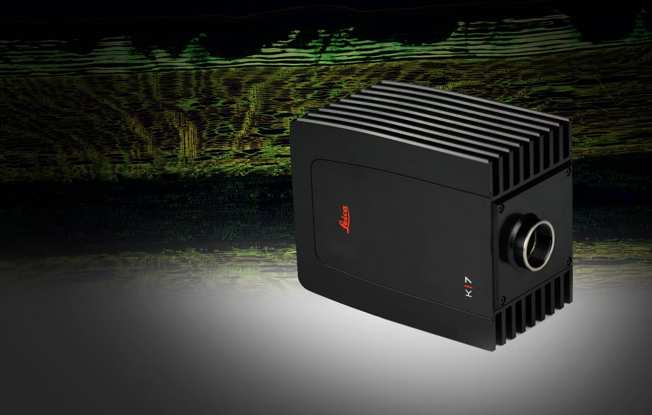
K7 Color CMOS Microscope Camera
Capture K7 color CMOS microscope camera images for fluorescence and brightfield microscopy, fast scan speeds, high temporal resolution, CMOS sensor. Get a quote!
K3C & K3M The Microscope Camera K3 series
Microscope Camera K3 series images challenging samples with precision. k3-m. Request a quote!