Leica LMD Systems Laser Microdissection Microscopes
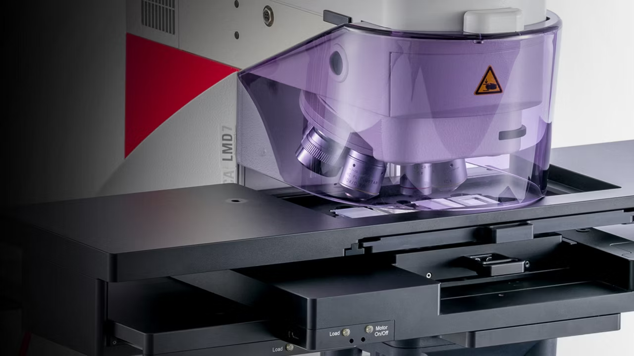
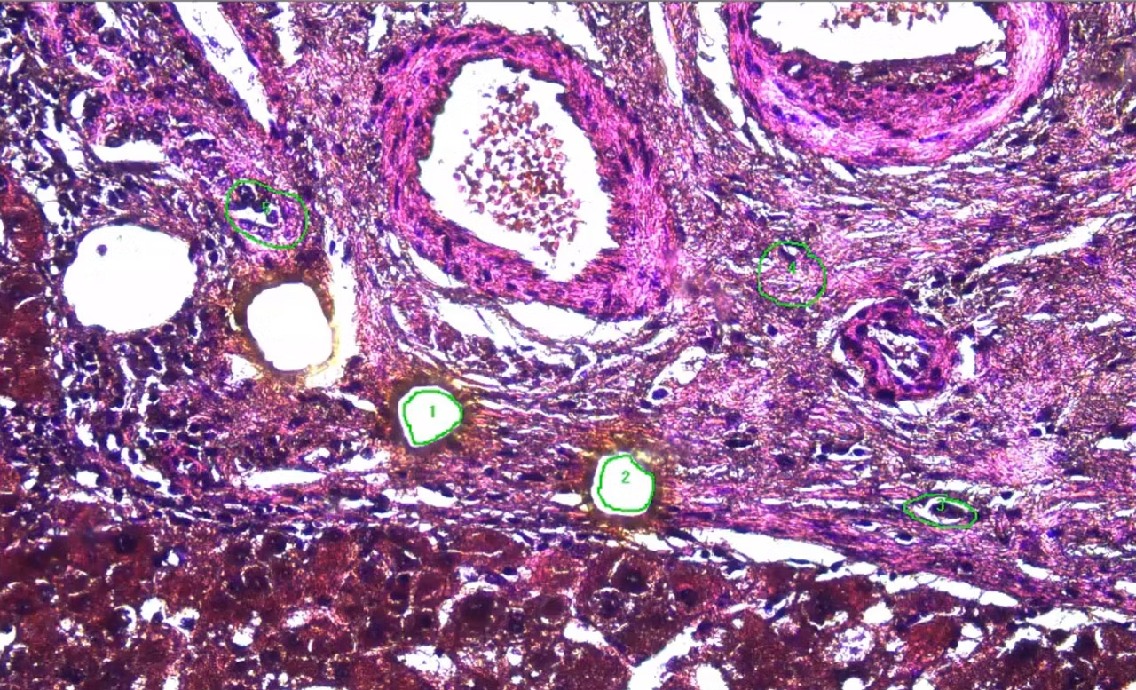
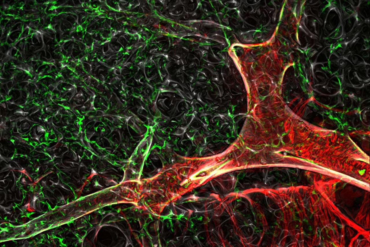
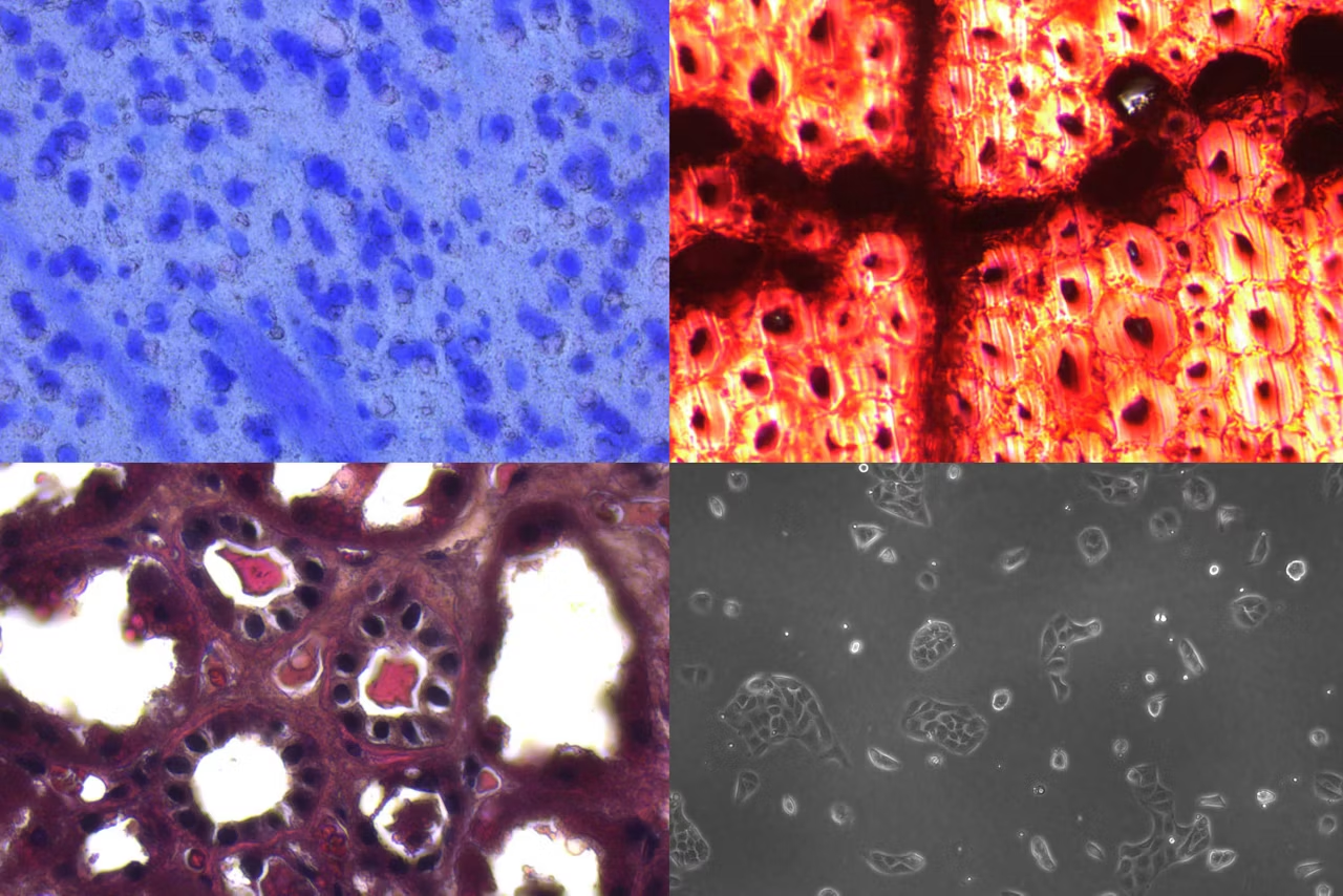
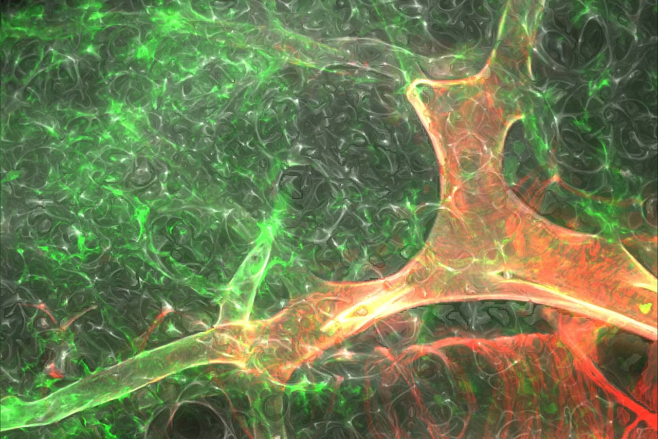
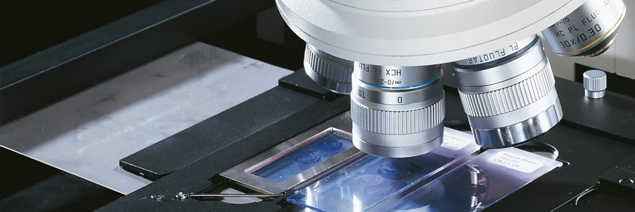
Laser Microdissection (LMD, also known as Laser Capture Microdissection or LCM) enables users to isolate specific single cells or entire areas of tissue. Leica LMD systems allow users to easily isolate Regions of Interest (ROI) from entire areas of tissue down to single cells or even subcellular structures such as chromosomes.
LMD is typically used in genomics (DNA), transcriptomics (mRNA, miRNA), proteomics, metabolomics, and even next generation sequencing (NGS). Researchers in neurology, cancer research, plant analysis, forensics or climate research rely on this method. LMD is a perfect tool for live cell culture (LCC), for cloning and re-cultivation, manipulation or downstream analysis.
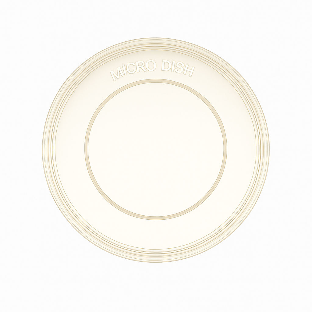
PEN Petri Dishes 4 µm (1 pack=20 pc)
Petri dishes with 4 µm thick PEN membrane bottom for cutting, pack of 20

PPS FrameSlide (4 µm), steel frames, 50
Metal frame slides with a 4 µm thick PPS membrane, pack of 50
PEN Frame Slide(4µm),steel frames,50pcs.
Metal frame slides with a 4 µm thick PEN membrane, pack of 50
POL FrameSlide, steel, 50pc RNase-free
Metal frame slides with a 0.9 µm thick POL membrane for RNA work, RNase-free, pack of 50
POL FrameSlide, steel frames, 50 slides
Metal frame slides with a 0.9 µm thick POL membrane, pack of 50
PET FrameSlide,steel frames, RNase-free
Metal frame slides with a 1.4 µm thick PET membrane for RNA work, RNase-free, pack of 50

PEN Membrane Slide(4µm),glass,50 slides
Glass slides with a 4 µm thick PEN membrane, pack of 50

µ-Slide w. membrane, 18well,ibidi,15 pcs
18-well Ibidi u-slide with 2 µm PEN membrane bottom, pack of 15
PEN MembranSlide, glass, RNase-free,50pc
Glass slides with a 2 µm thick PEN membrane for RNA work, Rnase-free, pack of 50
PEN WhiteCoat(2µm), glass, 200 pcs
Glass slides with a 2 µm thick PEN membrane, slide has writable area on one end, pack of 200
Gravity for Cleanliness
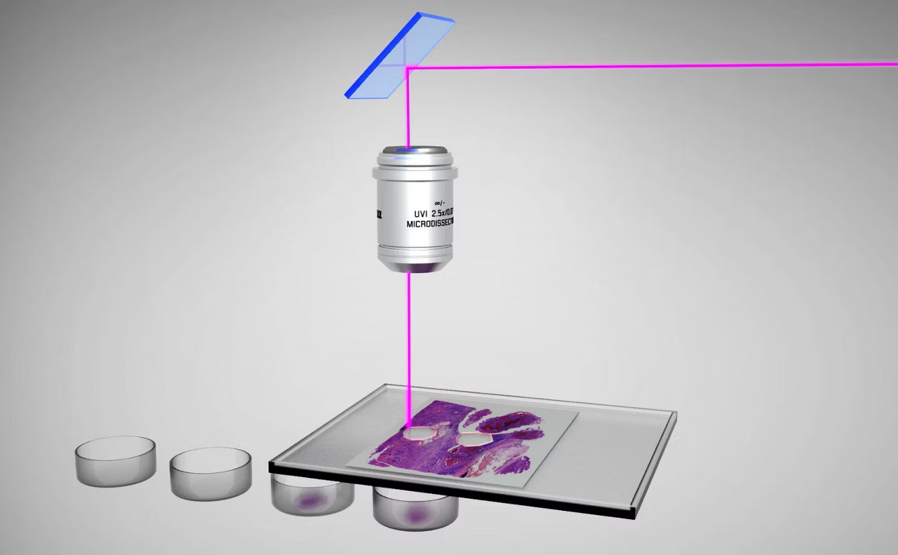
Your downstream analysis relies on contaminate-free isolates. This is why Leica LMD systems collect dissectates via gravity. Their unique laser-guidance based dissection method preserves the integrity of your isolates – contact-free and contamination-free.
1, 2, 3 – contamination-free sample!
- You choose the area of interest
- You move the laser along the area to be cut out
- The dissectate drops into the Petri dish and is ready for further analysis
The real asset: Gravity always works.
We Move the Laser, not the Sample

Try writing your name on a piece of paper by moving the paper instead of the pen. Hard to do? That’s why we move the laser, not the sample for laser microdissection.
Only Leica Microsystems uses high-precision optics to steer the laser beam along the desired cut line on the tissue with prisms. That means that the Leica LMD cuts perpendicularly to the tissue and leaves clean cuts of contamination-free isolates.
Precision. Always.
- Cut with highest possible precision and speed
- Cut directly, real-time with "Move and Cut"
Laser Adjustment for your Sample

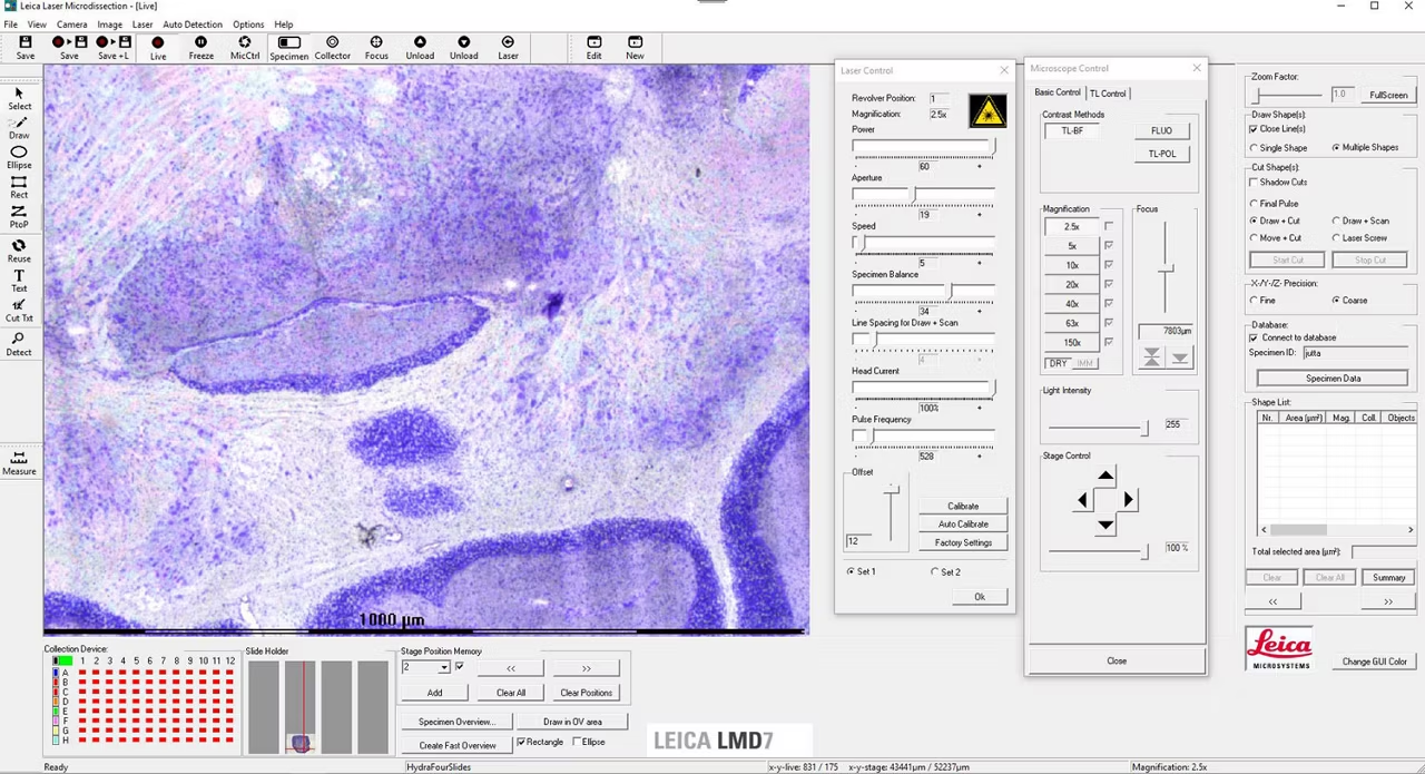
The LMD7's power, aperture, speed, head current, and pulse frequency settings are tunable to the needs of your sample. For example, thick plant sections require high power, while cutting around fragile neurons will require less power. Thin sections can typically use faster cuts than thick sections.
Advanced cutting modes add enhanced precision to how you will dissect your sample. For example you can draw first and cut afterwards (Draw + Cut), or cut in real-time by mouse click or with a PEN screen (Move + Cut). The Draw + Scan mode is made for ablation from glass, whereas the Laser Screw cuts thick specimen in consecutive rounds. The Final Pulse mode enables you to kick demanding dissectates into the collection vessel.
Leica Laser Microdissection Software

The Leica LMD software is an intutive application software designed to streamline the process of selecting settings so that you can focus on obtaining high-quality regions of your sample. Select, dissect, and visualize with ease!
- Control laser and microscope
- Guide the laser beam by mouse or pen screen
- Create a Specimen Overview for better orientation
- Detect and cut your samples automatically
The Leica LMD Software connectivity:
- Use your self-made pattern recognition software
- Import your ROIs from other devices e.g. slide scanners
Consumables

A) Specimen
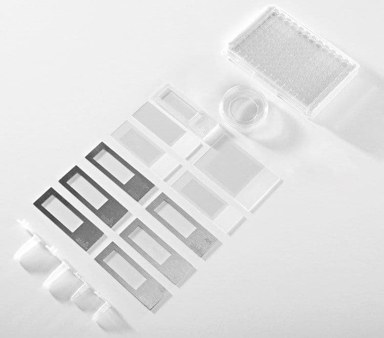
Leica Microsystems offers different slide options for laser microdissection.
- Use any kind of membrane slides for genomics and transcriptomics
- Use PET slides for certain applications in proteomics and metabolomics because PET is almost free of softeners.
- Choose DIRECTOR slides to work completely membrane free
Read how to choose the adequate consumable for Laser Microdissection.
B) Collection
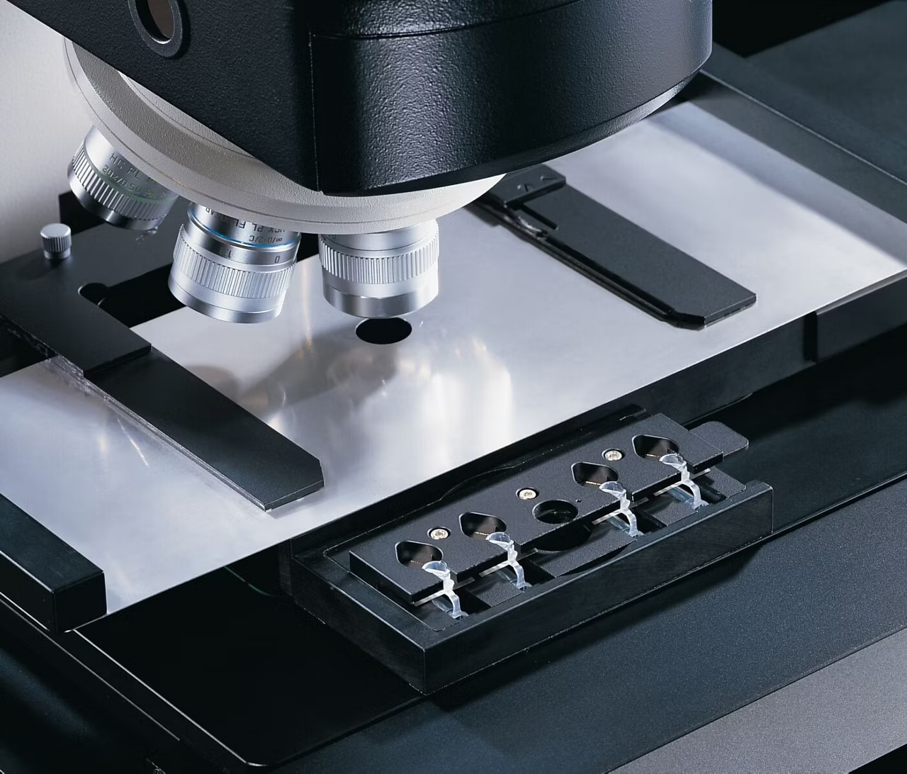
Since the Leica LMD systems collect dissectates simply by gravity, you can use standard devices for their collection. Just use common molecular biology reaction devices such as 0.2 or 0.5 ml tube caps – which you already have in your lab. Collection devices can be dry or filled with reaction buffer or culture media for the LMD
application.
Automation Options

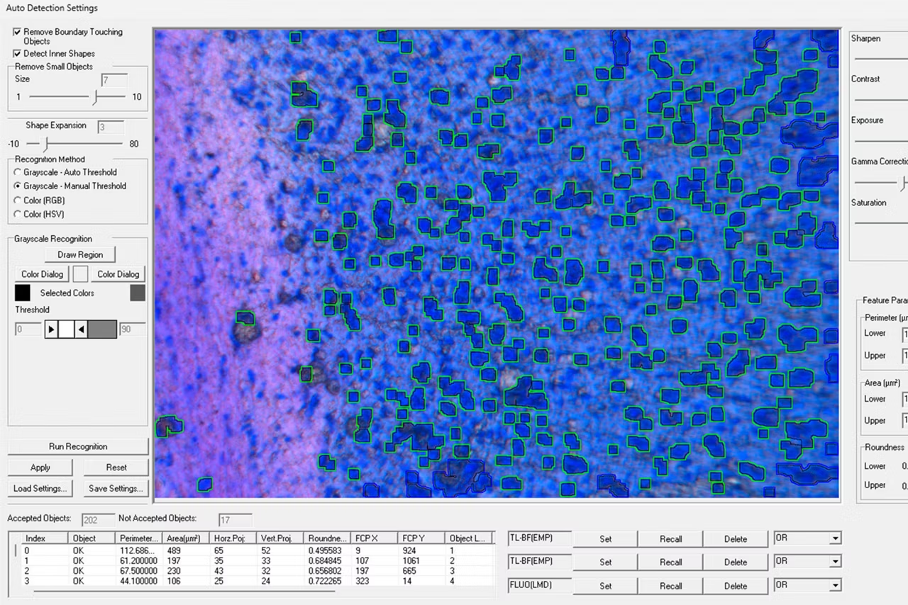
The ADM (Auto Detection Mode) is a software module which helps collect large amounts of similar dissectates. You just have to encircle a region you are interested in and the software will use this as a template for other regions. For example, Proteomics analysis requires a large amount of sample to be captured, and ADM has provided a great benefit to labs working with 100's-1000's of dissectates.
Autofocus can be used before each dissection process to keep the focus of hundreds of ROIs distributed over the slide. Moreover, you can automatically inspect and document all collection devices which were used.
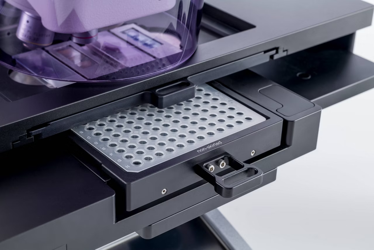
The systematic approach: A raster tool separates your Field of View into a given number of regions. With this function, you can systematically dissect your specimen and collect your dissectate into a variety of collection devices such as 96-Well plates.
High-Throughput Capability

Need more throughput? The LMD systems can be configured with different kinds of stages. The LMT350 ultra scanning stage provides the fastest, most silent and most precise navigation among them.
The LMT350 is capable of holding collector vessels starting from 0.2 ml tube caps, through 8 and 12 cap-strips up to standard 96-well and 386-well plates*. This enables you to do high-throughput experiments.
Because you don’t need any special material but use regular microtiter plates, these plates can be loaded directly into standard PCR machines.
*max.352 wells addressable
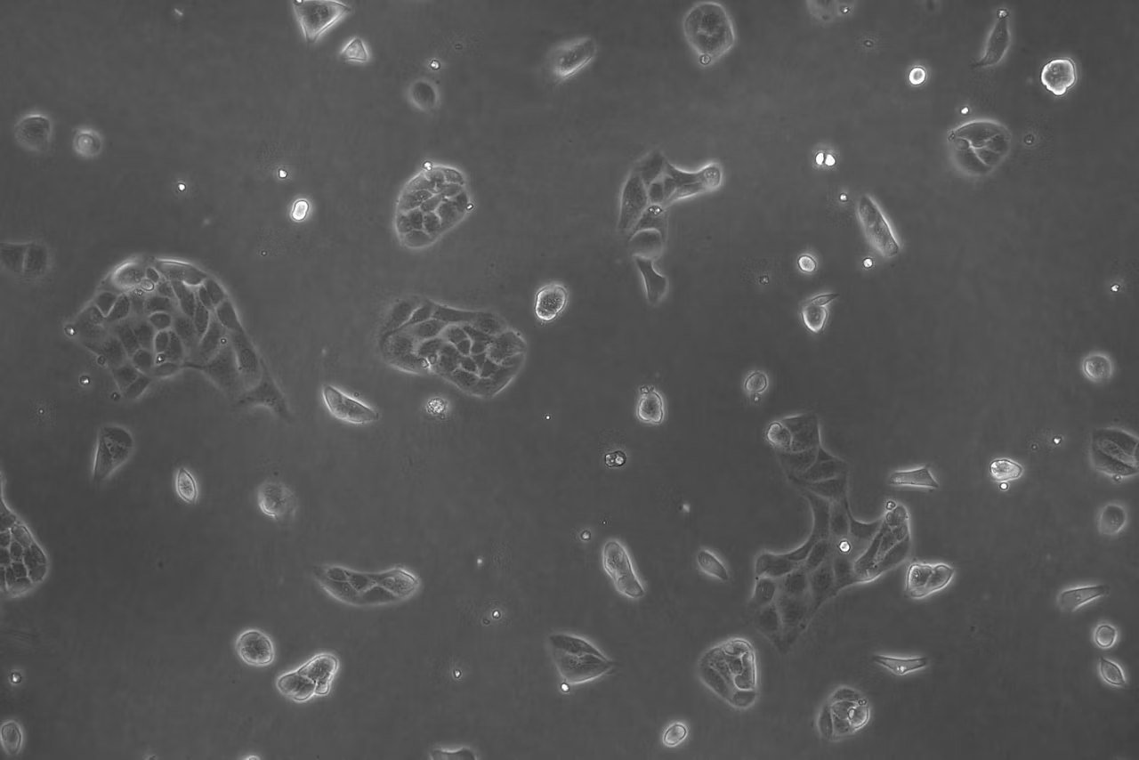
LCC – Live Cell Cutting

If you work with live cells, you are used to working with inverted microscopes. Live cell cutting works with our LMD systems, even though the Leica LMD systems are based on an upright microscope.
- You can dissect living cells in culture in order to re-cultivate, clone, or analyze single cells, colonies, or cell clusters
- You can attach a climate chamber to the LMD system
- You can grow cells on petri dishes with PEN membrane or multi-well ibidi slides
- You can collect the dissectate of living cell cultures into Petri dishes with or without PEN membrane, ibidi slides, or 8-stripe tubes for re-cultivation, or into collection devices such as PCR tube caps for analysis
Success is Objective

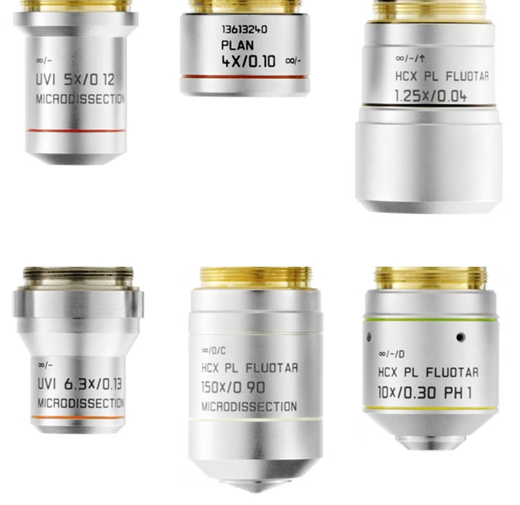
LMD objectives are designed for the task of enabling flexible laser settings for unique applications. Leica's development and manufacturing of optics has been among of our core competencies from the start in the 19th century, so you can rely on the performance of our dedicated LMD objectives.
- Choose from a range of dry objectives – from 2.5x to 150x
- Use the 150x SmartCut objective to go into detail with high magnification when required
- Get a large field of view to cut large parts of samples in one piece with low-magnification objectives
- Benefit from objectives with the highest possible transmission of laser light at 350 nm to cut tissue, bones, teeth, brain, plants, chromosomes and living cells – try them for your application!
The outstanding imaging performance our objectives provide goes without saying.
Same Principle, Two Systems

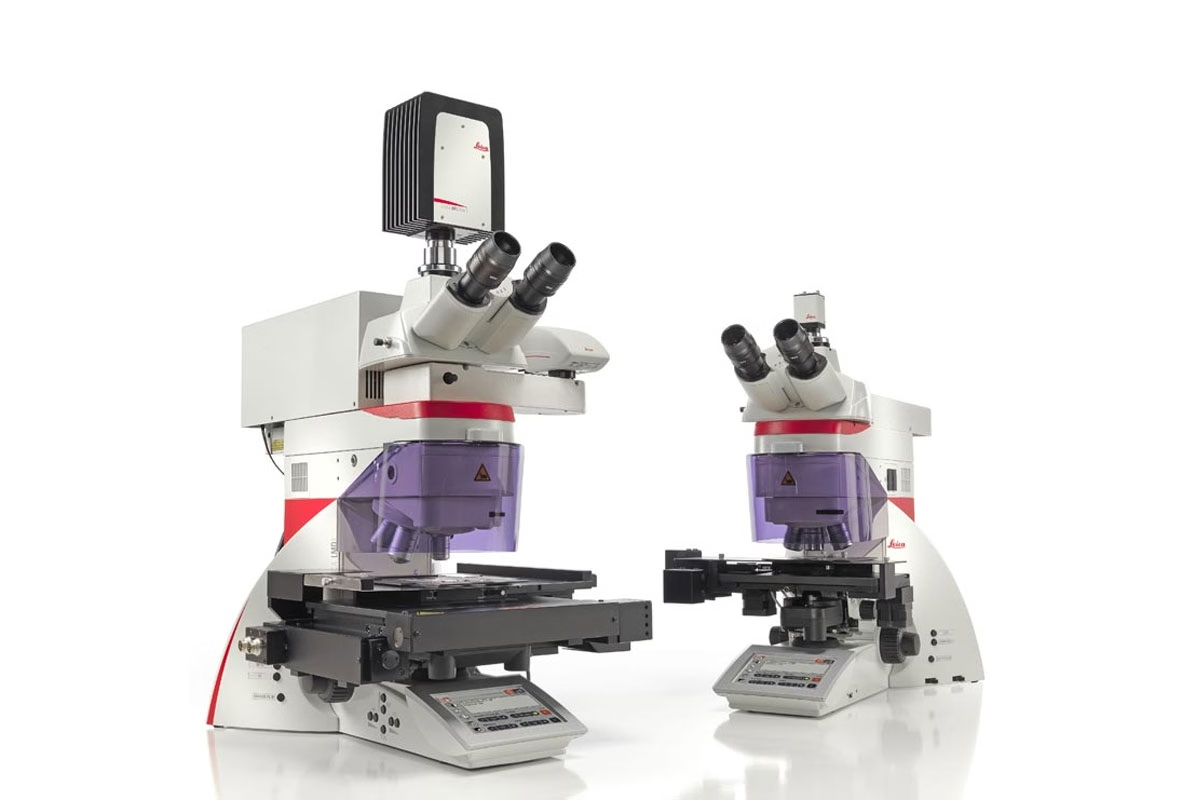
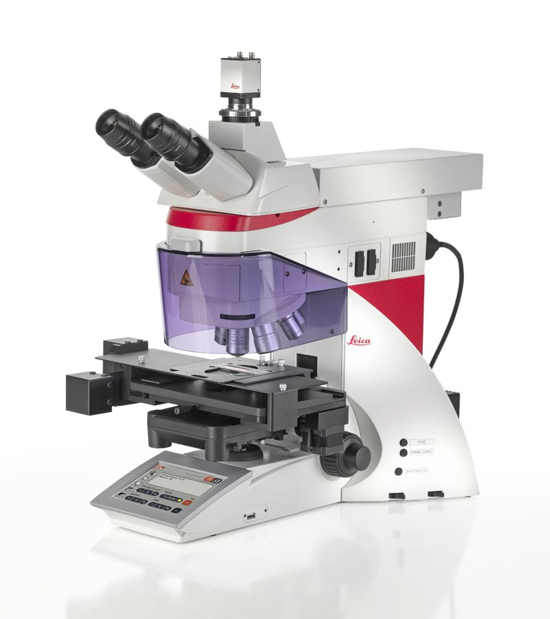
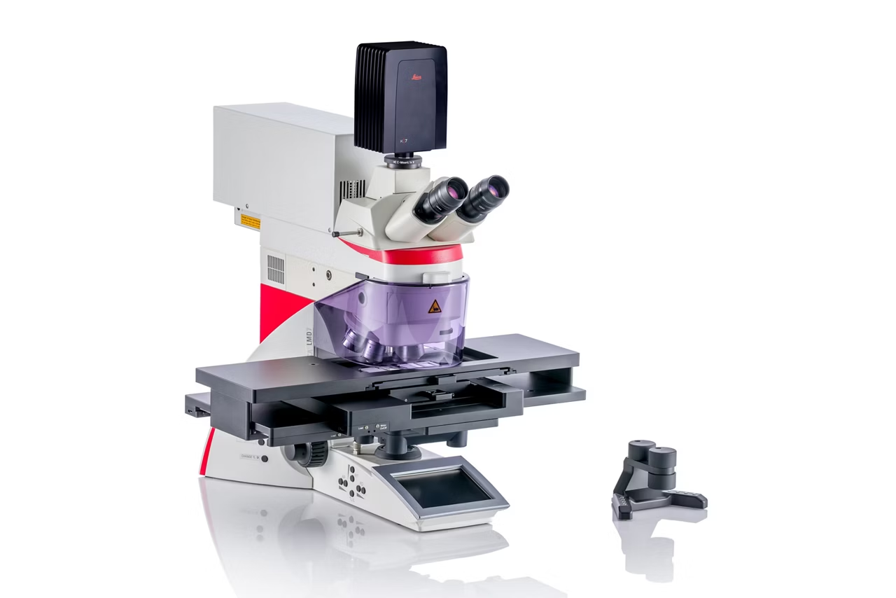
Choose your system: The difference between the Leica LMD6 and Leica LMD7 is the laser. The LMD7 provides higher laser power and more laser controls than the LMD6
This makes the Leica LMD6 the tool for standard applications dissecting soft tissues such as brain, liver, or kidney. And it makes the Leica LMD7 ideally suited to dissect any kind of tissue independent of its size or shape.
Two systems for your discoveries Leica LMD6 for standard tissue dissection Leica LMD7 for higher laser power and more flexibility
Interface for Artificial Intelligence

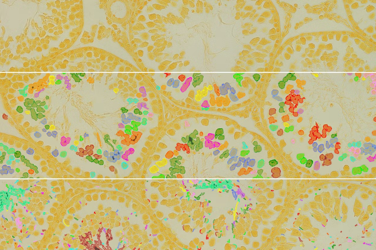
Aivia is our AI-based, image visualization, analysis and interpretation platform. With the help of AI-enhanced tools such as the Pixel Classifier, Aivia can be used to automatically define Regions of interest (ROI) which are destined for Laser Microdissection (LMD). ROIs can be detected by Aivia and imported directly into the LMD software for Microdissection.
Besides Aivia other external software can be used for automatic ROI detection. The LMD software simply needs an XML file containing the ROI information.
- Mund et al., Nature Biotechnology, 2022 https://doi.org/10.1038/s41587-022-01302-5
- Mitchell et al., JoVE, 2022 https://doi.org/10.3791/64171
Combination System: LMD System with THUNDER Imaging

The LMD6 and LMD7 systems can be combined with THUNDER. The base stand of the THUNDER Imager 3D Tissue and LMD systems are the same, thus such a combination offers:
- Lab space savings with one system for different tasks
- Laser Microdissection with the LMD without limitation
- Brilliant THUNDER fluorescence imaging using LAS X, even in multi-color and 3D (z-stacks) and visualization in the 3D viewer.
Dissection Perfection
We Move the Laser, not the Sample
Only Leica Microsystems uses high-precision optics to steer the laser beam along the desired cut line on the tissue with prisms
Gravity for Cleanliness

Your downstream analysis relies on contaminate-free isolates. This is why Leica LMD systems collect dissectates via gravity
LMD Software
The LMD software is an intutive application software designed to streamline the process of selecting settings so that you can focus on obtaining high-quality regions of your sample
Success is Objective

LMD objectives are designed for the task of enabling flexible laser settings for unique applications
LMD Software Laser Microdissection
Advanced laser microdissection software for precise cell isolation from FFPE, frozen & live samples. Supports genomics, proteomics, spatial biology. Get quote.