DM4 B & DM6 B Upright Microscopes
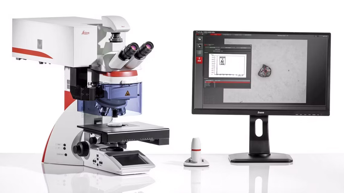
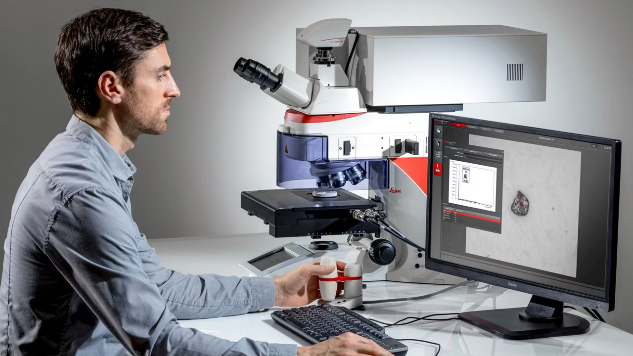
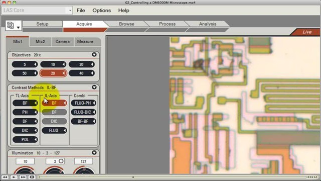
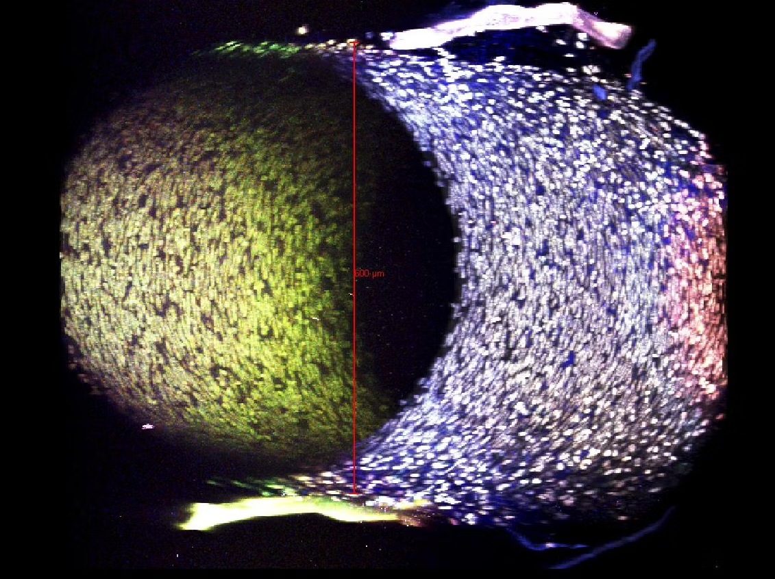
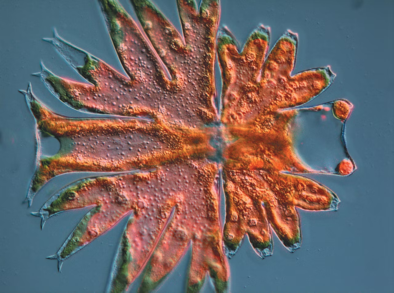
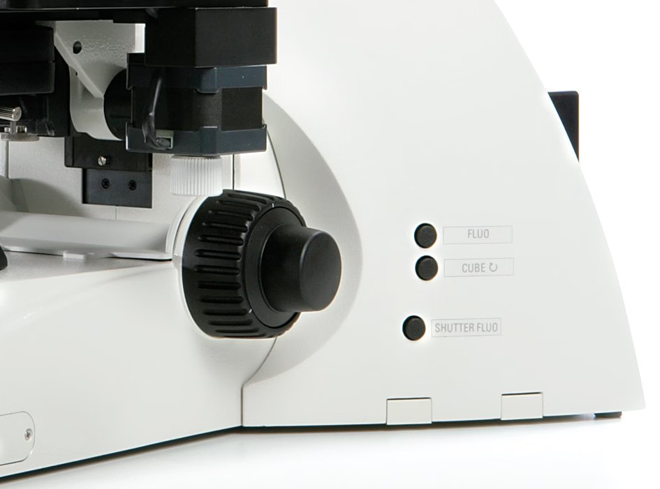
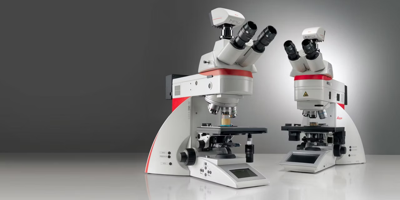
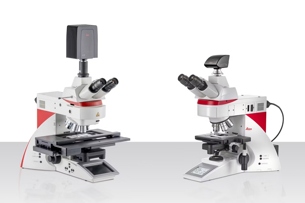
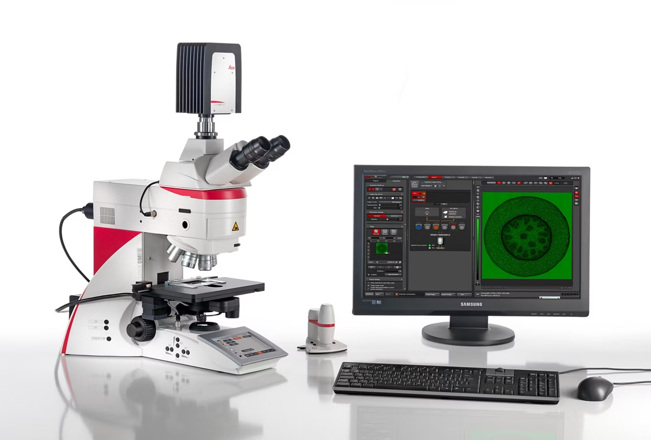
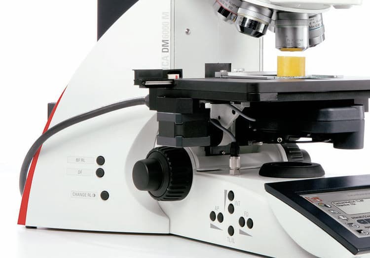
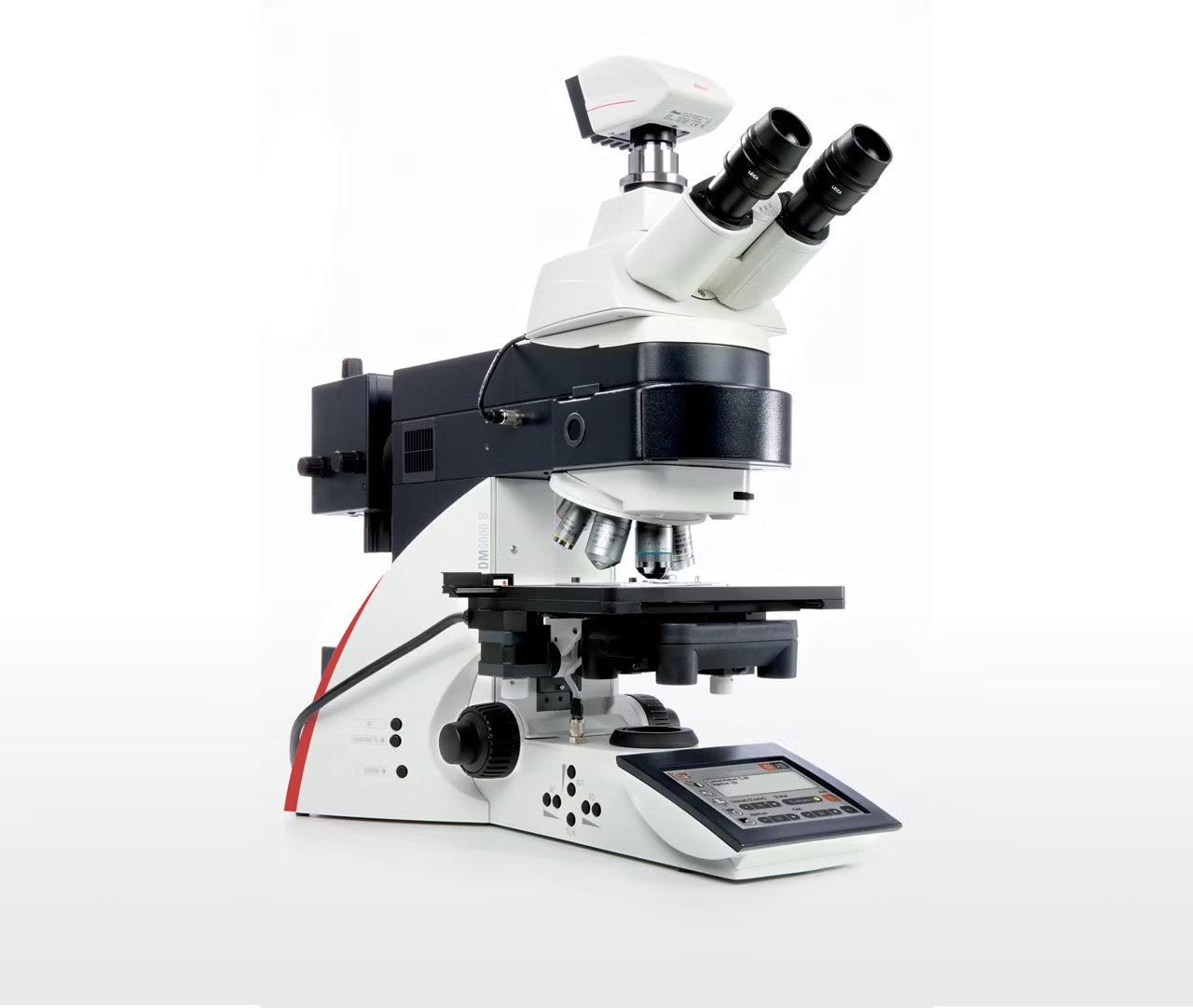
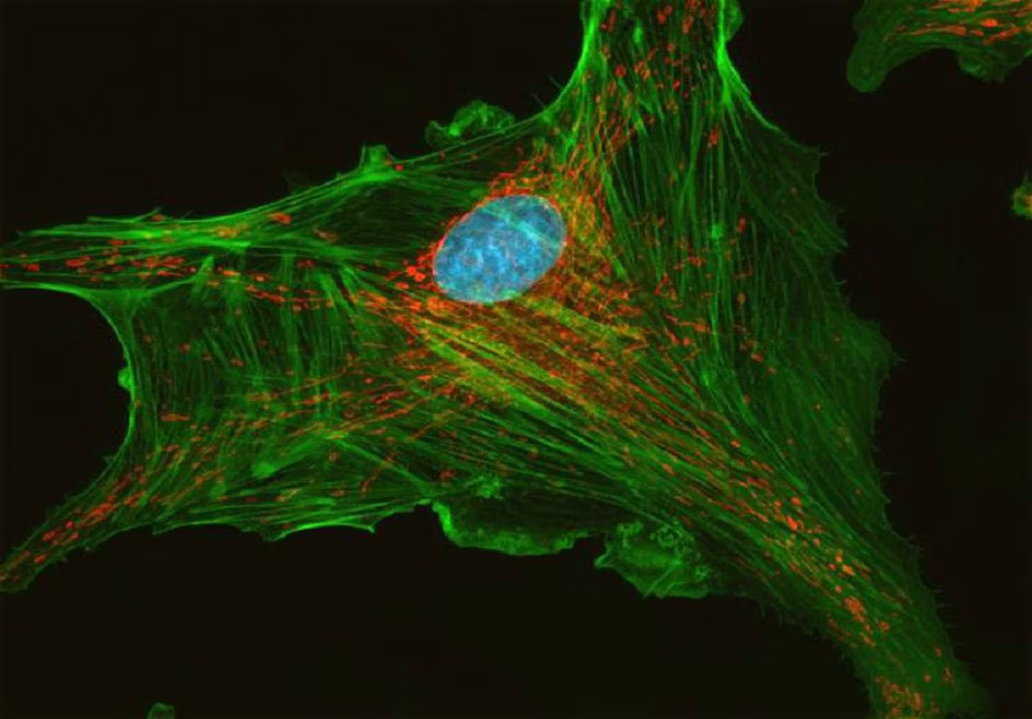
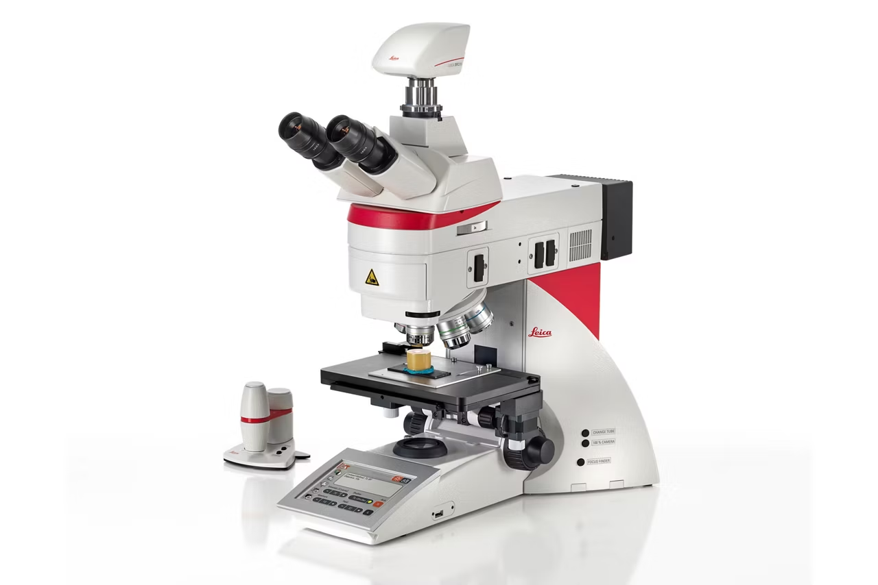
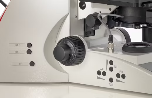


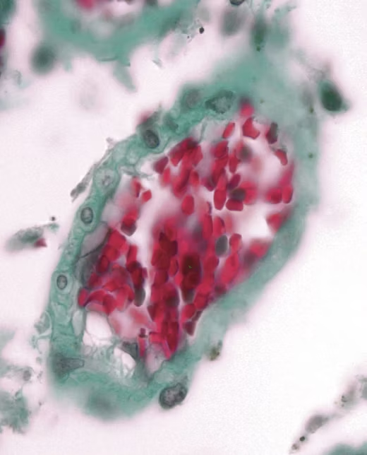
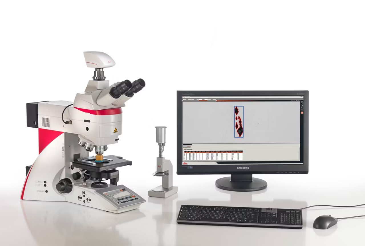
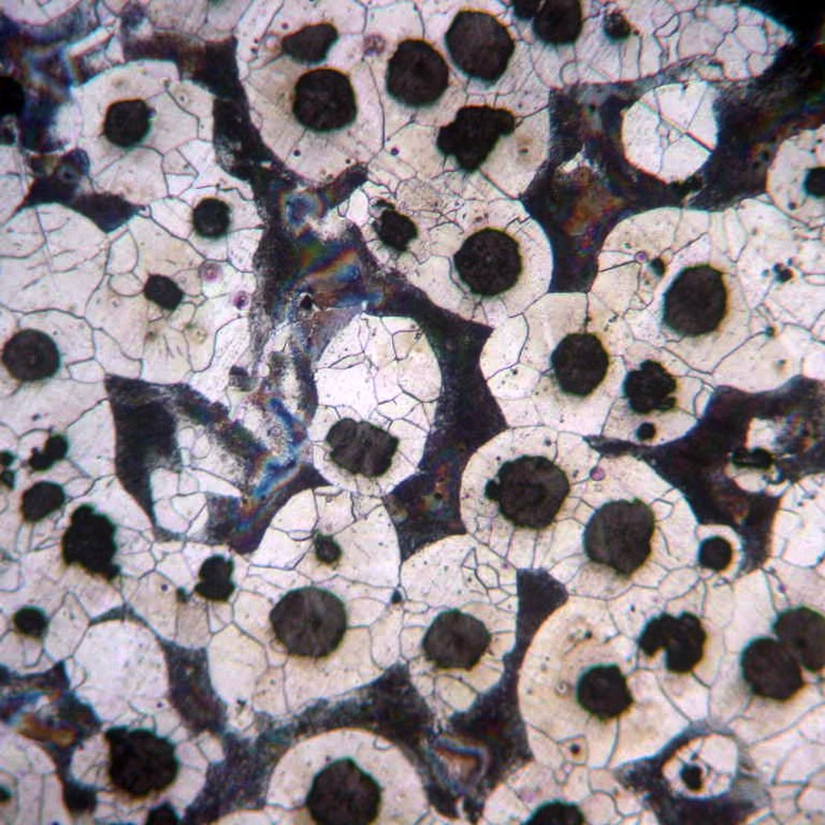

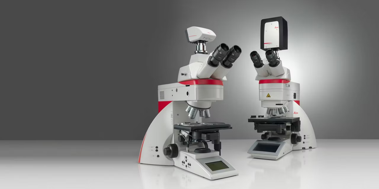
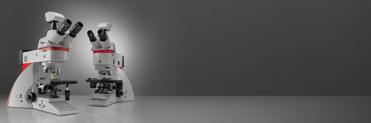
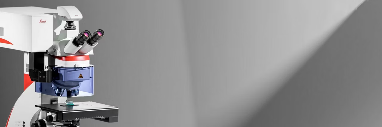

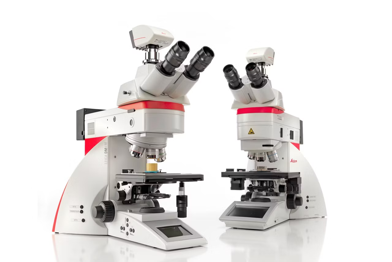
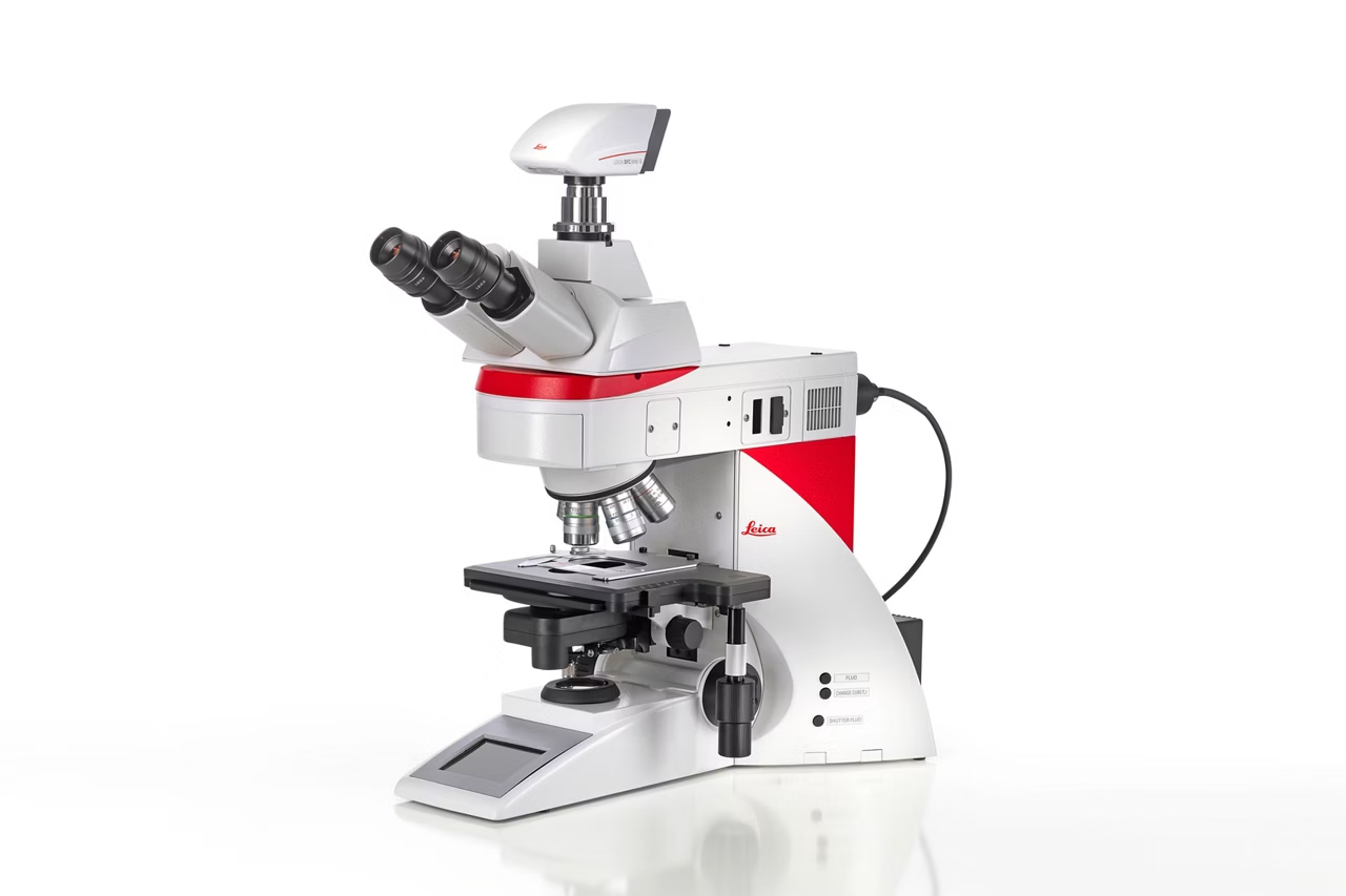
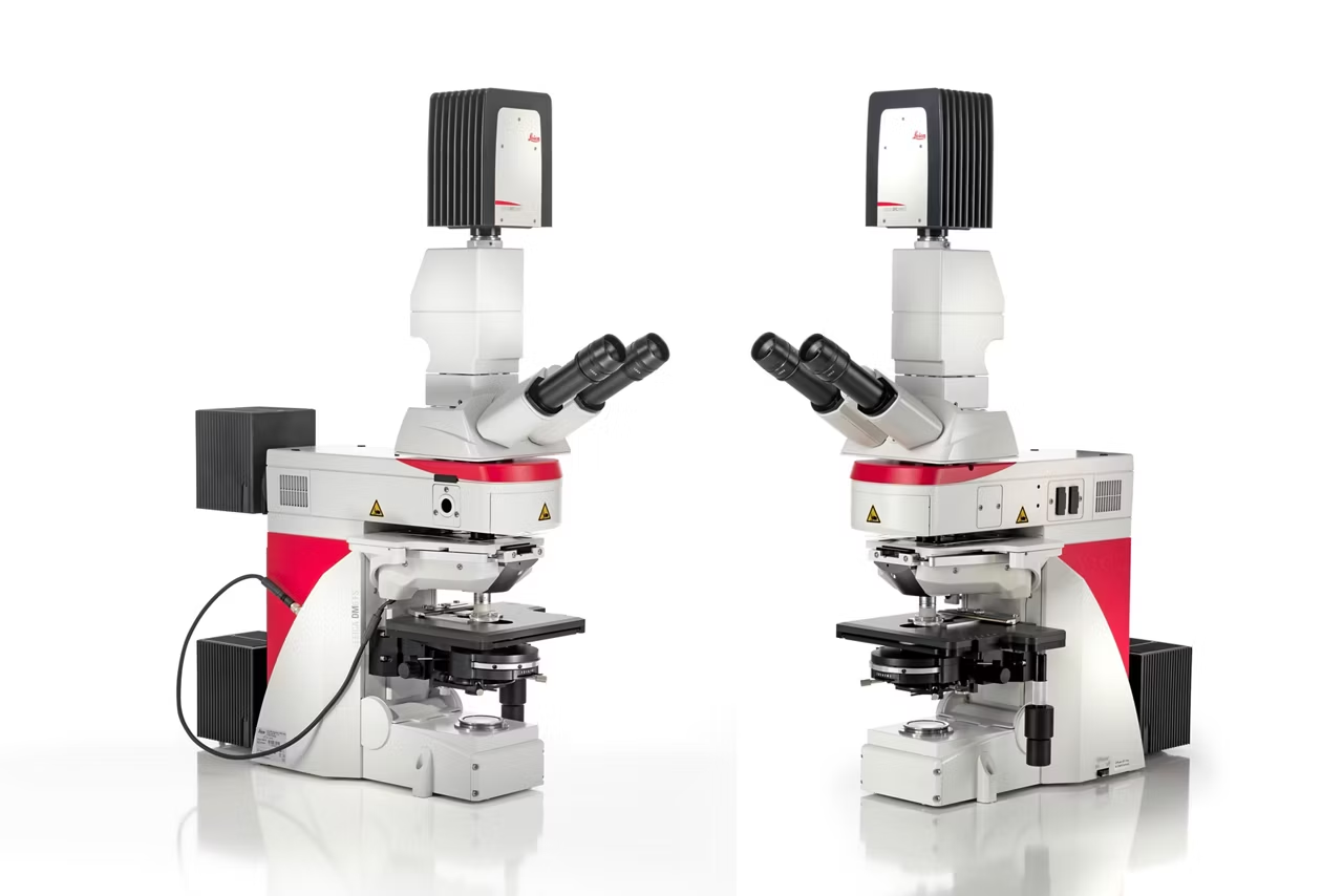
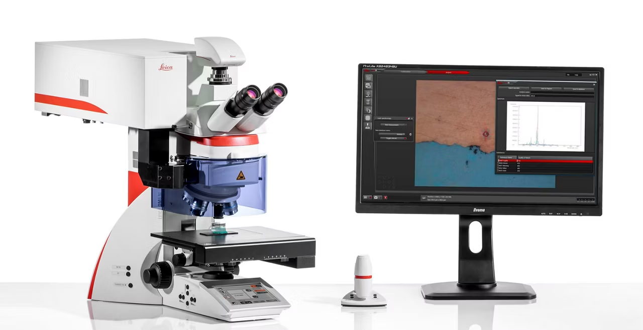
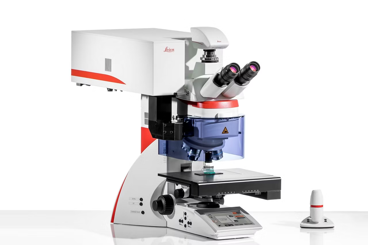
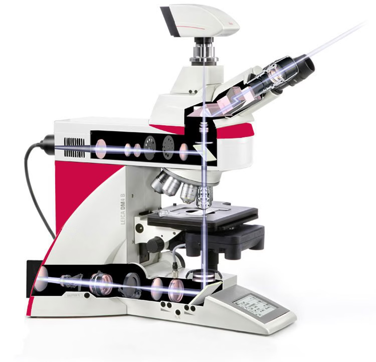
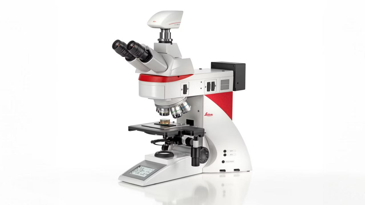
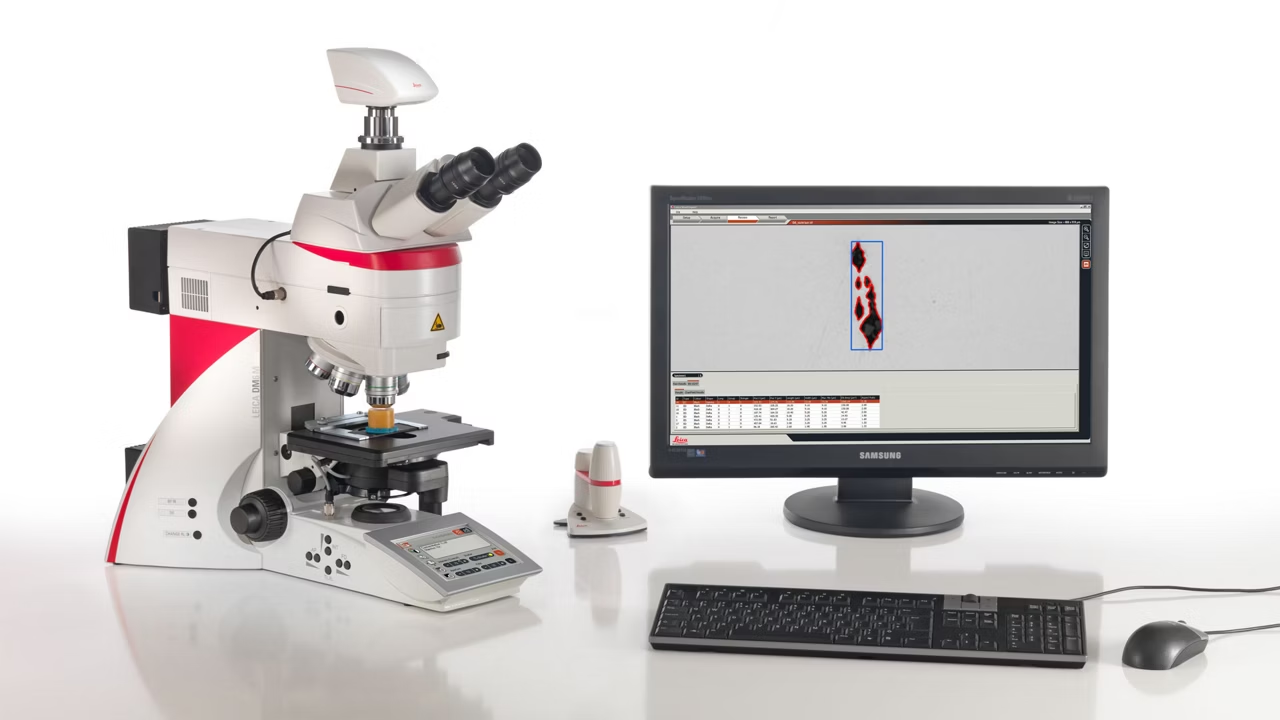
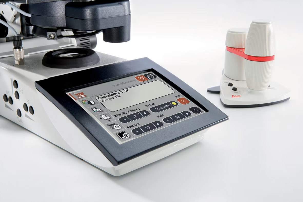
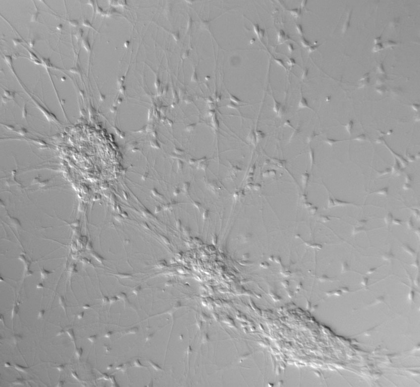
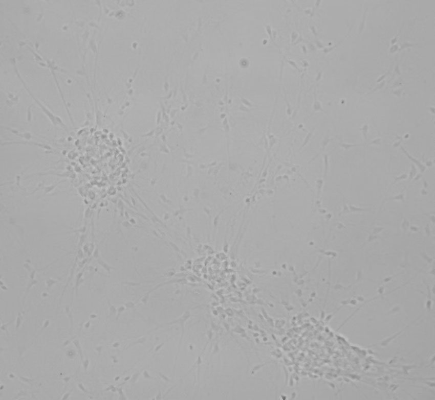
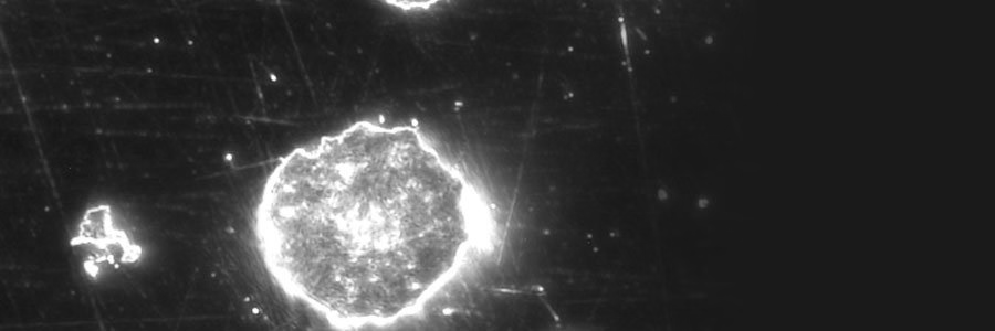
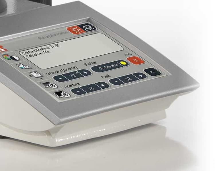

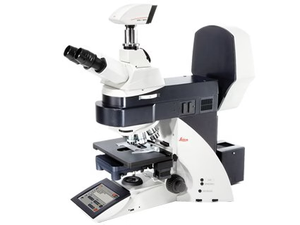
Enhance your life-science and clinical research with the DM4 B and DM6 B upright microscopes. Optimized for imaging tissue and plant specimens, as well as pathology, these microscopes are designed to accelerate and tailor your workflows. With flexible software solutions, they allow you to focus on experiments while reducing manual work.
Boost Productivity: focus on experiments, not managing the microscope
Increased speed and productivity by focusing on experiments, not manually managing the microscope:
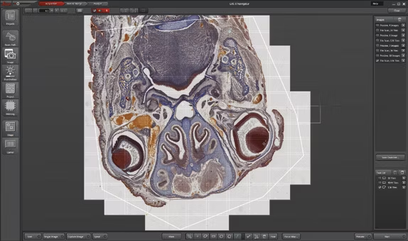
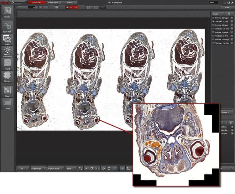
- Quickly and easily provide sample overviews and set up complex imaging experiments with Enhanced Navigator integration in LAS X
- Increase productivity by up to 2 times compared to traditional upright microscopes, thanks to Synapse™ technology, which synchronizes all microscope components
- Give your data spatial context throughout your workflow by switching from sample overview images to multi-channel view mode
Tailored workflows and flexible configurations for optimal results

Get optimal results by flexibly configuring the microscope to your specific workflow and application needs:
- Desired performance via customized workflows like slide scanning and fixed-stage imaging
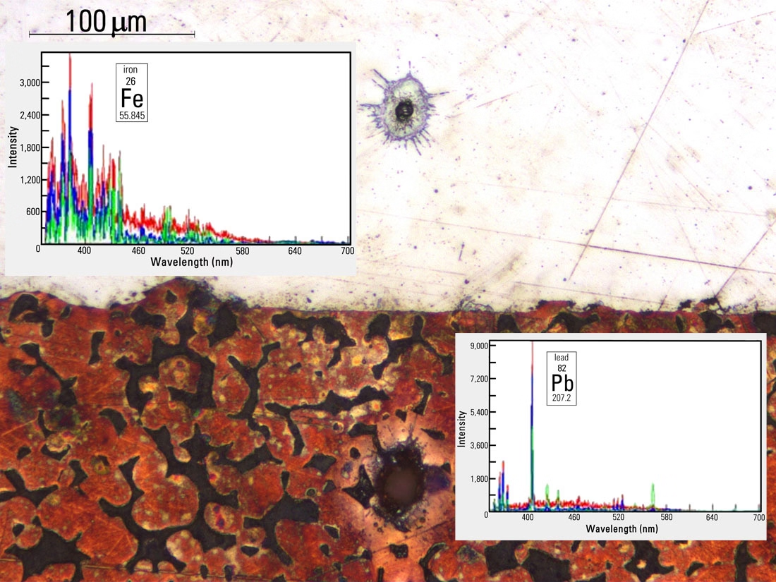
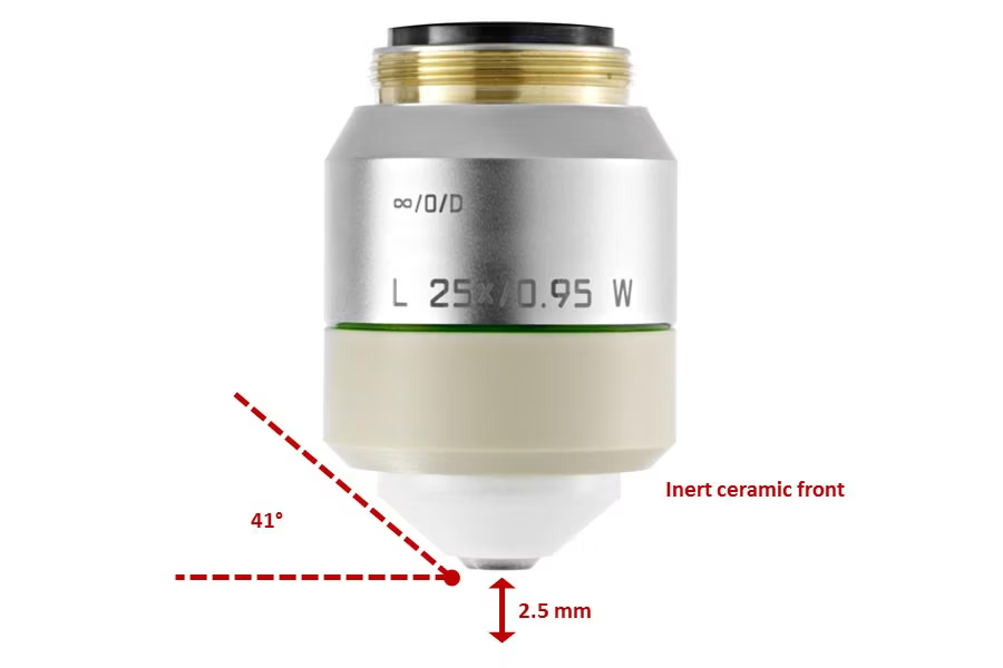
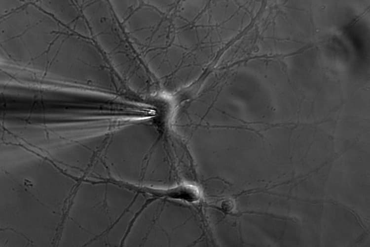
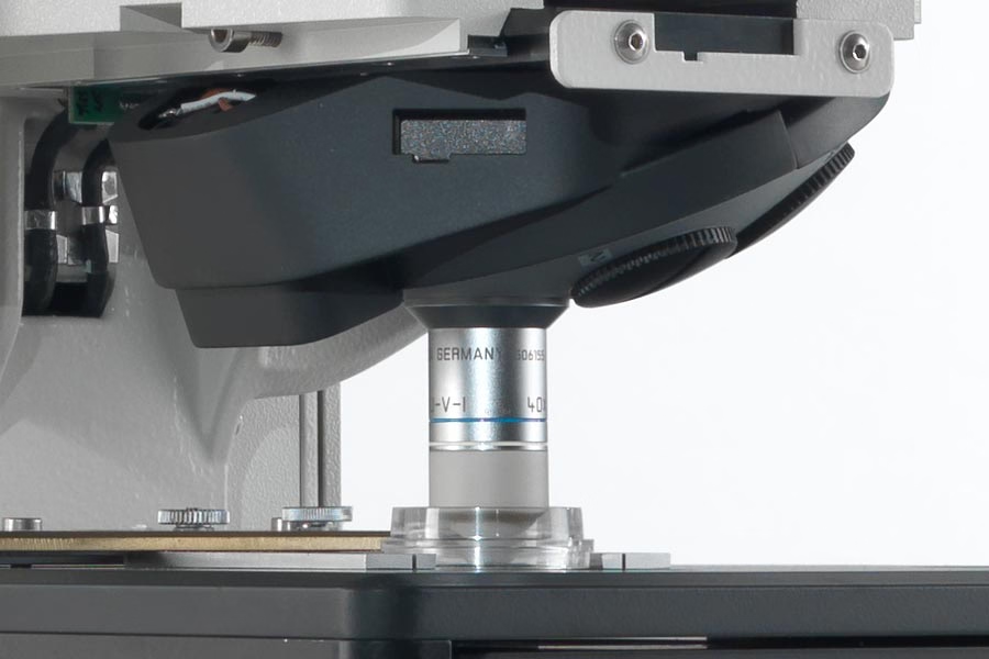
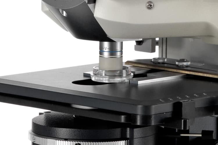
- Tailored solutions using configurations that provide contamination-free material collection using laser microdissection, high-quality images with computational clearing, and vibration-free fixed-stage electrophysiology
- Flexible configurations that meet the imaging needs of the sample by combining multiple contrast method
High-quality data and analysis for robust insights

Obtain valuable insights from high-quality data and analysis with Leica software solutions tailored to match your changing application needs:
- Improved insights thanks to software solutions like LAS X Navigator and AIVIA that align with your specific application requirements
- Increased Reliability with software solutions that provide consistent and dependable results, reducing the risk of error
- Consistency with software solutions that span the entire Leica life-science portfolio. No need to switch between separate software for imaging and analysis
Rapid results, customized workflows, and deep insights
Accelerate your workflows

Streamline image acquisition and reduce manual tasks to increase productivity.
Tailored workflows for your application needs
Tailored workflows enhance effectiveness by meeting your unique microscopy and application needs.
High-quality data and deep insights with adaptable solutions

Flexible software solutions which can be adapted to your changing needs to ensure consistent performance and reliable data.
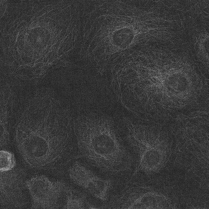
K8 Scientific CMOS Camera
Discover the K8 Scientific CMOS Camera for life science imaging. Achieve 95% quantum efficiency, low read noise and reduced phototoxicity. Request a quote!
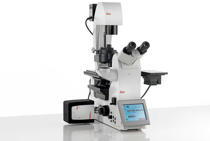
DFC7000 T and DFC7000 GT Microscope Cameras
2.8 MP cooled cameras for fluorescence and brightfield imaging. Detects DAPI, FITC to Cy7 including near infrared signals. Request a quote today.
LAS X Life Science Microscope Software Platform
Unified microscope software supporting confocal, widefield, stereo, super resolution, & light sheet systems. Save & recall settings for reliable results. Get quote.
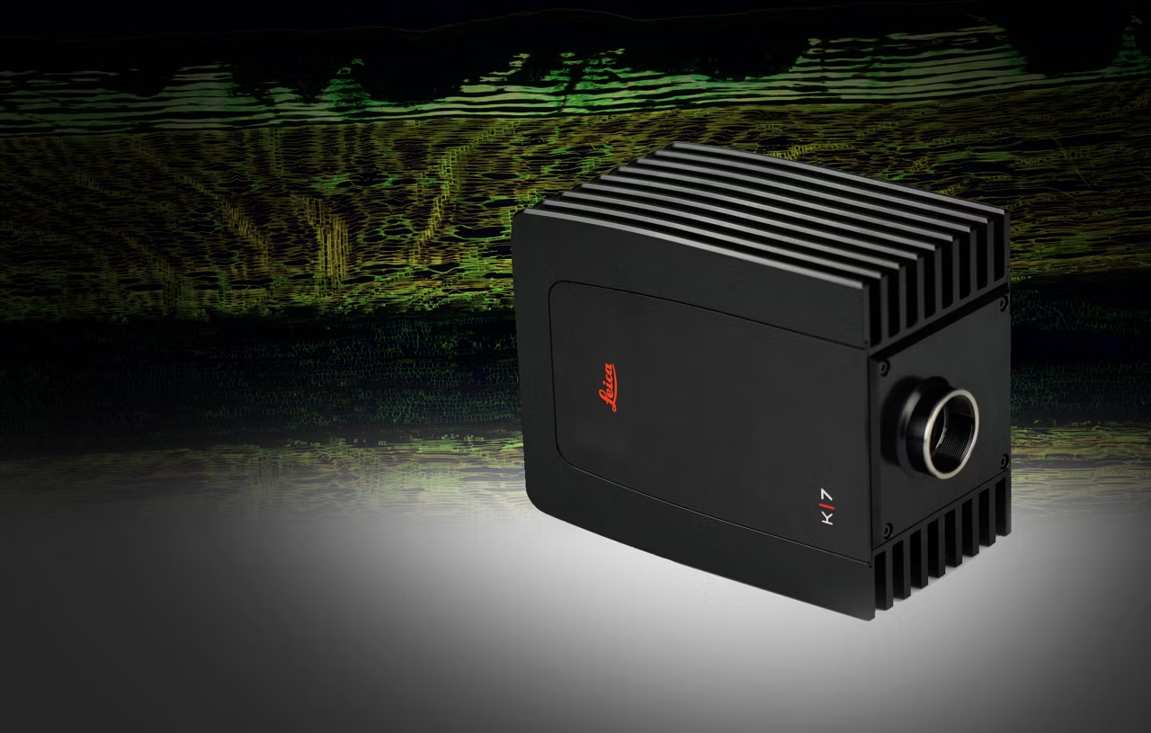
K7 Color CMOS Microscope Camera
Capture K7 color CMOS microscope camera images for fluorescence and brightfield microscopy, fast scan speeds, high temporal resolution, CMOS sensor. Get a quote!