JavaScript is disabled in your browser. Please enable JavaScript to view this website.

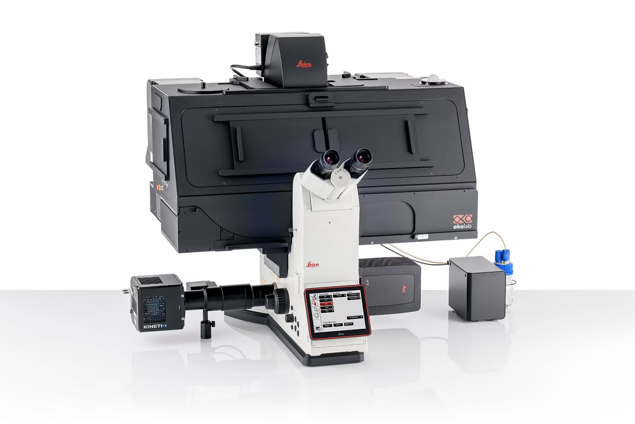
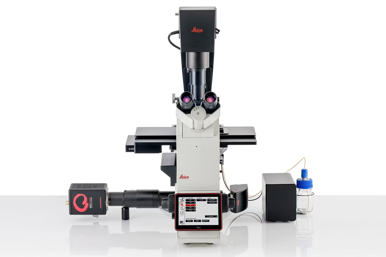
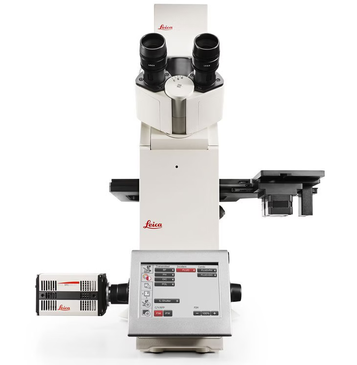
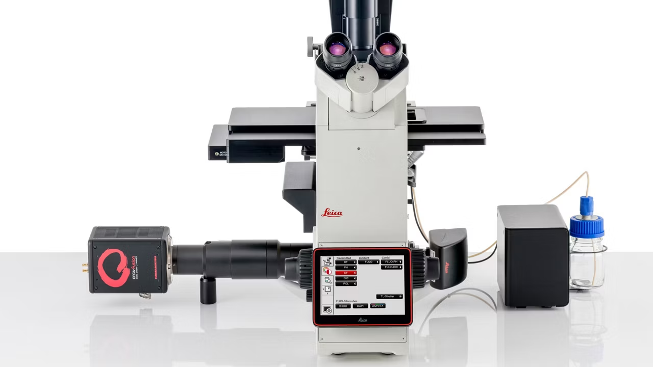
DMi8 Inverted Microscope
Dmi8 Inverted Microscope Stage V1 Kid21757783

DMi8 Multi Channel Mouse Brain Kid21757750

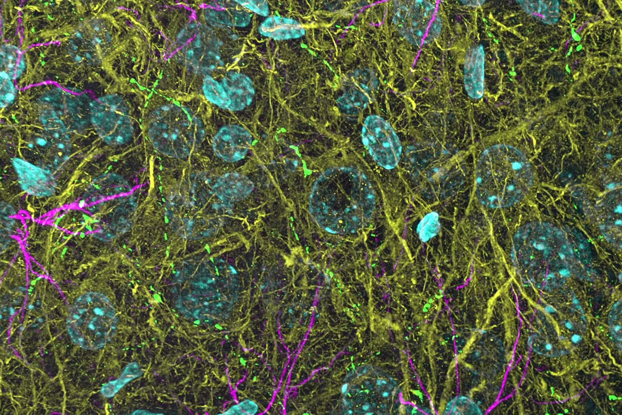
3D THUNDERed Spinning Disk Image Of Mouse Brain Kid21754304

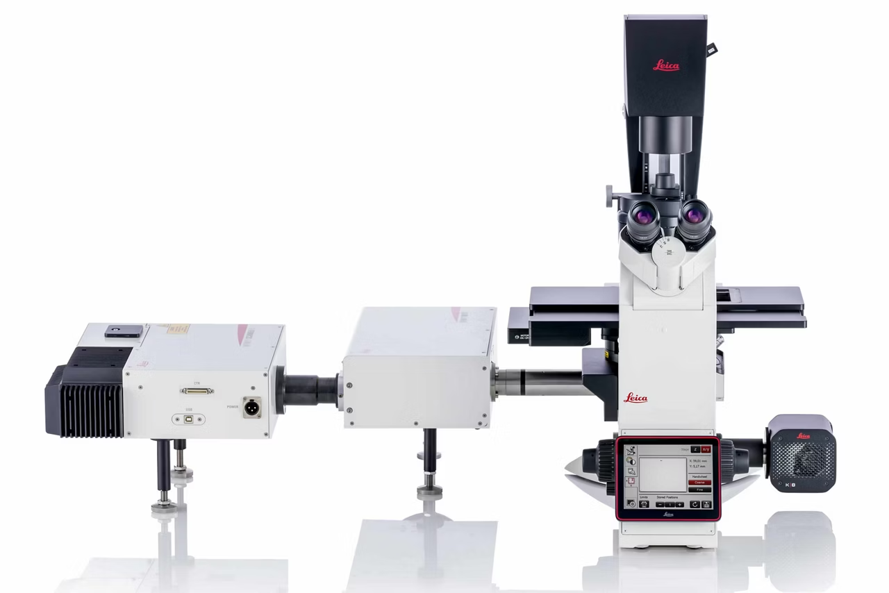
Dmi8 Full Configuration2 Kid21757779

Adaptive Immersion Extended2 1 1 Kid21755148

DMi8 Stopmotion Upgradability Kid21757761

DMi8 Navigator Zoom 1 Kid21757751

DMi8 Speed Comparison Video 1 Kid21757758

MotCORR Video2 Fig 8 1 Kid21767969

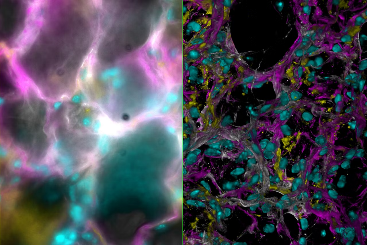
Mouse Lung Tissue Stained For A Study Of Type I Alveolar Epithelial Cell AT1 Biology Kid21768021

DMi8 S Blur Kid21757752

DMi8 Scanner TIRF K8 ThunderShooting 1070964 Web Kid21757755

Leica DFC9000 DMi8 02 Kid21765743

Leica DMi8 RFID Flyer En 1 Kid21765794

Leica DMi8 S 02 Kid21765795

Leica DMi8 StopMotion 11 V1 10

Leica DMi8 StopMotion 8 02

Dmi8 Bg Color Kid21757768

Dmi8 Inverted Microscope K3c Camera Kid21757782

Dmi8 Stage2021 Kid21757795

Highspeed Leica DFC9000 DMi8 W Kid21764968

Increase Microscope View By 10000 Kid21765231

Sample Over View Video 3 1 Kid21769130

DMI8 A Oben 04 Kid21757764 1

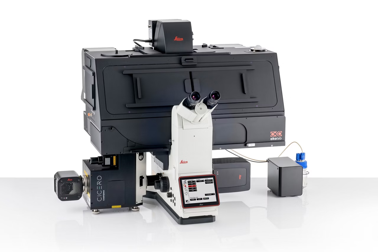
Streamline your complex microscopy workflows with the DMi8 inverted microscope. The platform enables you to reliably generate high quality data with solutions that you can tailor to your research requirements and budget.

Enhanced Navigator integration

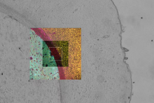
Easily switch from sample overview images to multi-channel view mode, giving your data spatial context throughout your workflow.

With enhanced Navigator integration, you can set up and perform complex multi-position experiments on a wide range of sample formats using templates for slides, dishes and multi-well plates.

Navigator combined with Adaptive Immersion technology allows you to seamlessly switch from overview images to high-resolution images using immersion objectives, without removing the sample from the stage.

Adaptive immersion technology

Patented and reliable Adaptive Immersion technology:

  • Employs an embedded sensor to monitor and maintain immersion throughout your experiments.
  • Enables you to use optimum water immersion objectives for live cell experiments under physiologically relevant conditions.  
  • Creates smoother workflows through hassle-free switching to immersion objectives.

Confidently gain more insights

Experience smoother and more efficient workflows with the DMi8

  • Seamless integration of hardware triggering for maximum efficiency
  • Faster scanning over large areas by combining 22 mm FOV optics with advanced cameras and the Quantum Stage

Convincing data from 3D samples

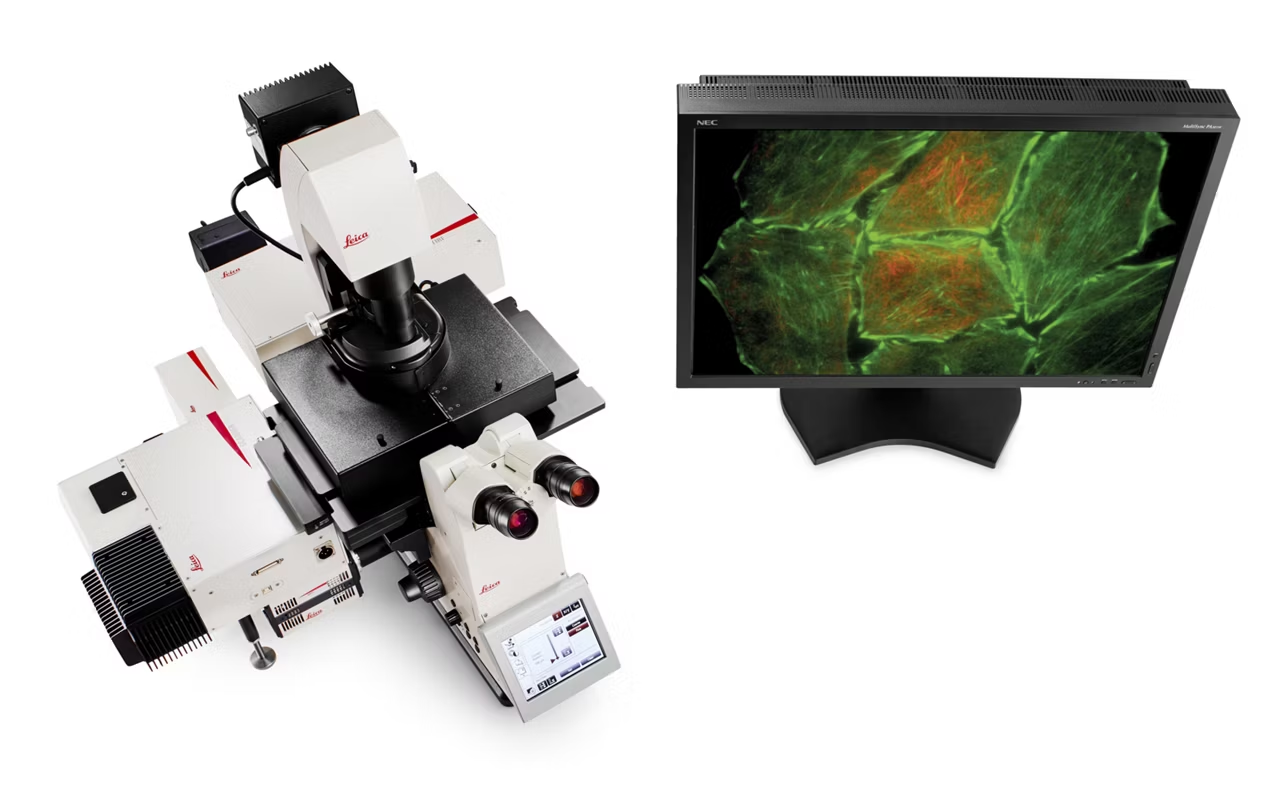
3D THUNDERed Spinning Disk Image Of Mouse Brain Kid21754304

Generate better quality data every time regardless of your experience level. Extract the most insights from your 3D samples with our advanced technology suite including

  • THUNDER
  • SmartCORR
  • Spinning Disk Confocal Scanner

Acquire precise and reproducible live cell data

Extract better data from your sample within seconds with SmartCORR, which optimizes your objective settings. This gives you the best possible data every time, with minimal effort.

Beyond deconvolution - See through the haze

Mouse Lung Tissue Stained For A Study Of Type I Alveolar Epithelial Cell AT1 Biology Kid21768021

Elevate your DMi8 image quality with THUNDER, an opto-digital technology that uses the Computational Clearing method to generate high resolution, high contrast images, by removing the out of focus blur inherent to widefield images.

A simple yet powerful real time image enhancement tool, you can use THUNDER on both widefield and spinning disk data sets and see stunning results. With THUNDER Live you can optimize parameters with immediate visual feedback, improving data quality and reducing time to optimal results.

Customizable and upgradable

Future-proof your experiments with this adaptable solution. The DMi8's modular structure allows you to tailor your system to fit your current needs and budget.

As your research evolves, upgrade your system by adding features such as

  • THUNDER
  • Adaptive Focus Control (AFC)
  • Climate control
  • Spinning disk modules

Get the most from your DMi8
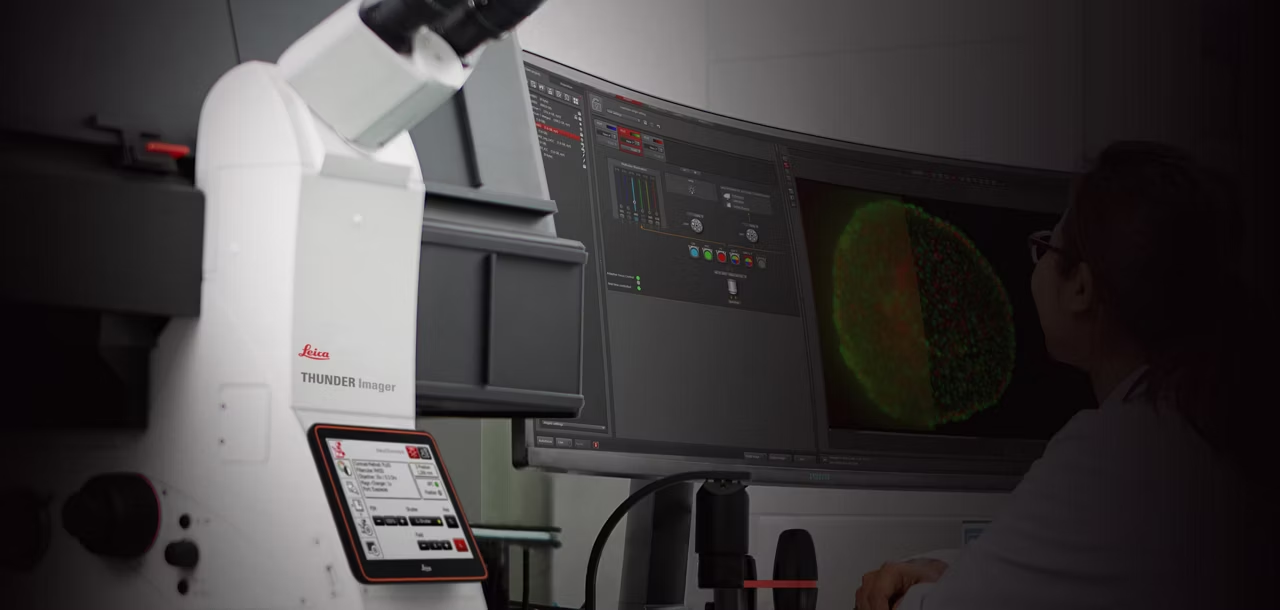
Csm DMi8 Jenny Nari Looking At Screen 1 1 7b03a44a50

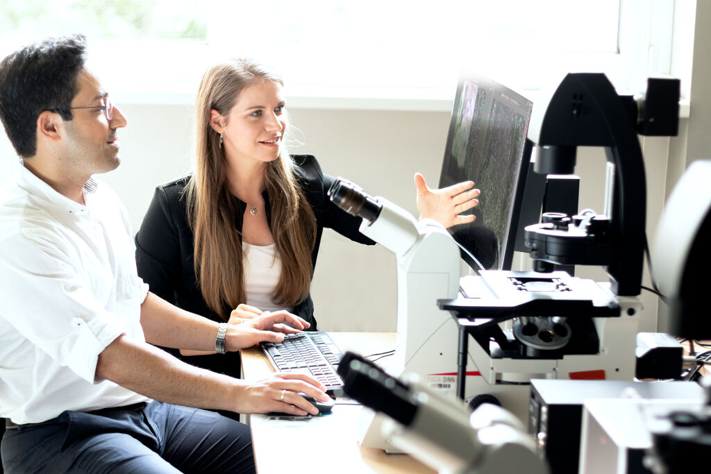
Maximize your investment through partnership with our Application Support Specialists throughout your system's lifetime.

Whether you are trying to get started or need to further tweak your system to address new workflows, our expert team will ensure you get the best possible data out of your system. 

Contextualize your data to see the full picture

DMi8 Multi Channel Mouse Brain Kid21757750

Efficiently set up your experiments with the DMi8’s intelligent automation, allowing you to focus more on your research questions. Gain relevant insights with:

  • Automated Sample Finder - Quickly gain a comprehensive view of your sample putting your data into context.
  • Fully Integrated Navigator Experience - Move through your experiments with ease.
  • Adaptive Immersion technology – Confidently use water immersion objectives for the most challenging live cell experiments.
  • 22 mm Field of View (FOV) – The throughput you need for enhanced data collection.

Automated Sample Finder

Simply place your sample on the stage and generate an in-focus overview of the entire sample at the click of a button with Automated Sample Finder.

  • Eliminate the time and frustration of searching for a focused image of your sample, reducing your time to result.
  • Gain context by understanding where specific data sets have been acquired from within your sample.

Advanced Imaging. Simplified.

Advanced automation to perform complex workflows

DMi8 Multi Channel Mouse Brain Kid21757750

Let your data speak, and focus on your experiment, not the system. Gaining deeper insights becomes simple with game-changing Adaptive Immersion technology.

Trust your results with higher quality image data and analysis

3D THUNDERed Spinning Disk Image Of Mouse Brain Kid21754304

Capture more convincing and reproducible data from demanding 3D and live samples with a fraction of the effort.

Flexible tailored solutions

Dmi8 Full Configuration2 Kid21757779

Customize your system to your needs and budget today, with upgrade options to support your evolving research requirements.

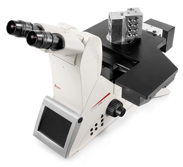
What is an inverted microscope?
An inverted microscope’s main distinguishing feature is the location of the optics. The optics are located below the stage. As a result, the sample is placed on top of the objectives. This means you will look at the sample from below it. Because the optics are underneath the sample, inverted microscopes offer ample working space to view samples of all shapes and sizes.
How big and heavy can samples be that I put on an inverted microscope?
Inverted microscopes are designed with the objectives and turret positioned below the stage. In theory, the sample could be as high as the ceiling – there is no height limitation. However, the maximum weight of a sample is limited to 30 kg depending on the stage used. This is ten times larger than can be accommodated by an upright microscope: samples can only be up to 80 mm high and weigh up to 3 kg.
What are the main differences between the Leica DMi1, the Leica DM IL, and the Leica DMi8?

The main difference is that they are designed for tasks of different complexity.

The Leica DMi1 is conceived for quick cell checks. You can perform cell and tissue checks quickly and efficiently in Brightfield and Phase Contrast.

The Leica DM IL enables you to perform more applications, for example involving fluorescence.

The Leica DMi8 is the fully modular high-tech instrument which can be equipped for almost every application.

What happens if I need to repair the microscope?
Please contact your local Leica sales representative. We will arrange everything for you.
What is an Infinity Port?
The Infinity Port gives users direct access to the fluorescence light path. Flexibly add illumination sources, without compromising imaging quality. The concept is unique and currently only available with the Leica DMi8.
Can I use the Leica DMi8 for in-vitro diagnostics (IVD)?
Yes, the Leica DMi8 is certified for IVD. Contact your local Leica Microsystems partner to find out more.
I want to use my microscope in a hood, are any modifications needed?
If required, the Leica DMi8 can be configured exclusively for digital imaging, often a great solution when working in a biohazard environment and when remote monitoring via the software is preferred.
How can I use the Leica DMi8 to build my own research solution?
Fully modular and flexible, the Leica DMi8 can be built exactly as you need. For advanced solutions, this is the platform of choice to integrate additional components such as photomanipulation devices, optogenetics, super-resolution, microspectroscopy, and many more. Homebuilt solutions can be attached via the Cage System 30 mm Infinity Port Connector.
Is there a solution for microinjection?
Applications that require micromanipulation such as transgenics, in-vitro fertilization, and more can be performed using the Leica DMi8 and micromanipulation solutions from Leica Microsystems, Narishige, Eppendorf, and other suppliers.
Can I do screening experiments?
Yes, the Leica DMi8 combined with the powerful LAS X software helps you design, set up, and perform screening experiments.
Can I do ratio imaging?
Yes. Choose options such as FURA objectives maximized for UV detection and a camera image splitter to simultaneously capture two fluorescent channels for ratiometric studies.
Can I do TIRF experiments?
Yes, you can. The Infinity Port provides direct access to the fluorescent light path, so that the Leica DMi8 is ideally suited to TIRF and similar high-end imaging applications. Choose the Leica AM TIRF system for high-performance TIRF applications, or adapt the Leica DMi8 to your system.
How can I use the Leica DMi8 for super-resolution?
The Leica DMi8 is a superb platform on which to build a super-resolution system. For a fully capable system, choose the Leica GSD 3D system, built upon a Leica DMi8. When you need a customized solution, rely on the Leica DMi8 as a stable and capable foundation to build up your own system.
I need to perform a high speed fluorescence experiment. Is the Leica DMi8 the right choice for that?
Definitely, yes. You can choose from a host of options to aid in capturing events as quickly as possible, such as fast external filter wheels, Adaptive Focus Control (AFC), compatibility with high speed sCMOS and EMCCD cameras, and control with the powerful LAS X software. Capturing a high speed multi-color experiment becomes less of a challenge this way.
Can I use the Leica DMi8 for live cell research?
The Leica DMi8 is an optimal microscope for live cell research. Build your system with a wide range of options for live cell imaging – from incubation chambers to keep your cells in an optimum environment, to time-lapse experiments with high speed cameras controlled by dedicated modules in the LAS X software, just to name a few.
What is a T-House?
T-House is the name for the modular component of the Leica DMi8 that the Infinity Port connections and interface for fluorescence illumination. In addition, this component provides the carrier for the stage and transmitted light devices.
Which software can I use with the Leica DMi8?
The Leica Application Suite X (LAS X) software is suitable for all applications with the Leica DMi8. The microscope software development kit, or SDK, can also be used to control the microscope via other software applications. For information about obtaining an SDK license, please visit our software page.
What is LID?
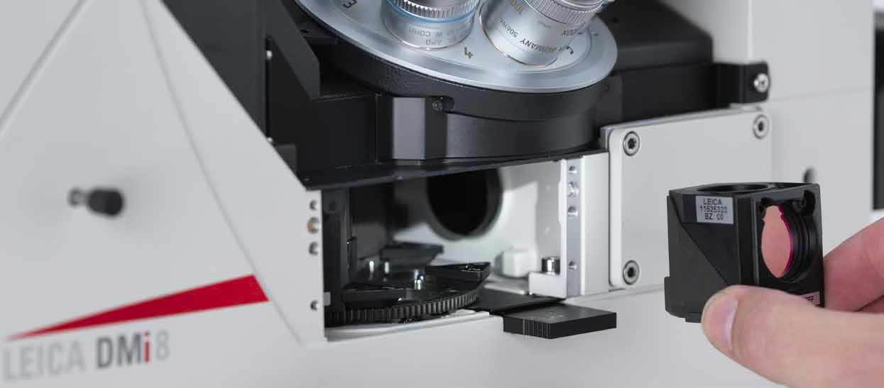
LID stands for Leica Radio Frequency Identification Device. All filter cubes for the Leica DMi8 are equipped with RFID technology, which can be used with the optional LID equipped fluorescence turret to automatically detect your filter cubes in the Leica Application Suite X (LAS X) software.
What is Adaptive Focus Control?
The Adaptive Focus Control (AFC) actively keeps the focus stable, even after changing environmental conditions. The systems utilizes an infrared LED to accurately measure the distance between the reflective surface at the image plane and the objective.
What is Closed Loop Focus?
Choose this option when high resolution imaging is required. By directly measuring the focus position, the optional Closed Loop Focus drive ensures highly accurate and reproducible positioning over the entire 12 mm focus travel range. The Closed Loop Focus uses an internal integrated measurement scale at the focus drive of the microscope to achieve precisely referenced positioning of the focus point. With repeated iterative measurements, a precision / reproducibility of better than 20 nm is obtained with the Leica DMi8 Closed Loop for a given value. Using this system to determine the focus position ensures that both gear slippage and hysteresis effects from different focusing directions are reduced to a minimum. Further, this precise reproducibility is independent of the focus step size, microscope optics or specimen used.
Can I upgrade my microscope after I purchase it?
Yes, you can. The Leica DMi8 is designed to adapt to your applications, now and in the future. Changing or upgrading most components is possible.
Why is modularity important?
You have full flexibility and great freedom to design the system that fits your research best. You can also upgrade the system any time your requirements change. Additionally, you can mix and match manual and motorized components to optimize the system to your budget. The modularity of the Leica DMi8 allows you to do all of this.
Which contrast methods can I use with the Leica DMi8?
All contrast methods are available with the Leica DMi8: Brightfield (BF), Darkfield (DF), Differential Interference Contrast (DIC), Integrated Modulation Contrast (IMC), Polarization, Phase Contrast, Integrated Phase Contrast(IPH), and Fluorescenceare all available.
I don’t need fluorescence. Do I have to have an Infinity Port?
No, the fully modular design means you can choose a less expensive configuration without fluorescent light, and thus also without an Infinity Port. If you need fluorescence capabilities later on, you can add them at any time.
How can I make use of an Infinity Port?
For routine fluorescence applications, attach either a manual or motorized fluorescence module, which include a liquid light guide connection and access to the field and aperture planes. For adding more advanced solutions, such as photomanipulation or DMD devices, several adapters for the infinity ports are available.
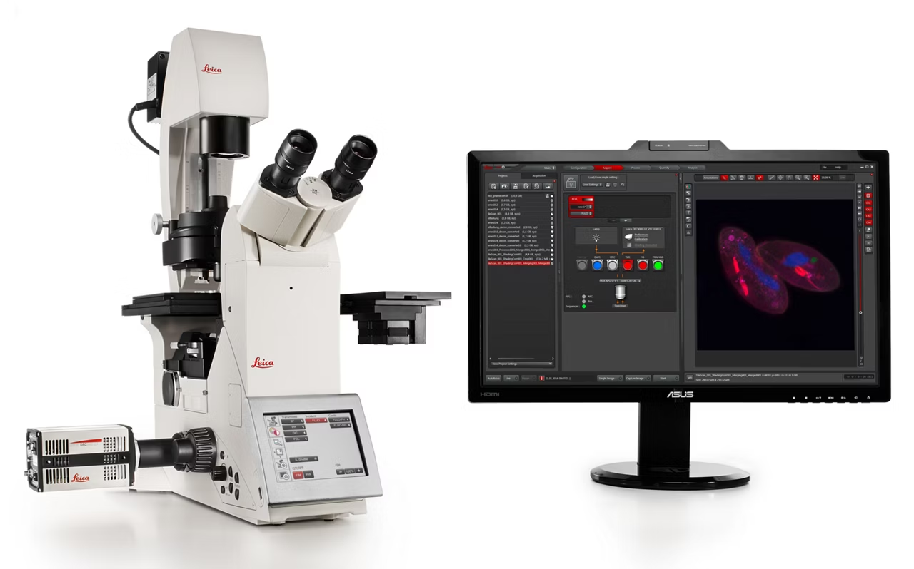
LAS X Life Science Microscope Software Platform
Software

LAS X Life Science Microscope Software Platform

Unified microscope software supporting confocal, widefield, stereo, super resolution, & light sheet systems. Save & recall settings for reliable results. Get quote.
K7 Color CMOS Microscope Camera
Instruments

K7 Color CMOS Microscope Camera

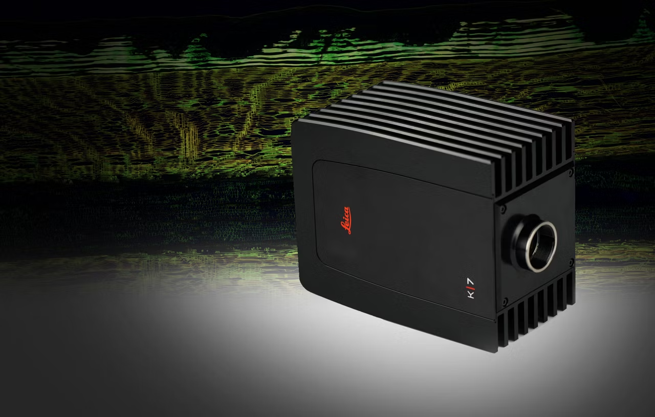
Capture K7 color CMOS microscope camera images for fluorescence and brightfield microscopy, fast scan speeds, high temporal resolution, CMOS sensor. Get a quote!
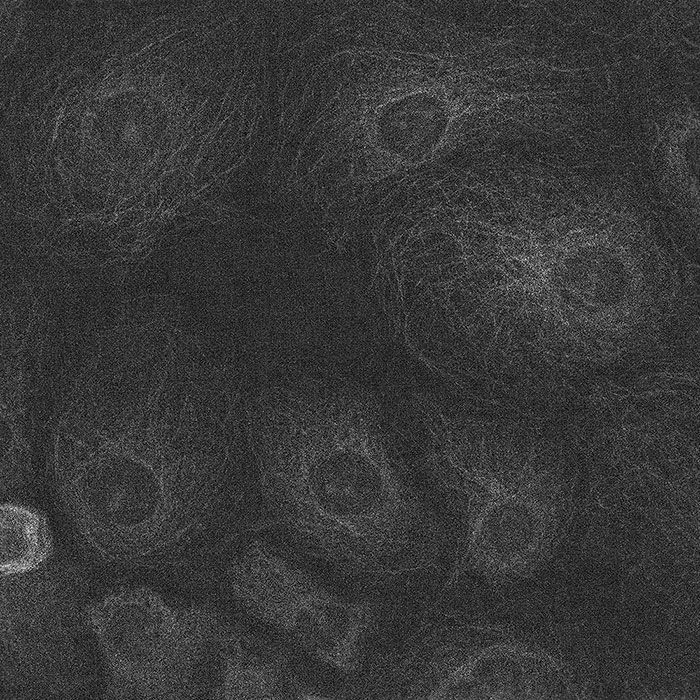
K8 Scientific CMOS Camera
Instruments

K8 Scientific CMOS Camera

Discover the K8 Scientific CMOS Camera for life science imaging. Achieve 95% quantum efficiency, low read noise and reduced phototoxicity. Request a quote!
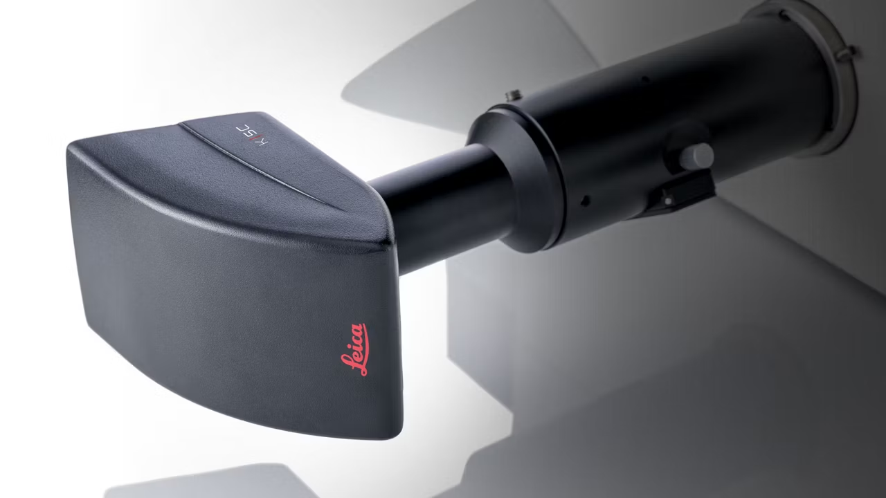
K5C The Color CMOS Camera
Instruments

K5C The Color CMOS Camera

Capture high-res 20 MP images with the K5C color CMOS camera. Features 32 fps live imaging, USB 3.0, TWAIN support, and LAS software compatibility.
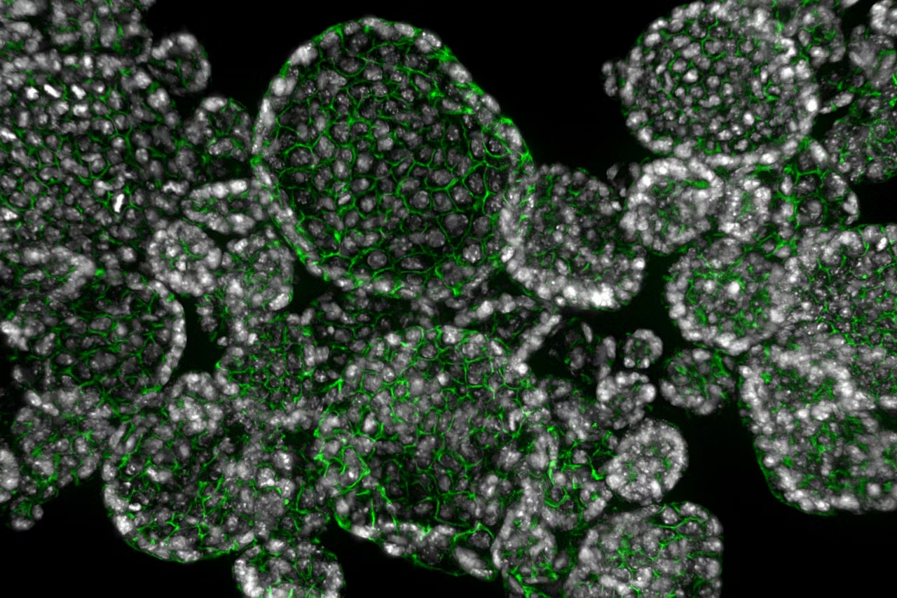
THUNDER Imager Cell
Instruments

THUNDER Imager Cell

THUNDER Imager enables precise 3D live cell imaging with low photobleaching, high-speed acquisition, and blur-free results in complex cell culture assays.
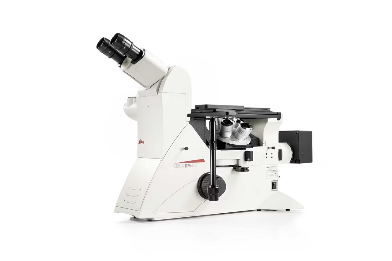
Leica DMi8 M / C / A Inverted Microscopes for Industry
Instruments

Leica DMi8 M / C / A Inverted Microscopes for Industry

Modular inverted microscope for metallography, medical device manufacturing, microelectronics. Supports multiple contrast modes. Request a quote!
THUNDER Imager Cell Spinning Disk
Instruments

THUNDER Imager Cell Spinning Disk

Combines Thunder technology with a spinning disk for clearer 3D sample imaging and higher quality data. Request a quote today.