THUNDER Imager Cell


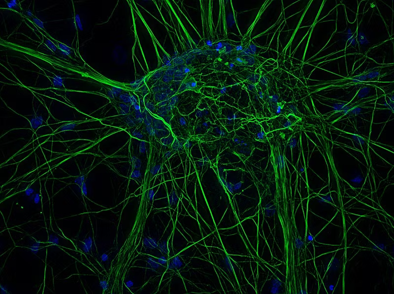


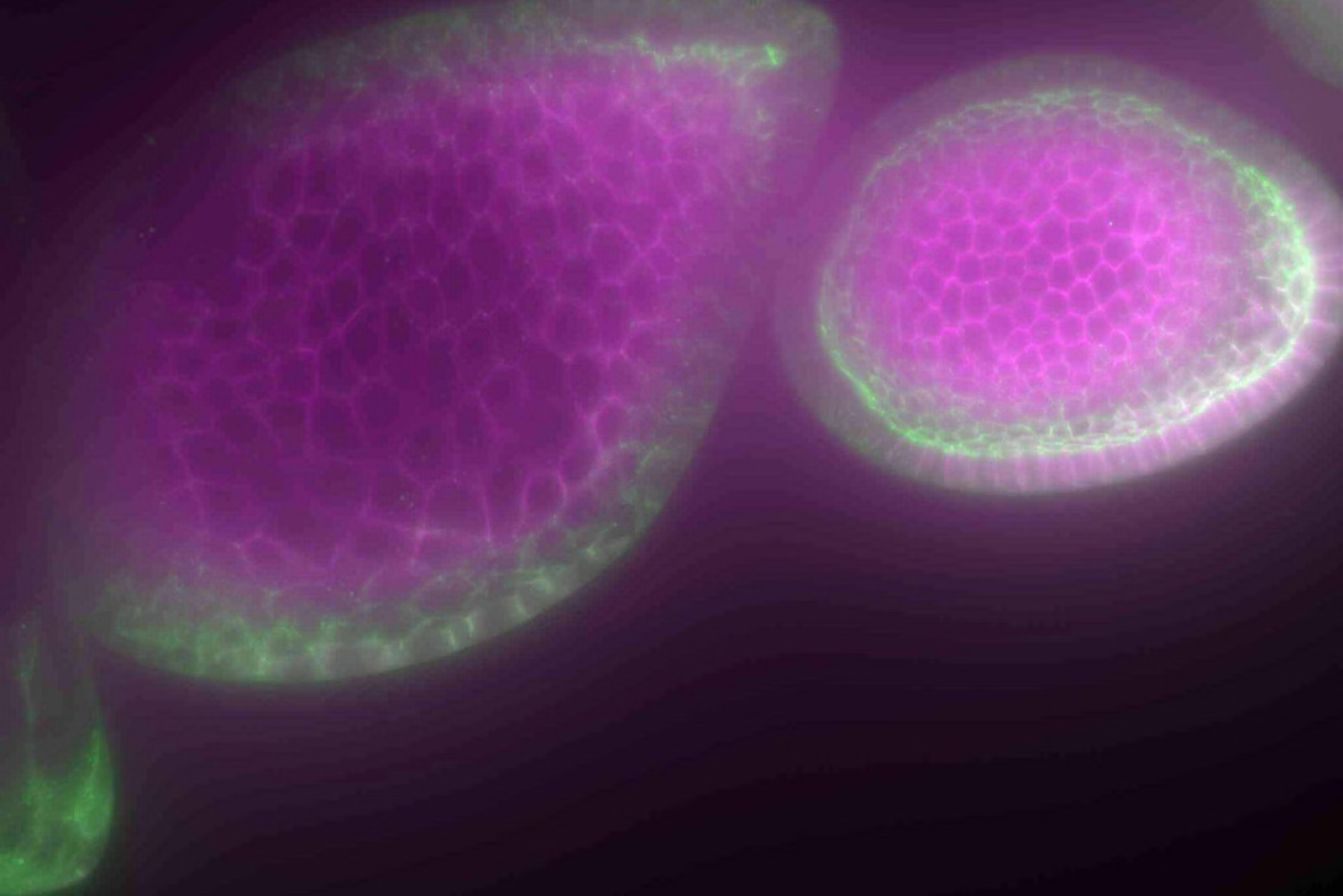
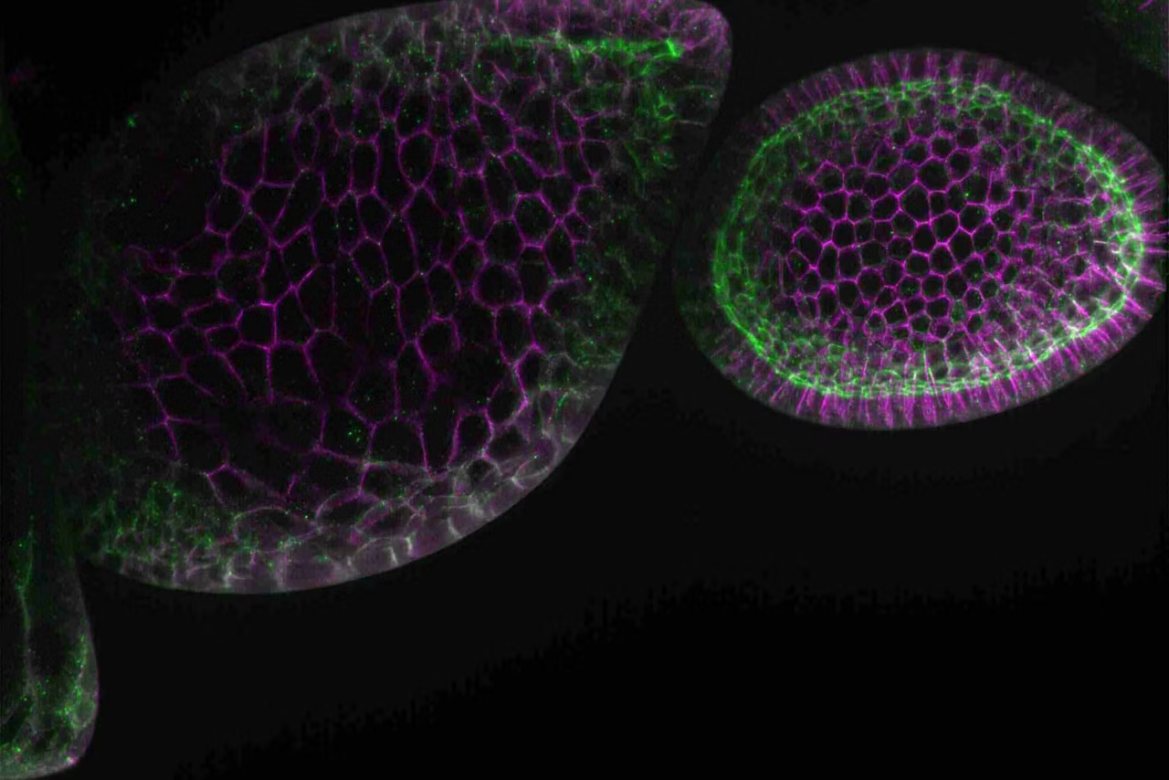

With THUNDER Imagers you begin every experiment with confidence. They are your solution for advanced 3D cell culture assays, whether you want to study stem cells, spheroids, or organoids.
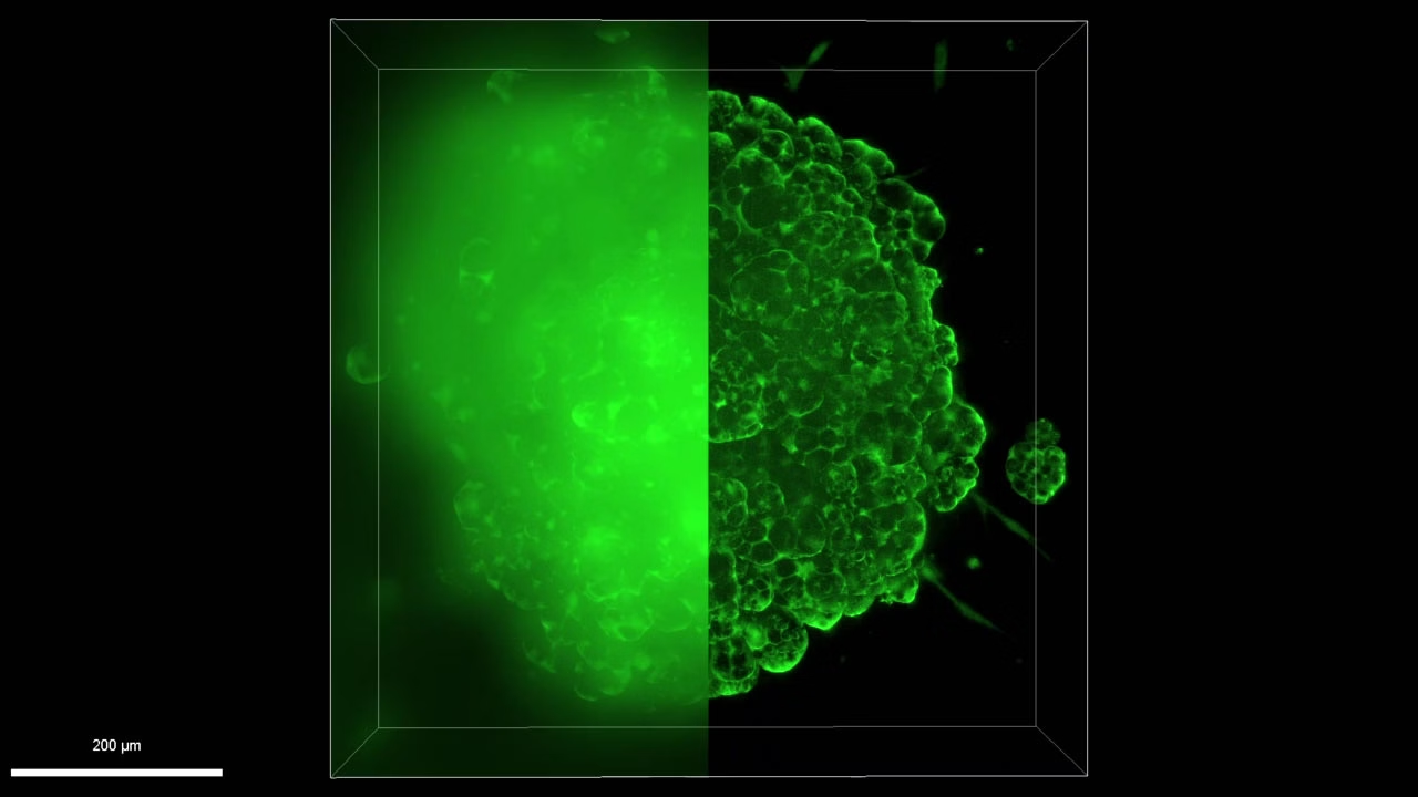
THUNDER Imagers feature innovative Leica technology Computational Clearing. It efficiently removes out-of-focus blur in real time, enabling the meaningful use of 3D specimens with camera-based fluorescence microscopes.
The high sensitivity of the system ensures low phototoxicity for physiologically relevant data sets. THUNDER systems offer a range of tools that assist you throughout your workflow from set up to data analysis.
*in accordance with ISO/IEC 2382:2015
Testimonial

THUNDER Imager Live Cell was for me the perfect solution to get very high-quality images and also spend little time because it's just so fast to acquire each individual slide. By using THUNDER’s Large Volume Computational Clearing, I was able to remove this typical widefield haze and distinguish between background and signal. And that gave me the perfect starting point for my downstream analysis
Yassin Harim, PHD Student, German Cancer Research Center (DKFZ), Heidelberg, Germany
High throughput for better statistics and workflow efficiency


Automate your 3D cell culture assays to study next-generation disease models efficiently. THUNDER enables you to image large sample volumes, such as lung organoids, at high speed. In addition, the automation minimizes user interaction even for complex experiments.
The outcome is:
- precise and reliable data in less time
- higher throughput
- improved analysis results for more reliable data
Optimal physiological conditions – minimum light exposure

THUNDER offers a range of solutions that allow you to acquire blur-free images of 3D samples with a single image.
This allows you to rapidly image over large areas, or at speeds of over 100 fps, at low light levels for physiologically relevant conditions, and gives you the freedom to set up a wide range of experiments.
Find the THUNDER Imager system that matches your needs

Whether you are looking for a dedicated high-end imaging system that excels in a given application, or a versatile solution for a lab running different kinds of assays with various samples, we’ve got you covered.
Here are some selected application examples that show you the strengths of THUNDER:
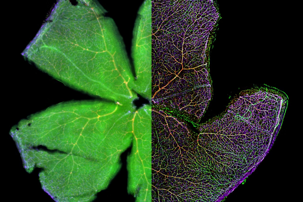
Mouse Retina.
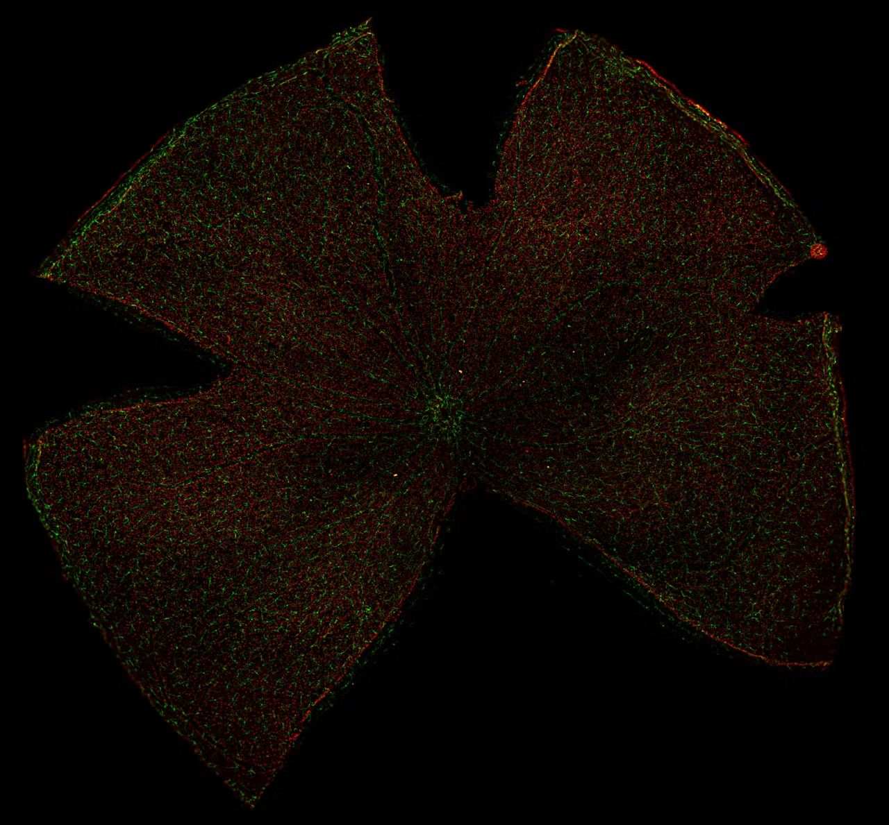
Reliable quantification of an entire mouse retina with THUNDER

Quantitative approaches to retina imaging analysis often require comprehensive descriptions of the retina morphology and function. Retina abnormalities as well as translational clinical applications need a reliable workflow to reproduce the transgenic target screening. Therefore, repetitive imaging of the morphology requires a system solution with accurate results which can be constantly reproduced. With the THUNDER Imager Cell you are able to clearly visualize the morphology and reliable count intracellular details, like single cell nuclei distribution in the retina.
With a THUNDER Imager Cell you have these advantages
- Immediate blur removal helps you visualize more intracellular details
- More utilizable depth with widefield approach
- Reliable quantification
- Ready-to-use for your particular workflow analysis
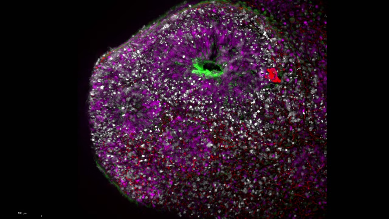
Brain Organoid.
Visualization of brain organoids with a THUNDER Imager Cell
Brain organoids as novel model systems are used to investigate human brain development and disorders. These self-assembled 3-dimensional cytoarchitectures are often characterized via multiple transgenic marker imaging. Classical challenges in these workflows are quantifying dynamics of molecules in time besides keeping physiological conditions and reaching sample depth at low signal levels. Therefore, the THUNDER Imager Cell is suitable to study the development of organoids close to physiological conditions, as our LED light sources help to minimize photobleaching. Furthermore, the timed detection of even though low protein signal levels is possible without the need to change the carrier.
With a THUNDER Imager Cell you have these advantages
- Possibility to observe your specimens in plastic bottom dishes makes your workflow more efficient
- Capacity to detect low signals of molecules with high QE camera
- Low bleaching and low sample perturbance with widefield approach and precise timed LED illumination
Explant Time-lapse.
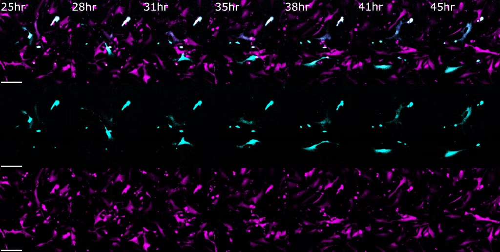
Low-phototoxicity in long-term live cell time-lapse imaging

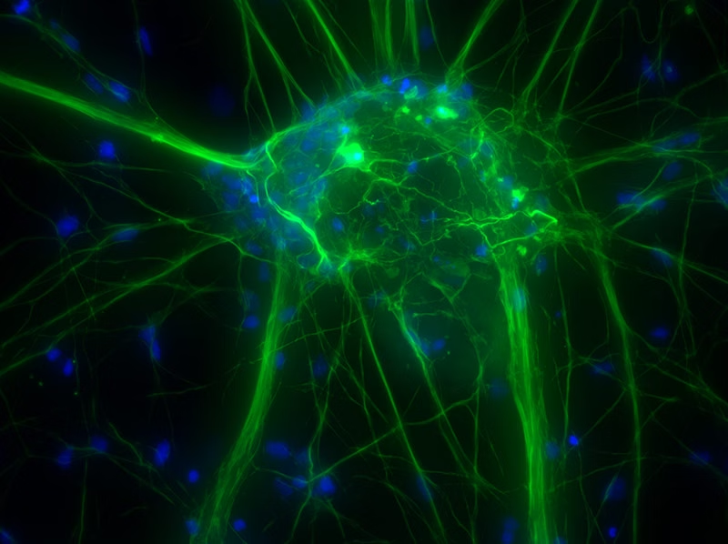
Explant cell cultures are oftentimes difficult to conduct imaging experiments with, as they require a stable cell culture environment and low-phototoxic imaging conditions. This example of explant cell culture imaging from Dr. Laura Shankman at the University of Virginia shows how abdominal aorta cells were able to be stably imaged over a 48 hour period. The THUNDER Imager Cell provides a complete microscopy imaging system for minimally invasive and precise live cell imaging experimentation. Sensitive cell culture experiments can be efficiently conducted thanks to fast and High-Quantum-Efficiency camera options, accurate stages, tunable LED light sources, Computational Clearing to reduce out-of-focus blur in widefield images, and an easy to use LAS X software to automate imaging and analysis workflows.
Advantages of the THUNDER Imager Cell
- Accurate live cell imaging experimentation allows tracking of fast cell movements
- Low phototoxicity keep sensitive cell culture alive, even in long-term experiments
- Speed-up your live-cell imaging workflow up to automated quantification and analysis
Multi position experiments: tracking cell changes
THUNDER Imager Cell offers both speed and reliability for your 3D cell culture multiwell experiments. For example, when tracking development of organoids, the system's speed and reliability allows you to obtain excellent results.
Accurate multi-position time-lapse experiments are aided by:
- Reliable focus drift correction with the Adaptive Focus Control (AFC)
- Software autofocus that compensates for changes in specimen position
- Reproducible Z-positioning with a precision of up to 20 nm (closed-loop focus)
The hardware triggered Quantum Stage moves rapidly and accurately to all positions with excellent repeatability (< ±0.25 µm).
Image at the speed of life – Cellular processes
Live cells contain a range of dynamic processes that require a system that can capture data with high temporal resolutions.
THUNDER Imagers only require a single fluorescence image to produce deblurred data. With THUNDER, you are only limited by the speed of your camera. THUNDER Imagers are offered with a range of cameras including the Teledyne Kinetix 22 allowing you to acquire data over 500 frames per second.
Decode 3D biology in real time*
Workflow efficiency

THUNDER Live helps you rapidly locate targets of interest for more efficient experimental set up
Improved analysis results

THUNDERed data sets enables improved object detection for better data analysis
Image at the speed of life

THUNDER offers simple, high speed 3D sectioning making it the ideal solution for imaging fast live process and large 3D samples
THUNDERed images are saved in the typical Leica file formats “lif”, “lof” and “xlef” (XLEF combined with TIF, JPEG, PNG, BMP or LOF). These established formats are compatible with image analysis software packages such as Aivia, and other analysis software that use open-source library bio-formats (I.e., FIJI and Omero).
Data can also be exported to the image formats TIF, JPEG, PNG and BMP and movie formats AVI, QuickTime, MP4 and WMV.
More about Thunder and Aivia.
Comparison to deconvolution: The core technology in all THUNDER Imagers is Computational Clearing. Computational Clearing removes blur in real time and extends the range of applications to thick samples compared to conventional Widefield techniques. THUNDER Imagers offer all the benefits of camera-based systems such as high acquisition speed, low light stress, high dynamic range, and high sensitivity, allowing imaging under physiological conditions. Image quality can be further improved by combining Computational Clearing with 3D deconvolution as offered in the methods Small Volume Computational Clearing (SVCC) and Large Volume Computational Clearing (LVCC). LVCC specifically benefits from Computational Clearing as it makes thick samples more accessible for deconvolution.
Comparison to other imaging techniques: another way to acquire optical sections with camera-based imaging systems is by using multiple-point illumination, such as a Nipkow disk or grid-projecting devices. The latter introduces artifacts whenever the grid cannot be projected sharply in the focal plane so they can struggle with thick specimens. In addition, they require multiple exposures for each image which slows down acquisition and may cause more photo damage to the sample. Disk-based systems, on the other hand, must deal with the finite distance between pinholes which introduces light contamination from out-of-focus planes at certain imaging depths.
The benchmark for optical sectioning of 3D samples are confocal point scanners which offer superior quality high-contrast fluorescence images. However, with point scanning confocal systems, imaging large samples can be time consuming.
THUNDER Imagers are integrated, well-established system solutions dedicated to specific applications. The THUNDER Imager family offers solutions for 3D tissue, 3D cell cultures, model organisms and CLEM applications.
THUNDER Imagers enable users to image thick samples that would be beyond the capabilities of conventional widefield systems by removing blur making thick samples accessible for analysis.
THUNDER imagers increase physiological relevance by using highly sensitive cameras that apply a minimal light dose to your sample. Furthermore, the statistical significance of the data can be increased as background independent quantification enables the detection and analysis of more objects in your images.
THUNDER Imagers minimize time to result by skipping the post processing step. 3D Widefield images require a deconvolution step prior to image analysis. This step is not necessary on a THUNDER Imager since the images are THUNDERed during acquisition and can immediately be used for analysis saving time.
From Organs to Tissues to Cells: Analyzing 3D Specimens with Widefield Microscopy - Bitesize Bio

Aivia AI Image Analysis Software
Explore AI Image Analysis Software transforms image processing with up to 74% faster analysis time. Achieve high-quality, reproducible results effortlessly.
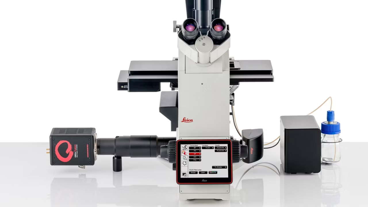
DMi8 Inverted Microscope
The DMi8 inverted microscope streamlines complex microscopy workflows delivering high quality data tailored to your research needs and budget. Get a quote!

Warranty Extension+
Full microscope coverage with annual maintenance, 3 business day on-site response, unlimited parts, and all labor included. Request a quote today!