T cells are the cornerstone of adaptive immune response, with direct and indirect mechanisms for recognizing and eradicating pathogens and tumors.¹ Their multifaceted defense mechanisms are further utilized in research and clinical applications, especially immunotherapy.² However, the success of these applications depends on efficient T cell activation and expansion. To achieve this, researchers must first isolate T cell populations from patients' peripheral blood samples. The quality and purity of T cells determine the viability and functionality of the T cell cultures.
Understanding T Cell Populations
T cells are central to adaptive immunity. Different T cell subsets are derived from naïve T cells upon activation by antigen-presenting cells, such as dendritic cells, co-stimulatory signals and cytokines. These subsets are distinguished by their surface markers and functions, primarily CD4+ helper T cells and CD8+ cytotoxic T cells.
CD8+ T cells directly kill infected or malignant cells by recognizing antigens presented as major histocompatibility (MHC) class I molecules. They form synapses with the target cells and release cytotoxic granules that induce apoptosis.³
On the other hand, CD4+ T cells support the adaptive immune response by releasing cytokines that activate CD8+ T cells and recruit B cells and innate immune cells. This helps maintain the intensity and consistency of the overall immune response. CD4+ cells further differentiate into subsets like Th1, Th2, Th17 and Treg, each with specialized roles.⁴
T Cell Isolation and Its Importance
Isolated T cells from blood samples give rise to scaled-up and engineered T cell cultures suitable for analysis, research and clinical applications, from T cell enrichment to CAR T cell therapies.²'⁵
Importance of Isolating Human Peripheral Blood Mononuclear Cells
Peripheral blood mononuclear cells (PBMCs) collected from donors are a rich source of T cells. Before further processing, T cells must be separated from the other lymphocytes and monocytes in the blood. Contaminants and other immune cell types in the culture might interfere with T cell activation and expansion.
Applications of Isolated T Cells in Research
Isolated T cells are expanded and engineered for a wide range of uses. Engineered and stimulated T cell populations are vital for immunotherapy, as they possess enhanced abilities to recognize and combat cancer and chronic infections.⁶ Furthermore, cell models generated from T cell isolates can be used as screening platforms for drugs that modulate or alter immune responses.⁷ From this perspective, T cell isolation is crucial for studying autoimmune disorders and transplant rejections, where T cell activity should be subdued.⁸'⁹
Featured Product
T Cell Isolation & Activation

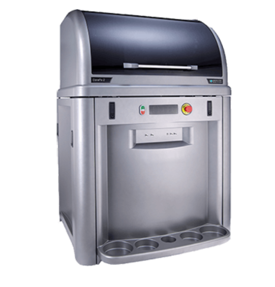
ClonePix® 2 Mammalian Colony Picker
A transformative cell line development workflow with automated clone screening and optional monoclonality assurance on day 0*
Overview of Common T Cell Isolation Techniques
Standard methods for extracting T cells from mixed cell populations include density gradient centrifugation and antibody-based separation. In density gradient centrifugation, cells are passed through a density gradient, where T cells can be separated from other cell types based on density differences. On the other hand, antibody-based separation relies on antibodies targeting surface markers specific to T cell subpopulations. Antibody-based separation involves magnetic- or fluorescence-activated cell sorting, yielding T cell populations with high purity and specificity.
Using Magnetic-Activated Cell Separation (MACS) for T Cell Isolation
MACS involves microbeads conjugated to antibodies that bind distinct T cell markers. The beads are passed through a magnetic column to separate the cells with the biomarkers from those without.
Flow Cytometry (FACS): A Method for T Cell Sorting
Instead of microbeads, FACS uses fluorescently labeled antibodies to label T cells, which are subsequently sorted via flow cytometry based on the number and composition of surface markers.
Commercial T cell isolation kits implementing the above techniques can accelerate isolation protocols and improve cell recovery and purity.
Activating T Cells in Culture
In vitro T cell activation aims to mimic the natural stimulation of T cells, triggering their proliferation and their regulatory or cytotoxic activity. Antibodies, such as anti-CD3, are combined with co-stimulatory signals (e.g., anti-CD28) and cytokines (e.g., IL-2) to trigger intracellular signaling pathways that lead to T cell proliferation and differentiation into effector and helper subsets. T cell markers, such as CD25, CD69 and CD71, which act as receptors for these signals, are particularly upregulated during the early stages of T cell activation.¹⁰
Culture Media Considerations for Cell Activation
Culture media composition and conditions determine T cell viability and functionality.
An optimum T cell media must contain a buffer system, proteins, vitamins, cytokines and nutrients. Commercial cell culture media, such as RPMI-1640 and AIM-V, are suitable for T cell culturing.¹¹ Although they lack the proteins and growth factors necessary for activation and growth, these can be supplied by adding fetal bovine serum to the medium.
There are many factors to consider when optimizing culture conditions:
- The culture should be kept at 37 °C to mimic the natural environment of the human body
- The CO2 levels must be kept at 5% to create the physiological pH levels
- The culture medium must be replenished continuously with nutrients
- Researchers must constantly monitor the cell culture for bacterial, viral and fungal contamination and frequently disinfect lab surfaces and equipment
Cell Expansion and Its Achievement
Clinical applications, such as adoptive T cell transfer and CAR T cell therapy, require large numbers of functional T cells, which can be achieved with robust T cell expansion protocols.
Understanding Cell Proliferation in T Cells
T cell proliferation requires combinatorial stimulation through T cell receptor (TCR) engagement, co-stimulatory signals and cytokines, mainly IL-2, IL-7 and IL-15.¹²'¹³ These signals mimic the natural T cell activation mechanism by triggering essential signal transduction and metabolic pathways. However, researchers need to pay attention to the duration of these signals, as prolonged stimulation may lead to T cell exhaustion.¹⁴
Using Expansion Kits for Enhanced T Cell Growth
Commercially available T cell expansion kits streamline and standardize in vitro T cell growth while eliminating risks of contamination and batch-to-batch variability. These kits typically include pre-coated beads or plates with anti-CD3/CD28 antibodies, optimized media formulations and cytokine cocktails.
Factors Influencing Cell Density in Culture
Cell seeding density is another crucial determinant of T cell growth. Research suggests that T cells need to come in contact with each other to induce growth. Therefore, a low seeding density leads to insufficient contact and hampers activation. On the other hand, overcrowding can lead to nutrient depletion and organic waste accumulation.¹⁵
Researchers should replenish the media with nutrients and adjust the culture volumes to accommodate the dynamic cell density throughout expansion.
Ensuring the Purity of Isolated T Cells
Although the density-based centrifugation and antibody-based cell sorting methods allow robust T cell isolation, the purity of the isolated cells should be monitored. Co-purification of B cells, monocytes or natural killer cells can interfere with the downstream analyses of T cell populations. Impurities can also reduce the efficacy of therapeutic T cell products.
Cell Purification Techniques for T Cell Samples
Post-isolation steps help ensure the purity of the T cell samples. Positive selection involves further antibody conjugation with magnetic beads or fluorescence tagging to target T cell surface markers like CD3. On the other hand, adverse selection separates unwanted immune cell types from the solution by labeling them with antibodies specific to their surface markers.
Depletion of Unwanted Cell Types
Targeted depletion involves antibodies against the surface markers, including CD14, CD19 and CD56, expressed on monocytes, B cells and NK cells.¹⁶'¹⁷ These cells are removed from the solution using magnetic beads or flow cytometry, ensuring that only the desired T cells remain.
Challenges Associated with T Cell Isolation and Activation
T cell isolation and activation can present technical challenges affecting yield, purity and functionality.
One common issue is low cell recovery, possibly due to suboptimal blood processing, prolonged handling times or improper centrifugation during PBMC isolation. Furthermore, incomplete T cell isolation can lead to contamination with non-T cells, affecting experimental outcomes.¹⁸
Weak stimulation during activation can result in low proliferation rates or incomplete activation. Weak activation could be attributed to degraded antibodies, incorrect bead-to-cell ratios or expired cytokines.¹⁵
Cell viability may decline during expansion if culture conditions are not carefully maintained. Issues such as pH shifts, nutrient depletion or temperature fluctuations can stress cells.¹⁹
To troubleshoot these problems, optimizing each step by using fresh reagents, validating antibody specificity and integrity and monitoring cell density and viability is essential. Commercial T cell expansion kits often help researchers bypass these steps by providing high-quality and pre-validated antibodies, reagents and culture media. Nevertheless, several parameters must be surveyed consistently, including but not limited to cell viability, size, volume, gene expression of activated T cell surface markers and metabolic activity.
See how Danaher Life Sciences can help
FAQs
What is a typical T cell isolation protocol and what reagents are required?
A typical T cell isolation begins with collecting peripheral blood and isolating PBMCs using density gradient centrifugation. T cells are then enriched via magnetic bead-based or fluorescent-based separation. Reagents include Ficoll for the centrifugation, MACS or FACS buffers and monoclonal antibodies.
How does T cell activation work and what are the key steps involved in the process?
T cells are activated by stimulating the TCR with anti-CD3 antibodies and co-stimulation via anti-CD28. IL-2, IL-7 and IL-15¹²'¹³ are added to promote proliferation. Activation induces surface markers like CD25 and CD69.¹⁰
How do different T cell activation protocols impact immune response?
Activation strength, duration and cytokine support affect T cell differentiation, memory formation and effector functions, influencing immune response quality.
How can T cell isolation and activation protocols be optimized for specific downstream applications?
Optimization involves adjusting cytokines, stimulation strength and purification strategies to suit applications like CAR T therapy, drug screening or immune profiling.
References
- Sun L, Su Y, Jiao A, Wang X, Zhang B. T cells in health and disease. Signal Transduct Target Ther 2023;8(1):235.
- Sterner RC, Sterner RM. CAR-T cell therapy: current limitations and potential strategies. Blood Cancer J 2021;11(4):69.
- Giles JR, Globig A-M, Kaech SM, Wherry EJ. CD8+ T cells in the cancer-immunity cycle. Immunity 2023;56(10):2231-2253.
- Xie L, Fang J, Yu J, Zhang W, He Z, Ye L, et al. The role of CD4+ T cells in tumor and chronic viral immune responses. MedComm 2023;4(5):e390.
- Titov A, Zmievskaya E, Ganeeva I, Valiullina A, Petukhov A, Rakhmatullina A, et al. Adoptive immunotherapy beyond CAR T-cells. Cancers (Basel) 2021;13(4):743.
- Ellis GI, Sheppard NC, Riley JL. Genetic engineering of T cells for immunotherapy. Nat Rev Genet 2021;22(7):427-447.
- Drijvers JM, Gillis JE, Muijlwijk T, Nguyen TH, Gaudiano EF, Harris IS, et al. Pharmacologic screening identifies metabolic vulnerabilities of CD8+ T cells. Cancer Immunol Res 2021;9(2):184-199.
- Tuomela K, Levings MK. Genetic engineering of regulatory T cells for treatment of autoimmune disorders including type 1 diabetes. Diabetologia 2024;67(4):611-622.
- Muller YD, Ferreira LM, Ronin E, Ho P, Nguyen V, Faleo G, et al. Precision engineering of an anti-HLA-A2 chimeric antigen receptor in regulatory T cells for transplant immune tolerance. Front Immunol 2021;12:686439.
- Mishra S, Telang G, Bennur D, Chougule S, Dandge P, Joshi S, et al. T cell exhaustion and activation markers in pancreatic cancer: a systematic review. J Gastrointest Cancer 2024;55(1):77-95.
- Watanabe N, Mo F, McKenna MK. Impact of manufacturing procedures on CAR T cell functionality. Front Immunol 2022;13:876339.
- Kim J-H, Lee K-J, Lee S-W. Cancer immunotherapy with T-cell targeting cytokines: IL-2 and IL-7. BMB reports 2021;54(1):21.
- Zhou Y, Husman T, Cen X, Tsao T, Brown J, Bajpai A, et al. Interleukin 15 in cell-based cancer immunotherapy. Int J Mol Sci 2022;23(13):7311.
- Baessler A, Vignali DA. T cell exhaustion. Annu Rev Immunol 2024;42(1):179-206.
- Ghaffari S, Torabi-Rahvar M, Aghayan S, Jabbarpour Z, Moradzadeh K, Omidkhoda A, et al. Optimizing interleukin-2 concentration, seeding density and bead-to-cell ratio of T-cell expansion for adoptive immunotherapy. BMC Immunol 2021;22(1):43.
- Wang X, Borquez-Ojeda O, Stefanski J, Du F, Qu J, Chaudhari J, et al. Depleting high-content CD14+ cells from apheresis products is critical for successful transduction and expansion of CAR T cells during large-scale cGMP manufacturing. Mol Ther - Methods Clin Dev 2021;22:377-387.
- Wang H, Tsao S-T, Gu M, Fu C, He F, Li X, et al. A simple and effective method to purify and activate T cells for successful generation of chimeric antigen receptor T (CAR-T) cells from patients with high monocyte count. J Transl Med 2022;20(1):608.
- Browne DJ, Miller CM, Doolan DL. Technical pitfalls when collecting, cryopreserving, thawing, and stimulating human T-cells. Front Immunol 2024;15:1382192.
- Amini A, Wiegmann V, Patel H, Veraitch F, Baganz F. Bioprocess considerations for T‐cell therapy: Investigating the impact of agitation, dissolved oxygen, and pH on T‐cell expansion and differentiation. Biotechnol Bioeng 2020;117(10):3018-3028.