What is STED Microscopy?
Stimulated emission depletion (STED) microscopy is a super-resolution microscopy technique that enhances resolution by selectively de-exciting fluorophores at the periphery of the focal point. In general, super-resolved microscopy methods enable visualization of nanoscale structures beyond the ~200 nm lateral and ~500 nm axial resolution limit imposed by the diffraction of light.¹ STED uses a spatially patterned depletion laser to break this barrier and minimize the fluorescence emission around the focal volume.²
The discovery of STED by physicist Stefan W. Hell was deemed a breakthrough because it allowed researchers to observe molecular structures and movement within living cells with superb detail. As such, it earned Dr. Hell the Nobel Prize in Chemistry in 2014.³
STED Microscopy Principle and Working Mechanism
The goal of STED microscopy is to confine fluorescence emission to achieve super-resolution. It sends two laser beams to the sample. The first is an excitation laser that briefly excites fluorophores within a diffraction-limited focal volume. Following this, a donut-shaped depletion laser beam is sent to selectively de-excite fluorophores in the outer regions of the excitation spot, leaving only the central region in an excited state.⁴
As a result, STED diminishes the microscope's point spread function (PSF), which is the three-dimensional image of point-like objects that light diffraction can form. In other words, STED reduces the effective fluorescence spot below the microscope's diffraction limit.⁵
STED variations include:
- Gated STED (gSTED) depletion beam is sent in pulses.⁶
- Continuous-wave STED (CW-STED) depletion beam is steady.²
- Fluorescence lifetime imaging (FLIM)-STED incorporates fluorescence lifetime measurements to separate signal from background.⁷
STED microscopy is particularly used for optical sectioning to minimize out-of-focus light and enhance contrast in thick specimens. Researchers gain precise control over the spatial extent of fluorescence emission, laterally achieving resolutions down to ~20–30 nm. Thus, they can directly visualize nanoscale molecular structures and arrangements far beyond the diffraction limit.⁶
Featured Product
STED Microscopy: An Overview


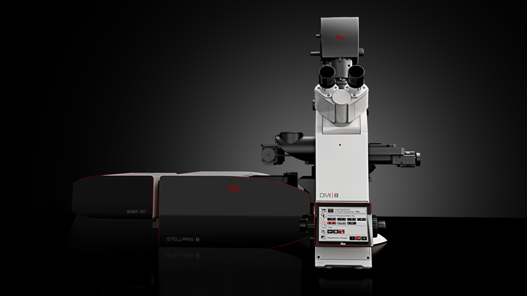
Leica Stellaris 8 STED Microscope
Our STED (stimulated emission depletion) technology joins the STELLARIS platform to provide you the fastest way of imaging beyond the diffraction limit. Obtain cutting-edge nanoscopy results in no time with astounding image quality and resolution, while protecting your sample. STED super-resolution allows you to study multiple dynamic events simultaneously, so you can investigate molecular relationships and mechanisms within the cellular context.
Components of a STED Microscope
A modern STED microscope integrates specialized optical components, lasers and detection systems to achieve nanoscale imaging resolution.
A STED microscope contains the essential pieces found in a confocal microscope, such as:⁸
- The excitation light source, which generates a light beam
- Dichoric mirrors that direct the light beam toward the objective lens
- The objective lens, which focuses the beam on the specimen.
- A scan unit moving the focal spot over the sample
- A detector that receives the fluorescence emitted by the sample.
The unique component is the STED light source, generating the STED beam that passes through a phase plate to assume a donut shape with zero central intensity. The STED source is designed to produce a wavelength that restores the ground state of the fluorescent molecules it encounters, leaving only the central molecules in their excited state.²
The fluorescent dyes used in STED microscopy must have high photostability, appropriate excitation/emission spectra and efficient stimulated emission cross-sections. Many commonly used fluorescent dyes, such as Atto 647N, Alexa Fluor 594 and Abberior STAR series, are suitable for STED microscopy. ²'⁹
Detector technologies and imaging software are crucial components of a high-quality STED microscope. Detectors with single-photon counting modules commonly used in confocal microscopy to reduce noise and improve sensitivity are ideal for STED systems. Furthermore, STED microscopy can be integrated with imaging software to synchronize laser pulses, scan the beam and perform real-time image reconstruction to enhance resolution and contrast.¹⁰
STED Sample Preparation and Imaging Workflow
The imaging workflow typically proceeds from fluorophore labeling to mounting and alignment, followed by laser power optimization, image acquisition and post-processing. During post-processing, deconvolution algorithms are used to maximize resolution and suppress noise. ¹⁰
Each workflow step must be carefully tailored to the cell types, research questions and resolution requirements.
As previously mentioned, fluorescent proteins and dyes must be photostable and compatible with the STED laser wavelength to withstand intense laser beams.⁹ Labeling density is also crucial to balance signal strength and specificity. The excitation and depletion lasers should be carefully synchronized. It is also crucial to fine-tune the phase plate to generate the donut-shaped depletion beam for uniform performance across the field of view.
Both fixed and live-cell samples can be imaged via STED microscopy, although each presents distinct considerations.
Fixed samples offer structural stability and reduced photobleaching, making them suitable for long acquisition times and high-resolution reconstructions. On the other hand, live-cell STED imaging presents challenges while enabling the study of dynamic processes. Phototoxicity caused by high laser intensities can alter cell behavior and survival, hindering long-term live-cell imaging capabilities. Therefore, live-cell imaging requires minimization of phototoxicity by using coverslips and optimum mounting media. Furthermore, time-gated detection and photostable dyes can help generate a strong contrast in low laser intensities. ¹¹
Applications of STED Microscopy in Biomedical Research
STED microscopy has empowered biomedical research by enabling nanometer-scale visualization of cells and biomolecular structures previously inaccessible with conventional light microscopy. Some examples include:
- Neuroscience: STED is used for resolving individual synaptic vesicles, mapping protein localization within synaptic clefts and elucidating the molecular organization of dendritic spines. Thus, it helps researchers correlate structural changes to learning, memory and neurodegenerative diseases.⁵
- Cell biology: STED reveals subcellular architectures and dynamic processes within the cell, crucial for understanding the fundamental mechanisms of cell signaling and intracellular transport. ¹²
- Biomedical diagnostics: STED microscopy can empower early disease detection by detecting subtle changes in biomarker distribution or nanoscale alterations in tissue architecture.¹³
- Drug discovery: STED enables visualization of drug–target interactions, therapeutics' intracellular trafficking, and drug-induced alterations in cell physiology. Thus, it can accelerate hit-to-lead optimization and uncover the drug's mechanism of action.¹⁴
Advantages and Limitations of STED Microscopy
The key advantage of STED microscopy is its ability to overcome the diffraction limit of light, achieving resolutions of 20–30 nm, compared to conventional confocal microscopy with ~200-250 nm resolution. Therefore, it is superior to confocal microscopy in resolving subcellular structures, protein complexes and molecular assemblies.²
Another advantage of STED microscopy is its capacity for real-time super-resolution imaging of live cell dynamics. STED is also highly versatile and compatible with multicolor imaging and fluorescence lifetime techniques, making it ideal for multiplexed biological studies.⁹
However, the method comes with notable limitations. STED systems are significantly more expensive and technically complex than standard confocal microscopes, requiring precise optical alignment and high-power depletion lasers. In addition, sample preparation can be cumbersome, limiting the choice of fluorophores to STED-compatible ones. More importantly, laser intensity should be carefully monitored, as high intensities can cause photobleaching or phototoxicity, obscuring the outputs of live-cell experiments.²
Despite these challenges, STED remains one of the most powerful optical imaging techniques, combining the high resolution of electron microscopy and the specificity of fluorescence-based imaging.
Combining STED with Other Super-Resolution Techniques
STED microscopy can be complemented with super-resolution methods to expand imaging capabilities and address complex biological questions. Combinations of STED include:
- STED + FLIM, which enables discrimination of signals based on fluorescence lifetimes,⁷
- STED + Structured Illumination Microscopy (SIM) for larger field-of-view imaging useful in dynamic cellular studies,¹⁵
- STED + single-molecule localization techniques, such as PALM (Photoactivated Localization Microscopy) or STORM (Stochastic Optical Reconstruction Microscopy), to observe molecular structures with varying levels of labeling¹⁶
- Hybrid workflows combining the strengths of each modality for increased versatility
Future of STED Microscopy in Life Sciences
Technological innovations are under development to make imaging with STED microscopy faster, smarter and more accessible.
AI-driven image reconstruction through deconvolution is a promising method for reducing noise, improving resolution and improving the accuracy of image analysis.¹⁷
Conversely, STED microscopes can be incorporated into automated imaging platforms to streamline workflows, minimize photodamage and protect cell integrity for long-term studies of dynamic biological processes. ¹⁰
Overall, STED microscopy will become increasingly important in precision diagnostics, biomarker discovery and drug development owing to its ability to visualize molecular interactions at the nanoscale and its integration with other imaging modalities and AI analytics.
See how Danaher Life Sciences can help
FAQs
Why is STED microscopy suitable for quantitative imaging?
STED provides nanoscale resolution beyond the diffraction limit, enabling precise measurement of cell molecular distributions and structural features. Its controlled fluorescence emission allows reliable intensity-based quantification.
How does STED work at the molecular level?
An excitation laser promotes fluorophores to an excited state, and a donut-shaped STED laser immediately induces stimulated emission at the periphery, leaving only a sub-diffraction central region fluorescent. This reduces the effective point-spread function.
How is sample preparation different for STED compared to conventional microscopy?
STED requires photostable, STED-compatible dyes or proteins, careful labeling to avoid excessive background and sometimes optimized mounting media to minimize photobleaching.
How does STED imaging help visualize live-cell subcellular structures?
By combining high-resolution imaging with time-gated detection and low laser intensities, STED allows observation of dynamic organelles, membranes and protein complexes in living cells.
What are common challenges in STED microscopy?
High system cost, technical complexity, photobleaching, phototoxicity and fluorophore limitations remain key obstacles.
References
- Abbe E. Beiträge zur Theorie des Mikroskops und der mikroskopischen Wahrnehmung. Archiv für mikroskopische Anatomie 1873;9(1):413-468.
- Lee D-R. Progresses in the implementation of STED microscopy. Meas Sci Technol 2023;34(10):102002.
- Hell SW, Wichmann J. Breaking the diffraction resolution limit by stimulated emission: stimulated-emission-depletion fluorescence microscopy. Opt Lett 1994;19(11):780-782.
- Xu Y, Xu R, Wang Z, Zhou Y, Shen Q, Ji W, et al. Recent advances in luminescent materials for super-resolution imaging via stimulated emission depletion nanoscopy. Chem Soc Rev 2021;50(1):667-690.
- Arizono M, Idziak A, Nägerl UV. Live STED imaging of functional neuroanatomy. Nat Protoc 2025:1-25.
- Jeong S, Kim J, Lee J-C. Suppressing background noise in STED optical nanoscopy. J Korean Phys Soc 2021;78(5):401-407.
- Torrado B, Pannunzio B, Malacrida L, Digman MA. Fluorescence lifetime imaging microscopy. Nat Rev Methods Primers 2024;4(1):80.
- Calovi S, Soria FN, Tønnesen J. Super-resolution STED microscopy in live brain tissue. Neurobiol Dis 2021;156:105420.
- Jeong S, Widengren J, Lee J-C. Fluorescent probes for STED optical nanoscopy. Nanomaterials 2021;12(1):21.
- Lukinavičius G, Alvelid J, Gerasimaitė R, Rodilla-Ramirez C, Nguyễn VT, Vicidomini G, et al. Stimulated emission depletion microscopy. Nat Rev Methods Primers 2024;4(1):56.
- Liu Y, Peng Z, Peng X, Yan W, Yang Z, Qu J. Shedding new lights into STED microscopy: emerging nanoprobes for imaging. Front Chem 2021;9:641330.
- Liu S, Hoess P, Ries J. Super-resolution microscopy for structural cell biology. Annu Rev Biophys 2022;51(1):301-326.
- Siegerist F, Campbell KN, Endlich N. A new era in nephrology: the role of super-resolution microscopy in research, medical diagnostic, and drug discovery. Kidney Int 2025.
- Yan J, Cheng L, Li Y, Wang R, Wang J. Advancements in Single-Molecule Fluorescence Detection Techniques and Their Expansive Applications in Drug Discovery and Neuroscience. Biosensors 2025;15(5):283.
- Chen X, Zhong S, Hou Y, Cao R, Wang W, Li D, et al. Superresolution structured illumination microscopy reconstruction algorithms: a review. Light Sci Appl 2023;12(1):172.
- Ghanam J, Chetty VK, Zhu X, Liu X, Gelléri M, Barthel L, et al. Single molecule localization microscopy for studying small extracellular vesicles. Small 2023;19(12):2205030.
- Ward EN, Scheeder A, Barysevich M, Kaminski CF. Self‐Driving Microscopes: AI Meets Super‐Resolution Microscopy. Small Methods 2025:2401757.