What is Cell Proliferation?
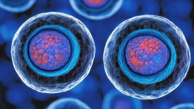
Cell proliferation is the process of expanding a cell population through the combination of growth and division. It is a fundamental biological process that influences early development, tissue and organ formation, wound healing, and, in the case of uncontrolled cell proliferation, cancer.
Cell proliferation plays a key role during cell line developement, which is carefully monitored and regulated. Cell line development is a critical step during the bioproduction of therapeutic proteins, gene therapies and vaccines in culture. Additionally, proliferative cell lines constitute a platform for disease research, drug discovery and cytotoxicity assessment.
Role in Cell Line Development
Cell proliferation plays an essential role in various biological processes, from embryonic development¹'² to tissue regeneration³ and cancer development.⁴'⁵ However, it also has transformative significance in life sciences and biotechnology.
In biotherapeutic production, cell line development starts with transfecting a construct of interest into a cell, followed by clonal selection based on successful DNA integration and product yield. Subsequently, proliferation becomes an important criterion for selecting and expanding top-ranked cells.
Cell proliferation remains fundamental throughout the maintenance and characterization of cell lines. Especially for longitudinal workflows used in disease modeling, drug evaluation, and cytotoxicity assessment, cells must be able to proliferate continuously to achieve consistency and continuity in research applications. To that end, researchers employ immortalized cell lines that either naturally harbor the capacity to proliferate indefinitely (e.g., HeLa derived from cervical cancer cells) or are genetically modified to display continuous proliferation (e.g., Human embryonic kidney 293 (HEK 293) cells or Chinese Hamster Ovary (CHO) cells).
Aberrant proliferation is observed in many cancers and is often targeted by anticancer drugs. Thus, cancer cell lines with highly proliferative phenotypes are convenient for examining the genetic mutations associated with proliferation and screening for drugs.
Key Regulators of Cell Proliferation During Cell Line Development
Understanding the mechanisms involved in regulating cell proliferation is necessary for maintaining candidates during cell line development.
The cell cycle is influenced by internal signals from various pathways that are initiated when growth factors, such as epidermal growth factor (EGF) and vascular endothelial growth factor (VEGF), bind to their specific receptors. Such binding events trigger signaling cascades that ultimately activate transcription factors involved in cell cycle progression.
The cell cycle is tightly controlled by cyclins and cyclin-dependent kinases (CDK), proteins determining whether the cell is suitable for proceeding from one phase to the next. Cyclin-CDK complexes regulate the synthesis of cell cycle-associated proteins during phase transitions, also known as checkpoints⁶. The regulation is strengthened by the activation of tumor suppressors, such as p53 and Rb, that prevent uncontrolled cell growth and division. Finally, small non-coding RNAs and miRNAs can strongly influence cell proliferation by modulating gene expression involved in protein synthesis, cell cycle progression, and cell death.
Cell proliferation is also influenced by external factors, such as nutrient and oxygen levels, which the cell needs to generate the energy necessary for growth and division. Considerations of cell type and culture conditions should influence the choice of bioreactor for optimal proliferation.
Importance of Bioreactors for Cell Line Proliferation
Bioreactors are functional platforms integral to the controlled proliferation of mammalian cell cultures for industrial applications. Bioreactors can be operated in three ways: batch, fed-batch, and perfusion methods.
Batch
A batch process is a closed system in which cells are suspended in a pre-defined culture medium without additional media and nutrients. Although batch processes are cost-effective and easy to operate and reproduce, they are susceptible to batch-to-batch variability and require precise control over parameters such as pH, temperature, and even distribution. Nevertheless, batch bioreactors provide a controlled environment and flexible design to allow cell proliferation for various cell lines.
Fed-batch
Fed-batch bioreactors combine elements of batch and continuous processes to ensure optimum growth, cell density, and product yield. Instead of adding the entire culture medium from the beginning, fresh medium is added throughout the process in a controlled manner, which mitigates nutrient depletion and starvation. Adding nutrients at specific time intervals also prevents the accumulation of metabolic byproducts that might interfere with cell proliferation. These advantages make fed-batch processes suitable for industry-scale cell line development.
Perfusion
Cell perfusion involves a continuous exchange of medium in a bioreactor. In addition to the continuous input of fresh media with nutrients, perfusion also encompasses the removal of cell waste and nutrient-deficient medium. The constant exchange enables a steady state with higher cell proliferation and productivity than fed-batch processes. However, perfusion is often more complex than fed-batch due to the need for sterilization and culture storage volume optimization.
Molecular Mechanisms in Cell Proliferation
Cell proliferation progresses through the following cell cycle phases:
-
The G1 Phase is associated with cell growth and metabolic activity, the synthesis of mRNA and enzymes required for DNA replication. Organelle duplication occurs.
-
The S Phase involves DNA replication
-
The G2 Phase is the final growth phase before cell division starts. The replication errors arising during the S phase are repaired.
-
The Mitotic (M) Phase involves four phases:
- Prophase: The nuclear membrane breaks down and releases chromosomes while the mitotic spindle forms.
- Metaphase: The released chromosomes align along the central plane of the cell, while the mitotic spindle binds to the centromere that links the sister chromatids
- Anaphase: The sister chromatids are segregated and pulled to the opposite sides of the mitotic spindle.
- Telophase: The chromatids reach the poles of the mitotic spindle. A nuclear envelope forms around the chromatids on each pole. The mitotic spindle disintegrates.
- Cytokinesis: Cytoplasmic division gives rise to two daughter cells⁷.
Understanding where in the cell cycle cultures are crucial for measuring productivity, viability and sustainability. Factors such as free nutrients, metabolic waste, dissolved oxygen and time between feeds can influence cell culture growth.
Measuring cell proliferation
Measuring cell proliferation is essential for evaluating the successful achievement of industry-scale cell lines. Furthermore, it is indispensable when screening drugs for efficacy and cytotoxicity.
A plethora of assays are employed in proliferation measurements by monitoring proliferation hallmarks, such as DNA synthesis and metabolism, using a variety of staining techniques. While some assays are suitable for directly monitoring cell proliferation, others measure it indirectly via cell viability.
DNA Synthesis-based Assays;
5′-bromo-2′-deoxyuridine (BrdU) assay
This assay uses the thymidine analog BrdU to detect cells undergoing DNA replication in the S phase. The assay works by introducing BrdU to the cell culture and incorporating it into the cellular DNA. As cells prepare for proliferation, BrdU replaces thymidine and is integrated into the replicated DNA. It is then detected using anti-BrdU antibodies that can be visualized or quantified via fluorescence microscopy, flow cytometry, or enzyme-linked immunosorbent assay (ELISA).
5-ethynyl 2´-deoxyuridine (EdU) assay
EdU is a nucleoside analog that works similarly to the BrdU assay. Instead of fluorophores, the newly synthesized DNA is labeled using click chemistry for fluorescent detection. EdU is more advantageous than BrdU, as it does not require DNA denaturation. EdU assay is also faster and less toxic than BrdU.
Nuclear Proteins for Cell Proliferation Measurements
Cell proliferation can also be directly measured by detecting cell cycle biomarker proteins. Antigen Kiel 67 (Ki-67) and phospho-histone H3 (PHH3) are the most common markers. Ki-67 is expressed during all cell cycle phases of proliferating cells (G1, S, G2, and M), while the phosphorylation of PHH3 on its serine-10 residue mainly takes place in the G2 mitotic phases. These proteins can be detected via immunohistochemistry, immunofluorescence, and flow cytometry with monoclonal antibodies and can be used for evaluating proliferation in cancer cell lines.⁸
ATP Cell Viability Luciferase Assay
ATP is a crucial marker of metabolic activity in live cells. This bioluminescence-based assay measures ATP amount indirectly by the amount used to convert Firefly luciferase to D-luciferin, which emits a flash luminescence. Luciferase/luciferin assays are particularly useful for real-time measurement of cancer cell proliferation⁹.
Dynamic Measurement of Cell Proliferation
Fluorescent dye is a powerful technique to capture cell populations undergoing division. While the agents used in this method are not fluorescent on their own, the products of their reactions in the cytoplasm emit fluorescence. 5(6)-Carboxyfluorescein diacetate N-succinimidyl ester (CFSE) is one such agent, which forms a fluorescent compound stably conjugating the amine groups on proteins in the cell. The fluorescence is passed onto the daughter cells, although its intensity is halved in every division. This allows the real-time tracking of cells as they divide while also enabling the measurement of the number of cell divisions. A similar reagent is Calcein-AM, which interacts with esterase to synthesize a green, fluorescent product. Additionally, staining can be used to detect dead cells to accentuate live cells. Propidium iodide and trypan blue are dyes that permeate only through dead cells and emit red fluorescence or are colored blue, respectively.
See how Danaher Life Sciences can help
FAQs
What is the role of growth factors in cell proliferation?
Growth factors are signaling molecules that stimulate cell proliferation by binding to specific receptors on the cell surface and activating intracellular signaling pathways that trigger cells to enter the cell cycle.
What are the key regulators of cell proliferation?
Key regulators include growth factors, cyclins and CDKs, tumor suppressors, oncogenes, microRNAs and the extracellular matrix.
What are some challenges associated with cell proliferation in cell line development?
There are several challenges that can obstruct protein production in proliferating cell lines.
- Overgrowth and clumping: Excessive cell proliferation can lead to overcrowding, nutrient depletion, and waste accumulation, which can negatively impact cell health and productivity. Furthermore, cells in excess may form clumps, which can hinder nutrient exchange and oxygen diffusion, further limiting growth and viability.
- Phenotypic Drift: Over time, cells in culture may undergo differentiation and become quiescent, losing their desired proliferative traits or functions that made them valuable for research or production. If uncontrolled, repeated cell divisions can also increase the risk of genetic mutations, which can lead to phenotypic variation and loss of functionality.
- Contamination: Bacteria, fungi, and other microorganisms can contaminate cell cultures, leading to cell death, product contamination, and compromised research results.
- Environmental factors: Factors such as temperature, pH, and nutrient availability can influence cell growth rates and create variability in culture conditions.
- Senescence and Apoptosis: Proliferating cell lines may eventually undergo senescence, a state of growth arrest that can limit their lifespan and productivity. Additionally, apoptosis, or programmed cell death, can occur in response to various stimuli, leading to a decline in cell numbers and hindering culture growth.
- Difficulties in Scale-Up: Cell growth rates may change as cultures are scaled up to larger vessels, making it challenging to maintain consistent conditions and productivity. Adequate oxygen and nutrient delivery can become more difficult in larger-scale cultures, potentially limiting cell growth and viability.
What are some common methods for measuring cell proliferation?
Common methods include counting cells, measuring DNA content, monitoring metabolic activity, tracking cell cycle markers, and using assays like BrdU incorporation or Ki-67 staining.
References
- Green RM, Lo Vercio LD, Dauter A, et al. Quantifying the relationship between cell proliferation and morphology during development of the face. Preprint 2023.
- Boehm B, Westerberg H, Lesnicar-Pucko G, et al. The role of spatially controlled cell proliferation in limb Bud Morphogenesis. PLoS Biology 2010;8(7).
- Jackson LN, Silva SR, Ueda J, Watanabe H, Evers B. PI3K/AKT activation is critical for hepatic regeneration after partial hepatectomy. Journal of Surgical Research 2006;130(2):301.
- Shapiro P. Ras-MAP kinase signaling pathways and control of cell proliferation: Relevance to cancer therapy. Critical Reviews in Clinical Laboratory Sciences 2002;39(4–5):285–330.
- Marei HE, Althani A, Afifi N, et al. P53 signaling in cancer progression and therapy. Cancer Cell International 2021;21(1).
- Malumbres M, Barbacid M. Cell Cycle, CDKs and cancer: a changing paradigm. Nature Reviews Cancer 2009;9:153-166.
- Schafer KA. The Cell Cycle: A Review. Vet Pathol 1998; 35:461-478.
- Aziz S, Wik E, Knutsvik G, et al. Evaluation of tumor cell proliferation by Ki-67 expression and mitotic count in lymph node metastases from breast cancer. PLOS ONE 2016;11(3).
- Teow S-Y, Liew K, Che Mat MF, et al. Development of a luciferase/luciferin cell proliferation (XenoLuc) assay for real-time measurements of GFP-Luc2-modified cells in a co-culture system. BMC Biotechnology 2019;19(1).
See how Danaher Life Sciences can help
Cell Proliferation
