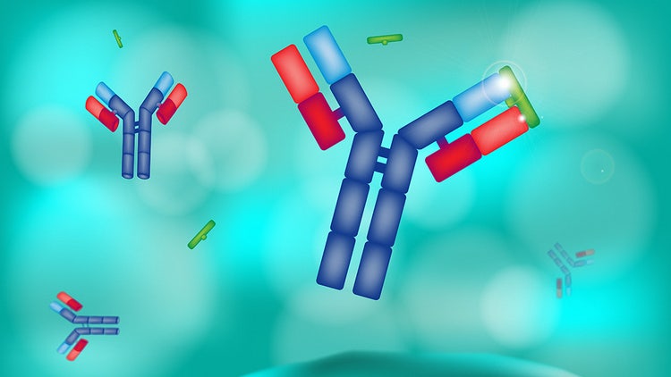
Antibody labeling is a critical technique in the biosciences field, enabling researchers to visually track specific antigens in complex biological systems. It hinges on attaching a detectable label to an antibody that binds selectively to its target molecule. This modification allows the labeled antibody to be visualized or detected by special methods, which provides valuable information about the distribution, abundance, and interactions of the target molecule in biological research. The importance of antibody labeling cannot be overstated—it transforms abstract biochemical interactions into tangible, visible phenomena, facilitating advancements in diagnostics, therapeutics, and our understanding of cellular processes.
Methods of antibody labeling
Direct labeling
In direct detection or labeling, the label and the primary antibody attach covalently. Generally, a fluorophore is covalently linked to a macromolecule, such as an antibody, and serves as a reporter molecule used to measure the presence of the macromolecule. These bioactive reagents are fluorescently labeled and are utilized in biological applications such as flow cytometry and immunofluorescence. Direct labeling has two main advantages: it requires only one incubation with the labeled reagent, which reduces the number of steps in the staining procedure and, most importantly, ensures minimal non-specific staining and less background.
Indirect labeling
An alternative method, indirect labeling, consists of an unlabeled primary antibody incubation followed by the addition of a labeled secondary antibody for detection. Any unbound antibody is removed, and the label is quantified. Factors such as cost, cell dysfunction, cell death, and immunogenicity are some of the limitations of indirect labeling.
Common labels used for antibody labeling
Fluorescent dyes
Antibodies labeled with fluorescent dyes and biotin are widely used in imaging, immune assays, flow cytometry, western blotting, and immunoprecipitation, among others. Fluorescein isothiocyanate (FITC) is an amine-reactive derivative of fluorescein and was one of the first derivatized fluorophores to be widely used. Derivatives of other fluorophores are also commonly used, such as rhodamine (TRITC, TAMRA), coumarin (available as amine- or carboxy-reactive derivatives), and cyanine (polymethine dyes that can be used at multiple wavelengths). All Alexa dyes and their conjugates are fluorescent and more photostable than their commonly used spectral counterparts listed above. Additionally, Alexa dyes are not sensitive in the pH range of 4-10¹.
Enzymes
Horseradish Peroxidase (HRP), a heme-containing oxidoreductase, is widely employed in biological applications, including western blots, ELISAs, and immunohistochemistry. Its ease of covalent addition, high enzymatic activity, adaptability, small size, stability, and cost-effectiveness have contributed to its 40-year-long use as the preferred antibody-enzyme conjugate.
Alkaline Phosphatase (AP), a hydrolase catalyzing phosphate group removal, is also commonly utilized in enzyme-linked immunoassays.
Transglutaminase (TGase) is a transferase that catalyzes the formation of covalent bonds between proteins or peptides and is utilized for site-specific protein conjugation, including antibody labeling.
Sortase (Srt), a transpeptidase, catalyzes the covalent attachment of a protein or label to a specific amino acid motif and is employed in site-specific protein labeling strategies.
Glucose Oxidase (GOx), an oxidoreductase, is used in antibody labeling as part of enzyme-linked immunosorbent assays (ELISA) and other biosensor applications. It catalyzes the oxidation of glucose, producing hydrogen peroxide as a byproduct, and is utilized in various immunoassays.
Applications enabled by antibody labeling
Enzyme-labeled antibodies play a crucial role in various analyses:
Western blotting detects proteins on membranes.
Classic ELISA detects antigens in samples using colorimetric or fluorescent reactions.
IHC (Immunohistochemistry) visualizes proteins in tissue sections using enzyme-catalyzed reactions.
Flow cytometry uses enzyme-labeled antibodies to quantify cell surface markers, aiding in cell characterization and sorting.
Quality control for antibody labeling
Quality control for antibody labeling involves several critical steps to ensure the reliability and reproducibility of biomedical research and diagnostic assays:
Verification of Specificity and Affinity
Conduct stringent testing to confirm that the antibody binds selectively and strongly to its intended target. Utilize techniques such as Western blotting, immunohistochemistry, and flow cytometry to assess cross-reactivity and binding behavior.
Optimization and Standardization of Labeling Process
Determine the efficiency of labeling, which is the proportion of antibodies successfully tagged with the label. Use absorbance measurements or signal intensity comparisons in assays like ELISA or fluorescence microscopy for quantification.
Assessment of Labeled Antibody Stability²
Evaluate the functionality and labeling efficiency of the antibody over time and under various storage conditions. Periodic re-evaluations using standard assays must be performed to ensure consistent performance.
Batch-to-Batch Consistency
Each batch of labeled antibodies should be tested for consistency in specificity, labeling efficiency, and performance. Routine quality checks must be implemented, and comparisons made between different batches.
Provision of a Detailed Certificate of Analysis
Essential information such as labeling efficiency, antibody concentration, and test results must be provided as proper documentation with each batch. This enables end-users to assess quality and suitability for specific applications.
Summary of methods, applications, and quality control considerations for antibody labeling
Antibody labeling is essential for the visualization of multiple antigens and offers advantages such as minimal non-specific staining. Enzyme labels such as bean peroxidase and alkaline phosphatase are common. Immunofluorescence is a powerful technique in a variety of applications. Quality control, storage, and continuous development of antibody-drug conjugates underscore the continued development and importance of antibody labeling in biomedical research.
The clinical development of protein therapies, such as antibody-drug conjugates, is increasing due to robust pipelines and successful outcomes in treating diverse diseases. Progress in monoclonal antibody development and conjugation chemistry has expanded the drug target space for antibody-based therapies, particularly in the case of antibody conjugates. These advancements are pivotal for the success of antibody conjugates.
See how Danaher Life Sciences can help
FAQs
What is antibody labeling, and what methods can be used for this process?
Antibody labeling involves attaching a detectable moiety, such as a fluorophore or enzyme, to an antibody to enable visualization or detection. Common methods for this process include chemical conjugation, enzymatic labeling, and biotinylation, each offering specific advantages depending on the experimental requirements.
What are the primary antibody labeling sites and available conjugation kits?
Primary antibody labeling sites include amine groups on lysine residues, thiol groups on cysteine residues, and sugar moieties on carbohydrate chains. Various commercially available conjugation kits are tailored for specific labeling sites, such as amine-reactive kits for lysine labeling or thiol-reactive kits for cysteine labeling.
What are some common reagents and enzymes used for antibody labeling and immobilization?
Common reagents and enzymes used for antibody labeling include NHS (N-hydroxysuccinimide) esters for amine labeling, maleimide for thiol labeling, and biotin for biotinylation. For immobilization, techniques involve physical adsorption, covalent coupling, or passive adsorption onto surfaces, with reagents like glutaraldehyde commonly employed in covalent coupling processes.
What considerations are important for quality control in antibody labeling?
Quality control involves defining antibodies with specific identifiers, providing detailed structural information, considering cross-reactivity data, and understanding the impact of matrix components. Storage conditions, including temperature and the use of stabilizing agents, are crucial for maintaining the activity of labeled antibodies over time. Continuous development, such as advancements in antibody-drug conjugates, underscores the evolving significance of antibody labeling in biomedical research.
How does indirect detection in antibody labeling work?
In indirect detection, an unlabeled primary antibody first binds to the target antigen, followed by washing away excess antibody. A labeled secondary reagent is then added, and the amount of label bound to the primary antibody is quantified. This method allows for signal amplification and facilitates various analytical techniques.
How does custom antibody labeling enhance experimental precision?
Custom antibody labeling allows a user to choose the most suitable label and conjugation method for specific experiments, minimizing non-specific staining and background. This tailored approach ensures optimal performance in a unique research context.
What is antibody fluorescent labeling, and how does it contribute to biological research?
Antibody fluorescent labeling involves attaching a fluorescent dye to an antibody, enabling its visualization under specialized methods. This process provides valuable insights into the distribution, abundance, and interactions of target molecules in biological research.
References
- Panchuk-Voloshina N, Haugland RP, Bishop-Stewart J, Bhalgat MK, Millard PJ, Mao F, et al. Alexa Dyes, a Series of New Fluorescent Dyes that Yield Exceptionally Bright, Photostable Conjugates. J Histochem Cytochem. 1999;47(9):1179-88.
- Hui Ma, Ciarán Ó’Fágáin, Richard O’Kennedy. Antibody stability: A key to performance - Analysis, influences and improvement. Biochimie. Volume 177. 2020
See how Danaher Life Sciences can help
Antibody Labeling