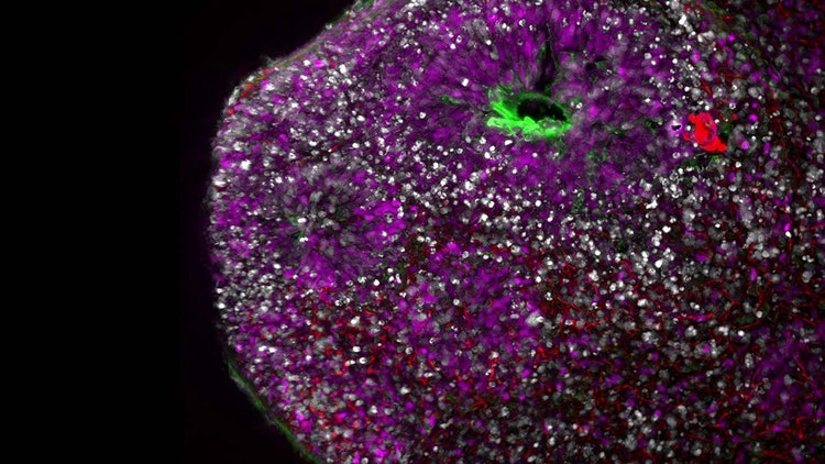
3D Analyses of Organoid Cultures

The increasing use of organoids in the preclinical validation of drug candidates requires advanced analytical tools to gather meaningful data on the biological processes and molecular interactions occurring within these 3D systems. Gathering accurate analytical information is vital as it ensures that drug interactions and physiological responses are understood across tissue, cellular and molecular levels, fostering the development of effective and safe therapeutics. Evaluation of molecule dosing through microscopy remains the principal method for assessing physiological responses. However, insightful analysis of organoid cultures still faces multiple technical challenges.
Imaging Acquistion
The current analytical methods for 3D organoid cultures rely mostly on morphological measurements (size, shape), fluorescent imaging and cell counting. These techniques play a pivotal role in phenotypic screening and gathering information on the impact of drugs on cell phenotypes.
However, the 3D nature of spheroid and organoid systems makes image acquisition challenging:
- Difficult image acquisition due to the depth and opacity of samples
- Frequent overexposure due to a need for long exposure times
- Ambiguous results that may be difficult to interpret
To address these challenges, hardware and software solutions that streamline organoid analysis are needed. For example:
- Combining an incubator with an automated imager minimizes the adverse effects of non-physiological conditions, allowing for the visualization of these 3D structures
- Leica Microsystems’ THUNDER computational image clearing enables high-quality imaging with reduced background noise and low phototoxicity
- Mica Microhub provides fully automated imaging, providing researchers with high-quality analytical imaging datasets from which to draw
Screening and Evaluation
Another challenge is the continuous evaluation of organoid growth and morphological characteristics. These dynamic tissues need constant monitoring, generating vast amounts of data. The Molecular Devices ImageXpress Confocal HT.ai High-Content Imaging System can help address this challenge. This automated solution allows high-content continuous screening of organoid cultures, reducing time and manual handling.
Manual imaging continues to be an important component of 3D cell culture analysis for evaluation screening. However, it presents several limitations:
- Researchers must manually select the organoid boundaries, which is time-consuming and subjective
- Observer variability during screening can lead to inconsistent results
- Large data volumes make manual information extraction and analysis unsustainable
Implementing tools based on AI/ML can address these challenges for organoid analysis during screening. While AI/ML tools will help interpret manually acquired images, these tools come into their potential when paired with large datasets.
Key benefits of AI-driven imaging and analysis include:
- Automated segmentation of complex 3D structures
- Faster phenotype quantification through IN Carta analysis software from Molecular Devices
- Removing subjectivity and increasing reproducibility in image analysis, Leica Microsystems Aivia AI Image Analysis Software offers researchers an analytical solution without requiring computational expertise
- Scalability for analyzing large batches of imaging data to eliminate laborious tasks and providing robust and reproducible segmentation results
As organoid research continues to evolve, implementing robust analytical tools is becoming imperative for streamlining the application of 3D cultures in preclinical testing. The Life Sciences companies of Danaher are committed to helping scientists automate, digitize and scale their complex 3D workflows and achieve their next big breakthroughs.
To learn more, visit our integrated Human Relevant Model and Spatial Profiling solutions or contact an expert today.

Bridging the Gap: Advancing Human-Relevant Models for Real-World Impact Webinar
