We See a Way to
train users 50% faster to image organoids and 3D cultures.
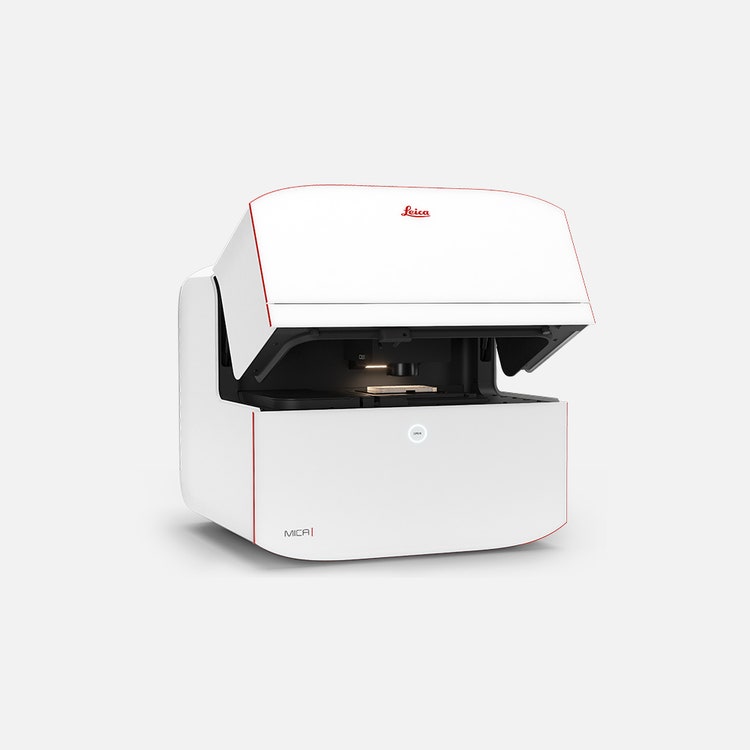
Leica Microsystems | Mica – The world’s first Microhub
Focus on Your Science, Not Figuring Out Your Microscope.


This changes everything.
More than a highly automated microscope , Mica unites widefield and confocal imaging in a sample protecting, incubating environment. With the simple push of a button , you have everything you need - all in one place - to transform your imaging workflows and get meaningful scientific results faster.
Access for all
Now everyone can leverage microscopy to make more discoveries. Mica provides a clear sample overview and allows observation conditions to be easily changed with just a few clicks.
- 85% fewer steps to the first image
- 33% less time to the first image
- 50% of the training time
Achieve physiological-like conditions throughout your experiments
Live cell experiments require cells to be in optimal shape. Typically, 3D cells in media require the temperature and the pH (via CO2) in the environment to be controlled. Stable nutrition and ion concentrations require the evaporation to be minimal. Some experiments even demand the O2 to be mimicked closer to physiological levels. Mica can provide the right conditions in the live cell configuration.
- Mica is an incubator : the entire encapsulated inner sample space can be climate controlled (temperature, CO2 and humidity regulation) and offers ideal conditions for short and long-term live cell observation.
- From dark to light : Mica also enables you to enjoy a brightly lit lab—freeing you from the constraints of sitting in a dark room for hours monitoring your experiment.
4x more data with 100% correlation
Mica enables you to simultaneously capture all 4 labels of different structures in a single acquisition for widefield or confocal, without ever moving your sample . This overcomes the spatiotemporal mismatch between labels of moving objects during sequential acquisition. All powered by patented FluoSync technology , a fast and gentle method for multicolor fluorescence imaging.
Select the right modality in real time
Mica unifies transmitted and fluorescence light imaging modalities . You can select from multiple imaging modalities all within one Microhub, including widefield, confocal, THUNDER imaging, LIGHTNING, Z-stacks, time-lapse and more.
This enables you to:
- Generate fast overviews with widefield at low magnification
- Gradually zoom in on the regions of interest
- Switch to confocal when and where needed without ever moving the Sample to a different system

3D Cell Culture, 7 day spheroid formation of U343 cells. tfLC3 EGFP and mRFP + DAPI + WGA Alexa 680. Objective: 20x/0.75 CS2 DRY. Acquired using Mica in confocal mode with LIGHTNING applied.
Radically simplified workflows
Intelligent automation and AI-supported analysis enables greater efficiency.
- Reduce over 60% of process steps through system intelligence
- Reduce time and effort from sample to insight by simplifying your entire workflow
- Enable 100% reproducibility and repeatability throughout your experiment
Speak to one of our leading life sciences experts
TTAE
TTAE
546006278
TTAE
add
Talk to an Expert
https://lifesciences.danaher.com/
https://lifesciences.danaher.com/us/en/solutions/mabs/cell-line-development.html