Leica M205 FCA & Leica M205 FA Fluorescence stereo microscopes
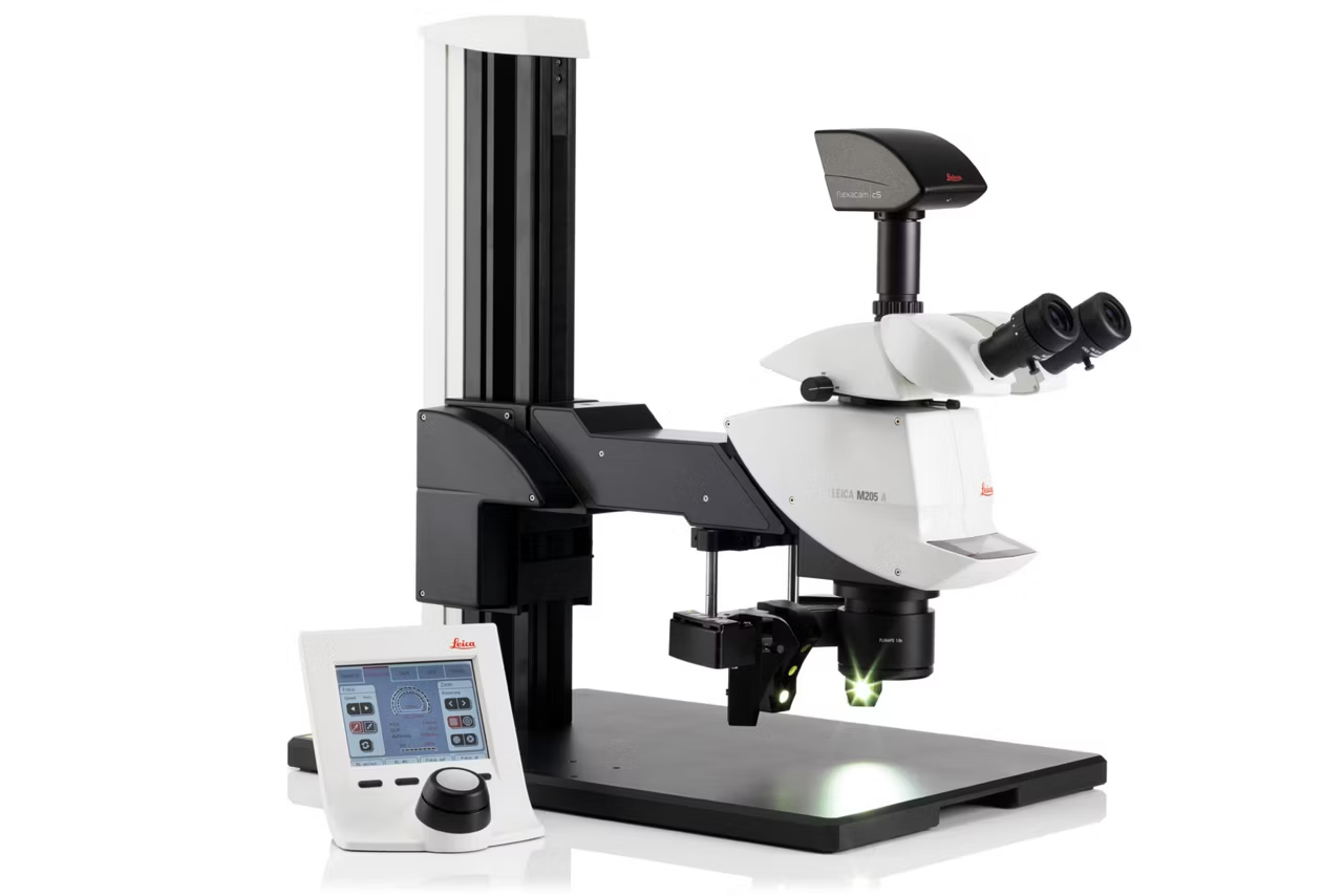
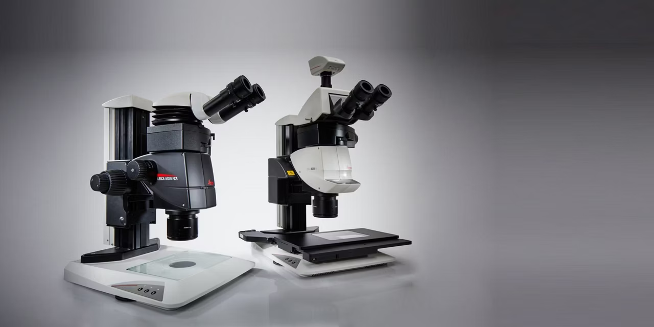
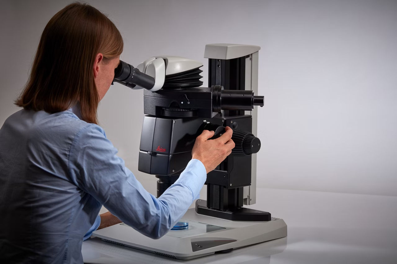
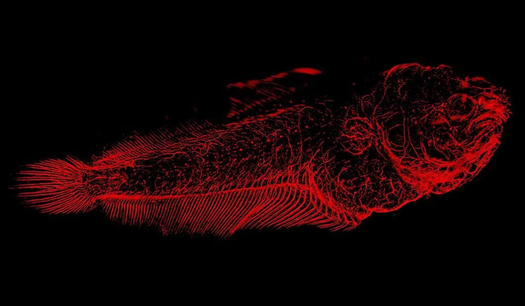
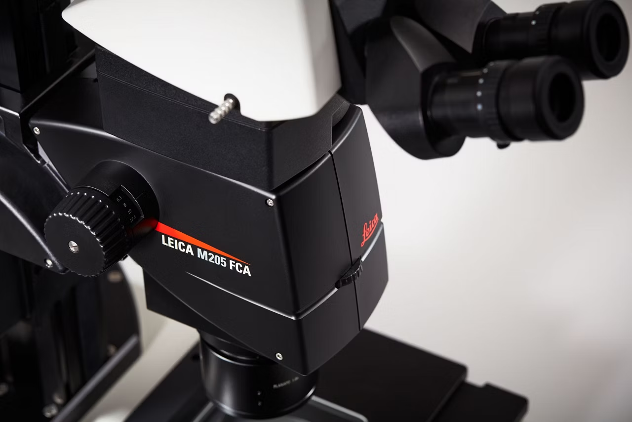

Do you sometimes feel like fishing in the dark because you are looking for fluorescence signals that are increasingly faint and weak? We have developed M205 FA and M205 FCA fluorescence stereo microscopes to enable you to detect transgenic expression like GFP and mCherry in early stages, allowing you to select the right sample to successfully base your studies on.
Discover the automated world of research

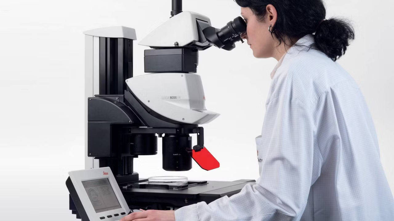
The Leica M205 FA opens up a new world of research in fluorescence microscopy, for instance when working in a sterile cabinet.
- Easily handle complex multi-channel fluorescence imaging procedures
- Execute demanding experiments with the motorized zoom, filter changer, Fluroescence Intensity Manager (FIM), and iris diaphragm
- Get high resolution at the stage level: the Leica LMT 260 scanning stage positions your sample in the sub-um range and holds life cell incubation equipment
Leica M205 FCA: semi-automated with manual zoom

Until now, you probably had to switch between two different systems: one for fast screening with a manual zoom that is intuitive to maneuver and a high-end solution to see and capture the faintest signals in an image.
The Leica M205 FCA fluorescence stereo microscope unites both worlds and combines super-fast manual screening with high-end imaging capabilities.
Now you can have it all:
- Get highly reliable and publishable results as all parameters are always auto-stored for you
- Eliminate interruption in your workflow with the four position coded filter changer
- Add a footswitch for easy change of filters, focus, and illumination adjustment, keeping both hands free for screening
Bright fluorescence signals – always.
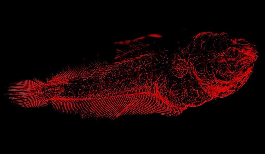
To detect even the faintest signals of your sample, you need an energy-rich excitation light to give a bright fluorochrome signal. However, the excitation light may cause reflections that blur the black background and compromise the detection of fluorescence signals.
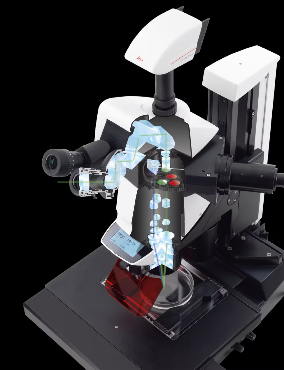
Leica’s TripleBeam technology completely eliminates this background “noise” by introducing a third optical zoom. This separates the fluorescence excitation light from the two observation channels and omits the need for a dichroic mirror.
The result is a clear and strong fluorescence signal against a noise-free, black background.
Finest details in 3D

Do you think that high resolution and maximum depth of field are irreconcilable opposites in microscopy? We prove they are not! Leica Microsystems' FusionOptics technology overcomes optical limitations by using the two beam paths for different tasks:
- The right channel delivers a high-resolution image at the largest possible numerical aperture.
- The left channel presents an image with high depth of field.
The result: you perceive an image with outstanding richness of detail and exceptional depth of field at the same time
Microscopic resolution in a stereo
Recognizing fine details is vital in research especially when working with small organisms. The Leica M205 FA and M205 FCA can achieve a resolution of 1279 lp/mm or 0.78 µm based on the resolution limit defined in ISO18221*.
The 2.0x PlanApo objective is an optical masterpiece. It has a maximum aperture of 0.35 – that’s the highest numerical aperture (NA) that has ever been achieved with a stereo microscope. This is smaller than a tenth of the diameter of a human red blood cell.
*M205 FA, 2x PlanAPO objective, camera adapter 0.63x, DMC4500 camera in full frame mode, Apple 5K with 27” monitor.
Speed matters – encoding helps
Image Coding provides convenient, reproducible settings for quick, easy documentation.
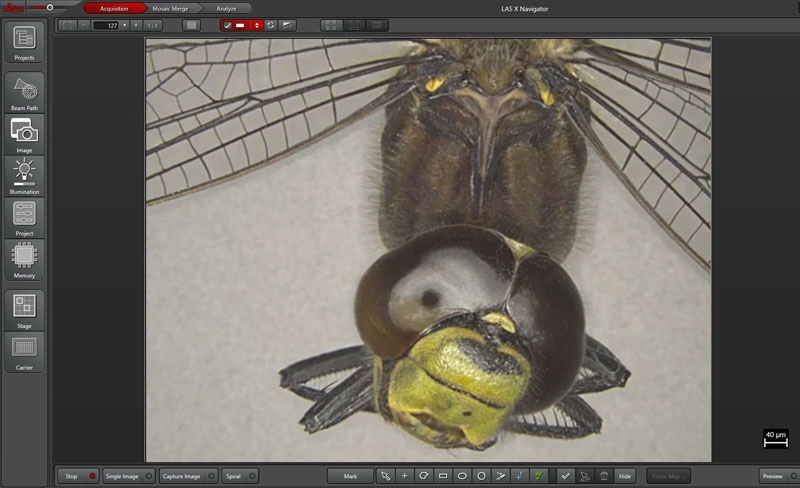
The integrated encoding transmits both the magnification and the position of the iris diaphragm to the software in real time. A scale bar is overlayed into the live image and updated when the magnification is changed. When an image is stored, all the settings are saved with the image and can be recalled at any time.
- Coded components offer ease of use and reliable results even for untrained operators
- The microscope system is intelligently linked to the software to allow you to change settings without having to manually adjust the calibration
- TL5000 Ergo transmitted light base adapts the aperture automatically to the zoom position to deliver optimal contrast
2 x CORR objective – making it clear!
With the Leica PLAN APO 2.0x CORR Objective, the refractive index can be adjusted – this way you will have pin-sharp images even if there is a water column of 5 mm between your sample and the objective. The objective enables users to observe and document samples as if the water did not exist.
When observing samples immersed in a watery solution, structures tend to blur especially in high magnification. This is due to the refractive index mismatch of air (index = 1) and water (index = 1.3). Potentially interesting or important structures may be easily misinterpreted because of spherical aberrations that may occur, as regular objectives are dedicated to the use with samples surrounded by air only.
Explore LAS X Software for Life Sciences

LAS X is the software platform for all Leica microscope solutions. Run complex fluorescent experiments with ease. LAS X guides you step-by-step through the entire analysis workflow.
- Live data mode: design experiment patterns and control your environment
- Extended depth of field (ED(O)F)
- Create unparalleled large overview images combining deconvolved z-stacks with XY
Full automation – full confidence: M205 A

Do you perform repetitive tasks at the microscope that require high precision and accuracy? Then you will appreciate the fully automated M205 A. Its intelligent automation makes you run your processes with a few mouse clicks - saving time while maintaining confidence in your results.
Streamline your daily workflow
Operate all functions of this high-performance stereo microscope using the user-friendly SmartTouch control unit or Leica software allowing you to work nearly hands-free.
Capture bright images – fast!
Discover the automated world of research

The Leica M205 FA fully motorized fluorescence stereo microscope opens up a new world of research in fluorescence microscopy
Bright fluorescence signals

With separated, yet synchronized beam paths, one for the fluorescence excitation light and two for observation, the M205 FCA delivers an evenly and fully illuminated field of view at any zoom position you choose
Finest details in 3D

FusionOptics technology overcomes optical limitations. Uniting high resolution and depth of field (DOF) it provides increased image brightness and facilitates orientation in 3D
Speed matters – encoding helps
Speed matters – encoding helps

Image Coding provides convenient, reproducible settings for quick, easy documentation
K5 Scientific CMOS Microscope Camera
Discover the K5 sCMOS Microscope Camera: 4.2MP resolution, 80% quantum efficiency. Ideal for fluorescence imaging, immunostaining, and 3D cell cultures.
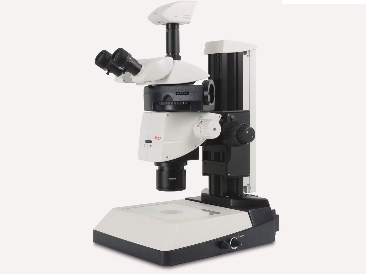
M165 FC Fluorescent Stereo Microscope
Explore the Leica M165 FC fluorescent stereo microscope, featuring 16.5:1 zoom optics that resolve structures down to 1.1 microns & enhanced image coding.
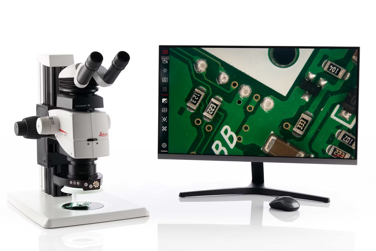
M125 C, M205 C & M205 A Encoded stereo microscopes
Leica M125 C encoded stereo microscope with encoding & FusionOptics, delivering calibrated & high-quality images. Check Leica M125 C price. Order now!
TL3000 Ergo & TL5000 Ergo Transmitted Light Bases
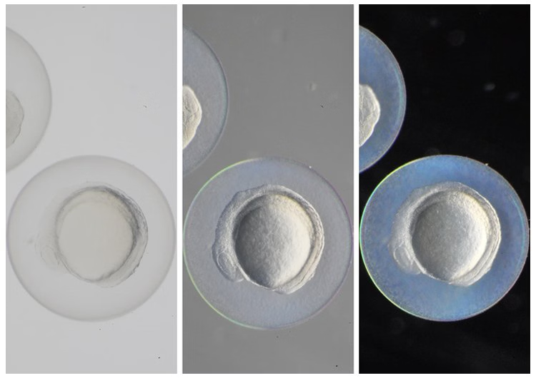
TL3000 and TL5000 Ergo transmitted light bases offer up to NA 0.9 resolution, high color fidelity, and contrast methods for detailed sample analysis. Get quote!
LAS X Life Science Microscope Software Platform
Unified microscope software supporting confocal, widefield, stereo, super resolution, & light sheet systems. Save & recall settings for reliable results. Get quote.
LED5000 RL Integrated LED Ring Light for M-series Stereo Microscopes
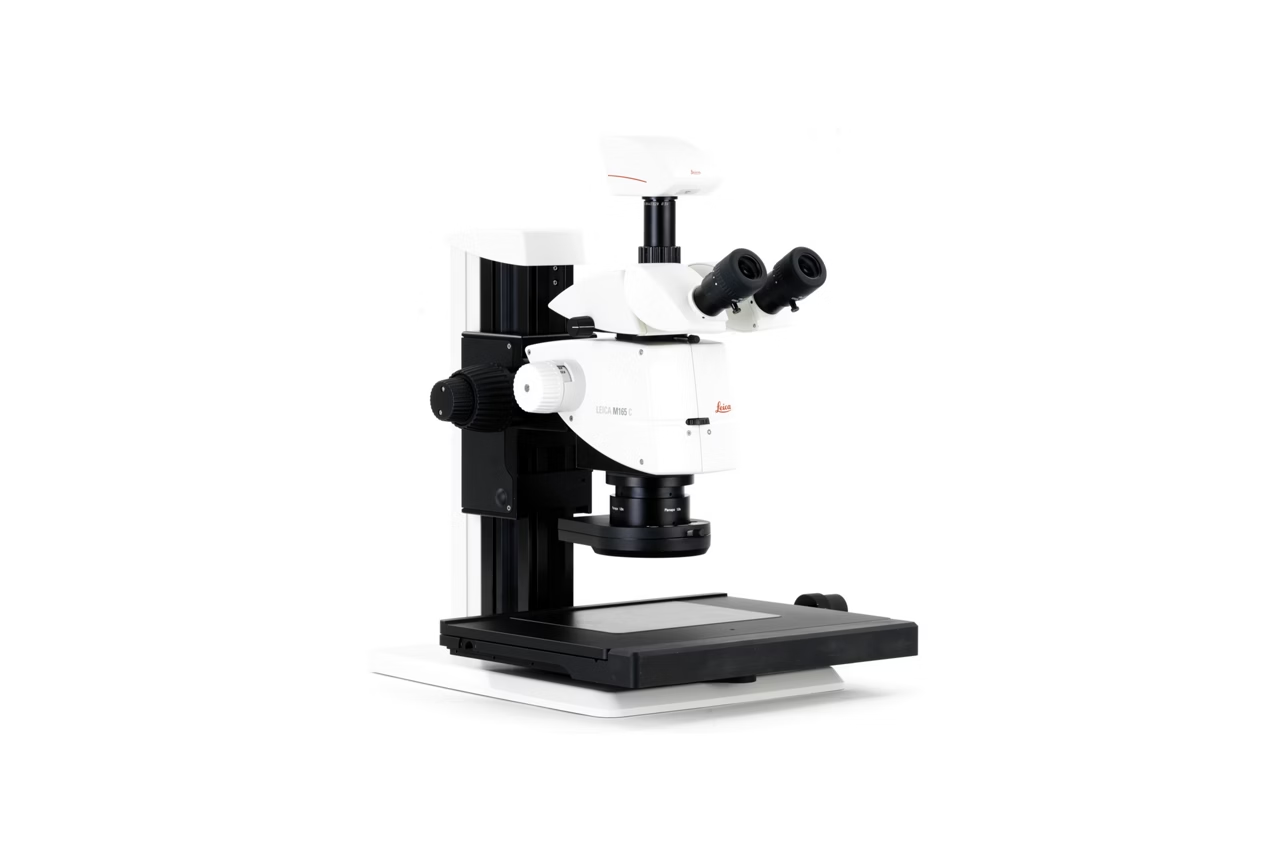
Ring light illumination with 40 LEDs, up to 50000 hours of flicker free daylight quality light and true color reproduction. Request a quote today.