JavaScript is disabled in your browser. Please enable JavaScript to view this website.

DMi1 Inverted Microscope for Cell Culture

$undefined
CellCheck 17 Cells Grow Divide Kid21756203 1

DMi1 21 Poster RGB HR Kid21757689

Leica DMi1 03 LabScene 07 Kid21765781

Leica DMi1 03 LabScene 18 Kid21765782

Leica DMi1 06 Workspace RGB HR Kid21765783

Dmi1 Expansion 05 Kid21757696

Dmi1 Objectives 07 Kid21757698

Dmi1 Sliders 07 Kid21757700

Dmil Lms Shopable Products Banner Images Kid21757802

Leica Dmi1 01 Kid21766210

Lms Dmi1 Feature Kid21766805

Lms Dmi1 Illumination Background Kid21766806

DMi1 Animation01 AK03 140516 23 Kid21757694

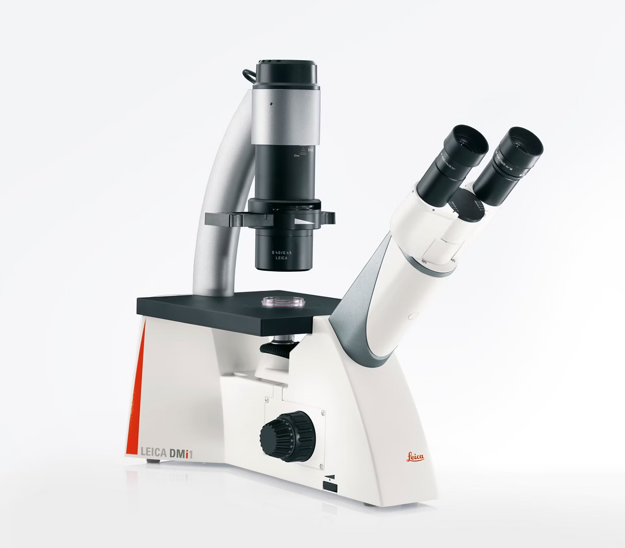
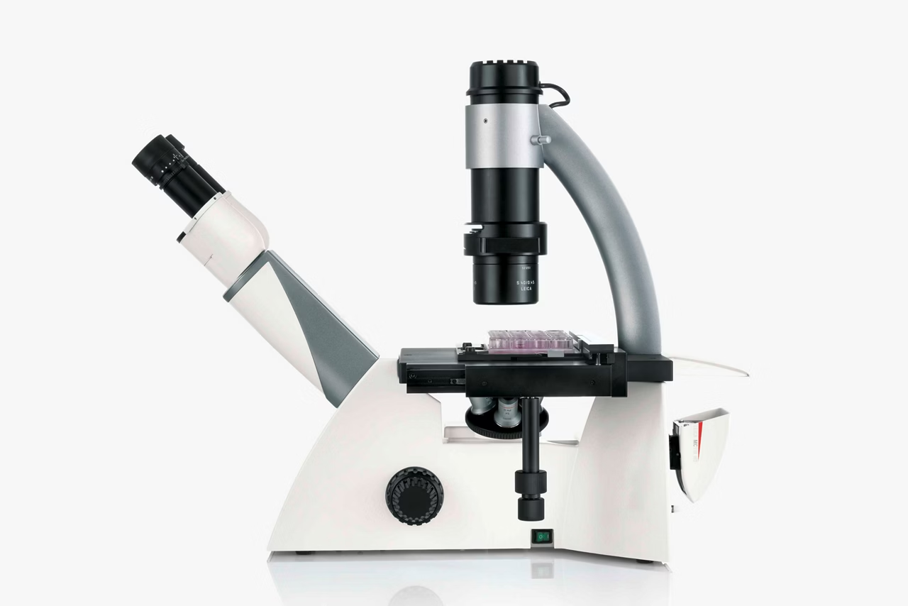
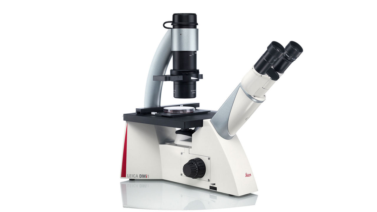
The DMi1 inverted microscope supports your specific work routine in your cell culture lab. Its operation is so intuitive, its handling so comfortable that you can fully concentrate on your work. Choose the functions you need, and, if necessary, you can easily add a variety of accessories that are important for your work.
DMi1 for Core Cell Culture

DMi1 for Core Cell Culture

The DMi1 for Core Cell Culture is a robust manual inverted microscope designed for routine tasks in cell culture labs. With precision optics and user-friendly features, this microscope delivers exceptional clarity and detail, making it an ideal tool for observing cell cultures and individual cells. Elevate your cell culture research with the DMI1's outstanding performance and reliable imaging capabilities.

DMi1 for Core Cell Culture

DMi1 for Core Cell Culture

The DMi1 for Core Cell Culture is a robust manual inverted microscope designed for routine tasks in cell culture labs. With precision optics and user-friendly features, this microscope delivers exceptional clarity and detail, making it an ideal tool for observing cell cultures and individual cells. Elevate your cell culture research with the DMI1's outstanding performance and reliable imaging capabilities.

DMi1 for Core Cell Culture

DMi1 for Core Cell Culture

The DMi1 for Core Cell Culture is a robust manual inverted microscope designed for routine tasks in cell culture labs. With precision optics and user-friendly features, this microscope delivers exceptional clarity and detail, making it an ideal tool for observing cell cultures and individual cells. Elevate your cell culture research with the DMI1's outstanding performance and reliable imaging capabilities.

DMi1 for Core Cell Culture

DMi1 for Core Cell Culture

The DMi1 for Core Cell Culture is a robust manual inverted microscope designed for routine tasks in cell culture labs. With precision optics and user-friendly features, this microscope delivers exceptional clarity and detail, making it an ideal tool for observing cell cultures and individual cells. Elevate your cell culture research with the DMI1's outstanding performance and reliable imaging capabilities.

Just Right For Your Live Cell Lab – Smart Choice For Quick Checks

DMi1 21 Poster RGB HR Kid21757689

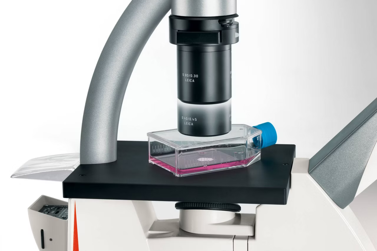
The microscope’s flexibility in accommodating an S40 condenser (40 – 50 mm working distance) or switching to an S80 condenser (80 mm working distance) is achieved by just a few hand moves. High-quality Leica objectives provide brilliant images.

The well-thought-out design and quality control elements enable you to work comfortably, even during long routines.

Smart features make your workflow smooth and efficient, saving time and resources. High-quality imaging technology from Leica Microsystems provides precise results and reduces the risk of errors.

The microscope’s LED illumination means low energy costs and minimal downtime. No need to replace lamps often, or wait for the right color temperature to develop.

The DMi1 is the smart choice for excellent quality.

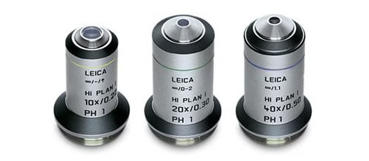
10x to 20x to 40x Phase Contrast With One Hand Move

Dmi1 Objectives 07 Kid21757698

The DMi1 is easy to use for phase contrast observation.

With 40x PH1 objective from Leica Microsystems you can switch from 10x to 20x to 40x phase contrast by simply swiveling the nosepiece. There is no need to change the light ring in the slider – the PH1 light ring matches all three objectives!

Cost Saving, Color-Safe LED Illumination

Lms Dmi1 Illumination Background Kid21766806

All DMi1 microscopes feature 5W LED illumination for constant color temperature through all stages of intensity.

The cool LED light provides a perfect climate for delicate live specimens. There are no distortions of the device due to heat, which provides unchanged focus.

Moreover, LED lamps make great economic sense. LED technology and a twohour auto-off function reduce energy costs. Up to 20 years life cycle (40 working hrs / week) per lamp under regular conditions means less downtime and low maintenance costs.

Auto Intensity

A smart sensor integrated into the slider makes sure that the light intensity automatically adjusts between brightfield and phase contrast. This feature protects the eyes, saves time, and adds comfort for the user.

Quick Change From 40 to 50 or 80 Millimeters Working Distance

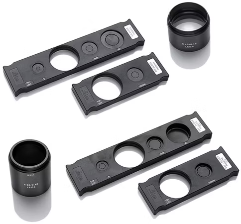
Dmi1 Sliders 07 Kid21757700

A wide range of working distances makes the DMi1 the perfect partner for your live cell laboratory. Use the S40 condenser for optimal resolution with slides, Petri dishes, multi-well dishes, and most types of flasks: The S40 provides an extra 10 mm of free space by simply moving it up.

When working with taller flasks, simply exchange the S40 for an S80 condenser with a few steps. There is no need to remove the whole illumination arm.

All-In-One Visualization And Data Storage

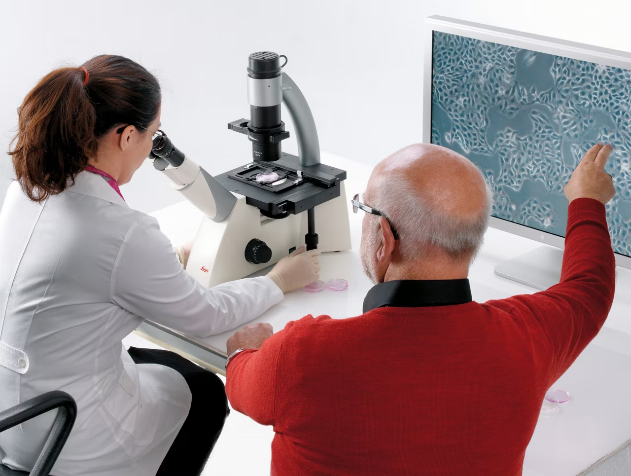
Leica DMi1 03 LabScene 18 Kid21765782

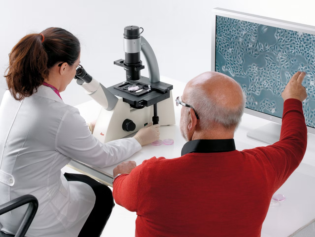
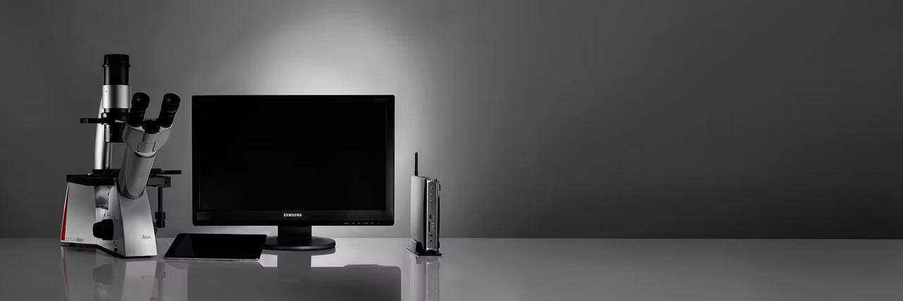
The DMi1 camera version is an all-in-one solution for image capturing and cell culture documentation. The Leica 12 megapixel Flexacam C1 camera transforms your microscope into a stand-alone digital imaging station with no need for a PC. Simply connect the camera to your microscope, preferred viewing device such as HDMI monitor, and network and start working! Save images directly on a USB stick or to your local network.

The camera is mounted at the back of the stand. No additional trinocular tube is needed, and you will have a clear view and easy access to the working area.

Solid Construction

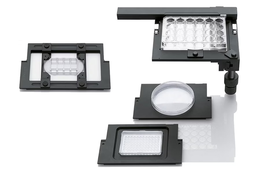
Dmi1 Expansion 05 Kid21757696

The Leica DMi1 is extremely stable due to its low center of gravity. The use of high-grade materials – almost exclusively metal – also helps avoid vibrations and resulting image blur. A scratch-resistant stage, high-quality optical components, and a wide range of accessories promote a long product life with low maintenance costs.

Modular Expansion

More flexibility and cost savings using existing Leica components across platforms. The object guide (accessory) allows the use of different holding frames for flasks, dishes, and multi-well plates.

Resources
All
Sorting
Newest
Resources
PDF
From Eye To Insight - Cleaning Of Microscope Optics
Guidelines for maintaining microscope optics. Covers considerations before cleaning, locating dirt, types of dirt, and step-by-step cleaning procedures.
PDF
From Eye To Insight - DMi1 – Designed For Routine Cell And Tissue Culture
Introduction to the Leica DMi1 microscope designed for cell and tissue culture, highlighting features like LED illumination, auto intensity, large working distance, and Full HD imaging.
PDF
Leica DMi1 Designed For Cell And Tissue Culture - Poster
Overview of the Leica DMi1 inverted microscope designed for cell and tissue culture, highlighting features like LED illumination, auto intensity, large working distance, HD imaging, and easy phase contrast.
PDF
Leica DMi1 Designed For Cell And Tissue Culture - The Intelligent Solution Brochure
The Leica DMi1 is an inverted microscope designed for live cell laboratory use. It offers flexibility in working distances, phase contrast observation, LED illumination, and options for digital imaging, making it a smart choice for cell viability checks.
PDF
Leica DMi1 Set Up
This quickstart guide provides essential setup and usage instructions for the Leica Microsystems DMI1 microscope.
PDF
Leica DMi1 Designed For Cell And Tissue Culture - Technical Data
Leica DMi1 designed for cell and tissue culture applications. Features transmitted light illumination, phase contrast, and digital HDMI camera.
What do I typically find out in a cell check?
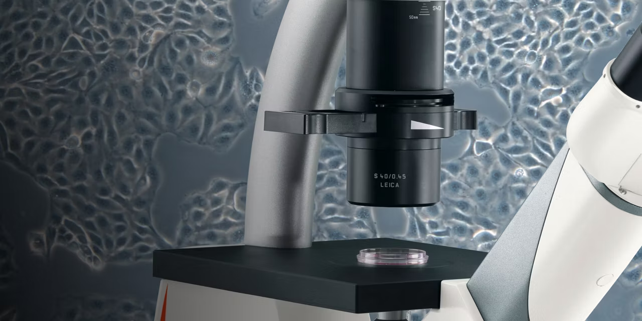
You can find out a lot with a cell check. You can: monitor cell fidelity during culture in general check the maturation and differentiation of cells (e.g. muscle cells, heart cells etc.) determine the confluence of cells before they are transfected with gene constructs (e.g. for GFP expressing experiments) count cells after adding particular selection media, drugs etc. check cells for contamination (yeast, bacteria etc.)
Who is the Leica DMi1 designed for?
The Leica DMi1 is designed for everybody in cell culture labs to facilitate their daily routine work.
What are the advantages of the Leica DMi1?
The main advantage of the Leica DMi1 is that it is focused on the necessary features. There is no nonsense about is, no gadgets, just exactly what you need to get work done. You can even operate it without a PC and software, if you want to. It is so small it can be placed almost everywhere in the cell culture lab, even directly inside the laminar flow box. It is designed for duration – a rugged instrument you can rely on every day. If you need a camera, there is the option to integrate one into the system and use Leica DMshare to make the camera wireless and transfer images to an iPad or Android tablet
Why is the Leica DMi1 particularly suited to cell and tissue culture?
The Leica DMi1 is designed for quick cell checks, for a smooth, fast workflow without time-consuming adjusting of the microscope. That is why it has only one phase ring slider for all objectives, for instance. You can monitor your cells in Brightfield and Phase contrast. When you switch from Brightfield to Phase contrast, the light intensity adjusts automatically. If you need documentation, a camera can be integrated into the microscope. And you can operate the Leica DMi1 with or without PC and software.
Which contrast methods can I employ with the Leica DMi1?
You can employ Brightfield and Phase contrast.
How do I benefit from LED illumination?
With LED illumination, you benefit from a constant color temperature. And the LED illumination of the Leica DMi1 has a practical auto-off function: After 2 hours with no interaction, the LED lamp switches off automatically.
What is the difference between a S40 condenser and a S80 condenser?
If you need a large free working distance we recommend the S80 condenser (80 mm free working distance), e.g. if you are mainly working with large culture flasks. In all other cases and especially if a higher numerical aperture which delivers higher resolution is more important to you, we recommend the S40 which has a NA of 0.45 with a free working distance of 40 to 50 mm. Both condensers are interchangeable.
What do I have to bear in mind when imaging live cells and tissues?
Most cell cultures, except for insect and yeast cultures, are based on mammalian cell lines with a temperature optimum of approximately 37° C. This means that after cells have been removed from the incubator, they should not cool down. You will find working in a non-contaminant or sterile environment conducive to your results. Although the cell culture medium usually contains antibacterial reagents, we recommend working in a laminar flow box, and gloves are a must. We also recommend cleaning the microscope stage and surfaces from time to time with 70% isopropanol. After transfection or drug treatment, cells are often sensitive. We recommend avoiding shaking or other mechanical irritation. You will find working in a working space that is cleared and barrier-free around the microscopes beneficial. You should avoid exposing GFP-expressing cells to bright light to avoid phototoxity effects, that is damage incurred by light.
Which contrast methods serve me best for my live cell research? Which contrast methods does the Leica DMi1 support?
For quick cell checks, you will do best with Brightfield and Phase contrast. They are cost-efficient and provide all information needed to evaluate the current status of your cell or tissue culture. The Leica DMi1 supports both contrast methods.
Which cameras can I use together with Leica DMi1?
You can order the Leica DMi1 with or without an integrated color camera. Camera option 1: Leica MC120 HD, a 2.5-megapixel color camera, HD format, interface to directly connect an HDMI monitor, slot for SD card, remote control. That is a good joice for basic documentation purposes. Camera option 2: Leica MC170 HD, a 5-megapixel color camera, HD format, interface to directly connect a HDMI monitor, slot for SD card, remote control. We recommend this camera for high resolution images.
How can I control my camera if no PC is connected?
You have three options: You can control the integrated camera with a remote control unit. Acquired images can be stored on the SD card of the camera. You can use an palm / footswitch to acquire images. You can use Leica DMshare V3 to make your camera wireless. Cells can be imaged live and shared with colleagues, even if they are in another room. The images are then stored on an iPad or Android tablet.
How can I document my results?
You can integrate a camera into the Leica DMi1, either a 2.5-megapixel or a 5-megapixel color camera. Control and acquisition work as follows: The camera can be connected to an HD monitor and acquires single images or movies on the SD card which is located in the camera head. The camera can be connected to a HD monitor and an additional imaging hub which makes the camera wireless and allows camera control and image acquisition with your iPad or tablet PC via the Leica DMshare V3 App. The camera can be connected to a PC and be controlled via Leica Application Suite (LAS) X software and a monitor.
Can I perform fluorescence imaging?
No, this is not possible. If you need fluorescence, we recommend the Leica DM IL inverted microscope which can be equipped with a fluorescence axis.
Can I perform Differential Interference Contrast (DIC)?
No- this is not possible. If you need DIC, we recommend the Leica DM IL inverted microscope which can be equipped for Integrated Modulation Contrast (IMC) or the Leica DMi8 inverted microscope which offers DIC.
Which accessories can I use with the Leica DMi1?
Leica offers a broad range of accessories for DMi1 ( like stage inserts, holding frames, filters). For more details please download the technical data sheet.
What is an inverted microscope?
An inverted microscope’s main distinguishing feature is the location of the optics. The optics are located below the stage. As a result, the sample is placed on top of the objectives. This means you will look at the sample from below it. Because the optics are underneath the sample, inverted microscopes offer ample working space to view samples of all shapes and sizes.
How big and heavy can samples be that I put on an inverted microscope?
Inverted microscopes are designed with the objectives and turret positioned below the stage. In theory, the sample could be as high as the ceiling – there is no height limitation. However, the maximum weight of a sample is limited to 30 kg depending on the stage used. This is ten times larger than can be accommodated by an upright microscope: samples can only be up to 80 mm high and weigh up to 3 kg.
What are the main differences between the Leica DMi1, the Leica DM IL, and the Leica DMi8?
The main difference is that they are designed for tasks of different complexity. The Leica DMi1 is conceived for quick cell checks. You can perform cell and tissue checks quickly and efficiently in Brightfield and Phase Contrast. The Leica DM IL enables you to perform more applications, for example involving fluorescence. The Leica DMi8 is the fully modular high-tech instrument which can be equipped for almost every application.
Enersight Microscope Software Platform
Software

Enersight Microscope Software Platform

Enersight simplifies image acquisition and documentation across industry, life sciences and clinical pathology with one consistent interface. Get a quote!

Flexacam c5 & i5 Microscope Cameras
Instruments

Flexacam c5 & i5 Microscope Cameras

Flexacam c5 and i5 microscope cameras deliver high resolution images with precise colors. Powered by Enersight software for easy use. Request a quote today.