Single Cell Cloning
Single-cell cloning is the process of isolating an individual cell from a diverse population and subsequently enabling it to proliferate to establish a uniform community of cells. The population of cells derived from single-cell clones are genetically indistinguishable, facilitating gene and protein investigations, biopharmaceutical manufacturing and drug discovery applications.
Single-cell cloning is a crucial method for generating monoclonal cell lines used in research and drug production. It involves dilution cloning and deterministic methods that use single-cell images to ensure monoclonality. In biotechnology, single-cell cloning plays a vital role in recombinant protein production from mammalian cells, allowing the expression of high levels of the desired protein. Moreover, single-cell cloning proves invaluable in drug discovery and screening, identification of drug targets, investigation of drug resistance mechanisms and development of personalized treatment strategies.
Single-cell cloning is predicated on building a genetically homogeneous population to minimize gene expression variables. The genetic homogeneity is also a regulatory requirement for producing uniform active pharmaceutical ingredients like recombinant proteins or monoclonal antibodies.
Methods of Single-Cell Cloning and Isolation
Single-cell cloning by limiting dilution is an established technique to derive a monoclonal population from a pool of cells. This method relies on the Poisson distribution to rapidly and conveniently isolate individual cells from diluted cell suspensions using hand-pipettes or pipetting robots. However, limiting dilution is inefficient, leading to wells containing either zero or more than one cell.
Microfluidics-based methods for single-cell cloning employ microfabricated devices that allow precise manipulation and isolation of single cells, enabling researchers to establish clonal cell lines with high genetic fidelity. Microfluidic platforms enable the parallel processing and analysis of many cells. These methods require small volumes of reagents and samples, making them suitable for working with limited or precious cell samples. They are also compatible with current workflows as single cells can be easily transferred from the microfluidic device to standard culture plates for further expansion using conventional micropipettes and microscopes.
Laser-assisted cell microdissection (LACM) method is used to isolate single cells from heterogeneous environments like cytological preparations. It enables the differentiation of normal and morphologically abnormal cells as distinct populations from complex mixtures, allowing precise investigation of subcellular profiles. The biomolecules extracted from dissected cells, including RNA, DNA, and protein, can be utilized in various downstream applications such as gene expression studies using polymerase chain reaction, proteomic analysis, etc. The method utilizes either ultraviolet (UV) cutting systems or infrared (IR) capture systems in combination with light microscopy and laser technology.
Manual cell picking in single-cell sorting and cloning is also used to isolate individual cells. It utilizes a combination of an inverted microscope and micropipettes that can be moved via motorized mechanical stages. This allows for precise positioning of the pipette for the isolation of single cells. Each isolated cell can be observed and photographed under the microscope, ensuring unbiased selection. Manual cell picking is particularly valuable for isolating live cultured cells or embryo cells. These isolated cells are then transferred to separate culture vessels, allowing them to grow and generate cell lines for further study or application.
Featured Product
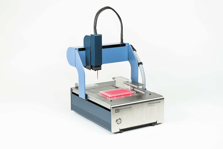
DispenCell™ Single-Cell Dispenser
Compact, automated cell dispenser for fast, easy and gentle single-cell isolation
Techniques for Single-Cell Identification and Selection
- Bright-field microscopy enables live cell observation without staining, using light absorption in regions of higher specimen density for contrast. Researchers can visually identify and manually pick individual cells using a micropipette for transfer into multi-well plates for clonal expansion.
- Fluorescence microscopy utilizes fluorescent dyes or markers to target specific cellular components, enabling enhanced sensitivity and specificity through short-wavelength light illumination, resulting in bright fluorescence against a dark background when observed through a barrier filter.
- Fluorescence-activated cell sorting (FACS) employs flow cytometry to sort fluorescently labeled cells based on their properties, allowing isolation into separate wells of a multi-well plate for clonal expansion.
- Magnetic-activated cell sorting (MACS) utilizes magnetic beads with specific antibodies to retain labeled cells based on surface markers, enabling efficient cell transfer into 96-well plates for clonal propagation.
See how Danaher Life Sciences can help
Challenges Associated with Single-Cell Cloning
- In single-cell cloning, preserving cell viability and survival rates stand as major obstacles. The isolation and culturing of individual cells can lead to cell damage or death due to mechanical stress or the delicate nature of certain cell types. To tackle this issue, researchers adopt gentle cell isolation techniques, ensuring suitable culture conditions and incorporating growth factors or nutrients into the culture medium.
- A significant challenge in single-cell cloning is genetic and epigenetic disparities among cells. Even seemingly uniform groups can exhibit variations due to genetic instability, environmental influences or random factors.
- Cell-cell contact is crucial for stimulating cell growth and proliferation. When cells contact one another, they can communicate through signals and molecules that control cell division, support growth, survival and overall health. In epithelial tissues, tight junctions and adherens junctions not only maintain the tissue's structural integrity but also facilitate essential cell signaling and communication processes. When cell-cell contact is lost, these crucial signals may be disrupted, potentially leading to apoptosis or programmed cell death. Thus, single cells frequently undergo cell death due to the loss of cell-cell contact.
Applications of Single-Cell Cloning
- Single-cell cloning has significant applications in cell fate determination and lineage tracing, particularly when studying human cells. By employing single-cell sorting and cloning techniques, researchers can isolate and clone individual cells to track their developmental trajectories, understand cell fate decisions and investigate lineage relationships.
- Single-cell cloning is vital for creating monoclonal cell lines used in research and drug production. Dilution cloning involves reducing cell concentrations to create low-density cell suspensions (0.25 to 1 cell/culture) that form single-cell-derived colonies when plated. Multiple cloning rounds enhance monoclonality chances and can identify stable clone populations. To eliminate repetitive cloning, deterministic methods using single-cell images to ensure monoclonality have been developed.
- Recombinant protein production in mammalian cells aims to quickly identify the most productive clone exhibiting highest yield and quality protein for research or therapeutic application. Ensuring a starting monoclonal population facilitates consistency in protein isoform expression and titer.
- Single-cell cloning is a valuable technique in drug discovery, especially for studying intratumoral heterogeneity, identifying drug targets, investigating drug resistance mechanisms and developing personalized treatment approaches. Single-cell cloning allows researchers to isolate and analyze individual tumor cells, gaining insights into their distinct features and drug responses.
Validation and Confirmation of Clonality
Genotyping and sequencing can validate clonality by analyzing the genetic landscape to determine if the cells share identical genotypes. Techniques like PCR or RFLP analysis can detect specific markers or mutations unique to the clonal population. Functional assays and phenotypic analysis determine cell clonality by assessing functional characteristics capable of identifying aberrant phenotypes from normal ones.
Scaling Up Single Cell Clones
Subcloning and expansion strategies involve isolating individual cells from the clonal population and further propagating them to generate multiple subclones, each originating from a single cell to study variations and ensure clonal stability.
Moving single clones from multi-well plates to fed-batch cultures in flasks and bioreactors is a crucial step in biopharmaceutical production. A single clone is selected based on desired characteristics and transferred to a larger vessel for expansion.
Quality control and monitoring are essential to ensure the uniformity and consistency of clonal populations. Complying with regulatory guidelines requires production cell lines to originate from a monoclonal progenitor and be thoroughly assessed for growth rate, productivity and final product quality.
Cryopreservation and long-term storage methods are employed to preserve clonal populations by freezing them in liquid nitrogen or at ultra-low temperatures. This process is referred to as cell banking.