Introduction to mass spectrometry
Mass spectrometry is a foundational analytical technique for identifying and quantifying the molecular composition of laboratory samples. It involves ionizing the sample to break the molecules into charged particles, which are sorted in the electromagnetic field and detected based on their mass-to-charge ratios. It is a cornerstone of analytical science with applications in proteomics, drug discovery, environmental science and forensics.
Principles of mass spectrometry
How does mass spectrometry work?
The molecules within a sample are bombarded with an electron beam, which fragments them into positively charged particles. As a result of this process, called ionization, a range of charged particles with distinct mass-to-charge ratios (m/z) is created. The ions pass through an electric or magnetic field, where their deflection patterns are determined by their m/z. In other words, ions with similar m/z will deflect similarly. Furthermore, the level of deflection is proportional to the particle mass and charge intensity. Finally, the ions are detected by an electron multiplier, generating a mass spectrum, a plot representing the relative abundance of ions against their m/z. The mass spectrum provides a molecular fingerprint, which researchers can analyze to calculate the molecular masses of the compounds and identify them by comparing them to theoretical molecular mass values. Thus, researchers can identify and quantify the analytes.
Key parameters in mass spectrometry
A successful mass spectrometry-based separation depends on the following parameters:
Mass resolution: Mass resolution denotes the ability of a mass spectrometer to distinguish between ions with similar mass-to-charge ratios. A higher resolution means the instrument can separate ions with smaller m/z differences.
Mass accuracy indicates how close the measured mass is to the theoretical mass of a compound. A mass spectrometer must boast high accuracy for correct compound identification, which involves analyzing samples with unknown compounds.
Ion stability: Vacuums in mass spectrometers prevent ion collisions, ensuring that positively charged ions can traverse and be sorted seamlessly.
Featured Product
Mass spectrometry
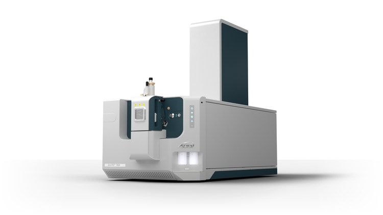
ZenoTOF 7600 System
A high-resolution mass spectrometry solution that combines powerful MS/MS sensitivity, fragmentation technology and a step-change in data independent acquisition.
Components of a mass spectrometer
Sample introduction
The sample is introduced into a reservoir called the sample inlet. From here, the molecules travel to the ionization chamber.
Ion source: methods of ionization
The ion source converts molecules into ions for electromagnetic manipulation and detection.
- Electrospray ionization (ESI): A liquid sample is sprayed with a high-voltage capillary, producing charged droplets containing the ionized particles. A desolvation gas evaporates the droplets and releases the ions into the gas phase. Thanks to its ability to generate large ion molecules in the gas phase, ESI is ideal for analyzing large macromolecules like proteins and peptides.
- Matrix-assisted laser desorption ionization (MALDI): This technique involves co-crystallizing the sample with a matrix molecule and irradiating it. This allows the analyte to pass into the gas phase and ionize with minimal fragmentation. MALDI is ideal for analyzing large biomolecules like proteins in proteomics and biomarker discovery.
- Electron ionization (EI): EI is a hard ionization technique where molecules in the gas phase are bombarded with electrons, which extensively fragment the molecule to create positively charged ions. It is ideal for the structural analysis of volatile organic molecules that rapidly pass into the gas phase.
- Chemical ionization (CI): A reagent gas is subjected to electrons and ionized. Instead of direct contact with the electron beam, the sample interacts with the ionized reagent to generate ions. The intermediary use of reagent gas reduces fragmentation, which explains why CI is considered a soft ionization technique. It is used for structural analysis and differentiation between isomeric compounds.
Mass analyzer
The mass analyzer is tasked with sorting ions by mass-to-charge ratio.
- Quadrupole mass analyzer: It contains four parallel rods generating oscillatory electric fields to filter ions based on m/z.
- Time-of-flight mass spectrometry (TOF-MS): Instead of an electric field, the TOF method accelerates ions such that the lighter ions reach the detector faster than the heavier ones. TOF is often combined with MALDI to enable rapid, high-throughput analysis necessary for proteomics and biomarker research
- Fourier transform ion cyclotron resonance (FTICR): It determines the m/z of ions by fixing them in a magnetic field, exciting them with an electric field and measuring their rotational frequencies. Due to its excellent accuracy in mass determination, FTICR is ideal for analyzing complexes, e.g., those containing isotopes.
Detector: ion measurement and data acquisition
As the final component of the mass spectrometer, the detector captures ions exiting the mass analyzer and converts their impact into measurable signals. An ion produces an electrical signal upon striking the detector. The intensity of this signal generated from an ion correlates with its concentration. The detector uses this correlation to convert raw data into a plot illustrating the abundance of each ion at a specific m/z.
See how Danaher Life Sciences can help
Chromatography coupled with mass spectrometry
Complex mixtures may need preliminary separation before mass spectrum analysis for accurate measurements, which is where chromatography enters the picture. GC-MS and LC-MS are the two main types of mass spectrometry coupled with chromatography.
Gas chromatography-mass spectrometry (GC-MS)
GC is used to distinguish between volatile and thermally stable compounds, which are ionized through EI or CI, respectively, for m/z measurements. GC-MS is ideal for identifying small molecule toxins and pollutants in a mixture and is applied extensively in forensic toxicology and environmental analysis.¹'²
Liquid chromatography-mass spectrometry (LC-MS)
Liquid chromatography separates constituents of mixtures based on their physicochemical properties and interactions with the column. After the preliminary physical separation, the components are transferred to the ion source, mainly ESI or MALDI. LC enhances the accuracy of spectral analysis in mass spectrometry and is efficient at separating proteins, peptides and metabolites based on size and polarity. Therefore, it is used in proteomics, clinical diagnostics, pharmacokinetics and food safety analysis.³
Comparison between GC-MS and LC-MS techniques
Applications of mass spectrometry
Mass spectrometry is central to molecular content analysis across several scientific fields.
Forensic science
MS analysis reveals significant information that can inform criminal investigations. Forensic scientists can decipher the structure and amount of toxins and illegal substances in biological samples. In arson and homeland security investigations, fire debris and surfaces can be analyzed to determine the presence of ignitable liquids or explosive compounds.⁴
Pharmaceutical and biomedical research
Pharmacokinetics employs LC-MS, triple quadrupole mass spectrometry or quadrupole-trap mass spectrometry (QTRAP Systems) to analyze blood and urine content.⁵'⁶ Researchers can measure the dose of drugs circulating in the blood or excreted via urine, gaining insight into how it is metabolized. Furthermore, more in-depth analysis can be performed to understand the drug's mechanism of action, proteome-wide specificity and binding sites.⁷
Mass spectrometry-based protein characterization methods, such as tandem mass spectrometry (MS/MS), help researchers explore the protein expression levels and post-translational modifications (e.g., glycosylation) in biological samples.⁸ It supports biomarker discovery studies by identifying aberrantly expressed or modified proteins in diseases like cancer.⁹'¹⁰
Troubleshooting and challenges in mass spectrometry
Despite its analytical advantages, mass spectrometry can be hindered by technical challenges that affect data quality.
Common issues and solutions
- Ionization efficiency problems: Low signal intensity may point to an issue in the ion source settings.
- Loss of sensitivity: Weak or noisy signals may also indicate sample contamination, which may be caused by gas leaks. Researchers can resolve this by checking the gas filter and the weldment or installing a leak detector.
- Lack of peaks: No peaks in the mass spectrum data may be due to the sample ions not reaching the detector properly. Checking the columns and the injection apparatus can help identify the source of the issue.
Mass spectrometry troubleshooting tips for beginners
- Regular calibration is vital in mass spectrometry, as calibration drifts may disrupt m/z measurement and mass assignments. Standard compounds, such as perfluorotributylamine (FC-43) for GC-MS and sodium cesium iodide (NaCsl) for LC-MS, are recommended for calibration.
- It is essential to ensure proper maintenance of high vacuum systems to establish ion stability.
- Although manufacturer settings are an ideal starting point, optimizing electric and magnetic field settings helps researchers fine-tune ion focusing or improve resolution.
See how Danaher Life Sciences can help
FAQs
What is mass spectrometry?
Mass spectrometry (MS) is an analytical technique used to measure the mass of molecules. It identifies compounds based on their mass-to-charge ratio (m/z), allowing scientists to determine molecular weight and structure.
What is the principle of mass spectrometry?
MS works by ionizing chemical compounds to generate charged molecules or fragments. These ions are then separated by their m/z using electric or magnetic fields and detected to produce a mass spectrum.
What is mass spectrometry used for?
MS is widely used in chemistry, biology, forensics and pharmaceuticals. Applications include identifying unknown substances, analyzing complex mixtures, studying proteins and detecting toxins or drugs.
What is the mass-to-charge ratio (m/z), and why is it significant?
The m/z value represents an ion's mass divided by its charge. It's crucial for identifying ions, as MS instruments separate and analyze ions based on their unique m/z values.
References
- Gould O, Nguyen N, Honeychurch KC. New Applications of Gas Chromatography and Gas Chromatography-Mass Spectrometry for Novel Sample Matrices in the Forensic Sciences: A Literature Review. Chemosensors 2023;11(10):527.
- Newton SR, Bowden JA, Charest N, Jackson SR, Koelmel JP, Liberatore HK, et al. Filling the Gaps in PFAS Detection: Integrating GC-MS Non-Targeted Analysis for Comprehensive Environmental Monitoring and Exposure Assessment. Environ Sci Technol Lett 2025.
- Chen CJ, Lee DY, Yu J, Lin YN, Lin TM. Recent advances in LC‐MS‐based metabolomics for clinical biomarker discovery. Mass Spectrom Rev 2023;42(6):2349-2378.
- Evans‐Nguyen K, Stelmack AR, Clowser PC, Holtz JM, Mulligan CC. Fieldable mass spectrometry for forensic science, homeland security, and defense applications. Mass Spectrom Rev 2021;40(5):628-646.
- Modi SJ, Tiwari A, Ghule C, Pawar S, Saste G, Jagtap S, et al. Pharmacokinetic study of withanosides and withanolides from Withania somnifera using ultra-high performance liquid chromatography-tandem mass spectrometry (UHPLC-MS/MS). Molecules 2022;27(5):1476.
- Zhou H, He Y, Zheng Z, Xing J, Liu Z, Pi Z, et al. Pharmacokinetics and tissue distribution study of 18 bioactive components in healthy and chronic heart failure rats after oral administration of Qi‐Shen‐Ke‐Li formula using ultra‐high‐performance liquid chromatography/triple quadrupole mass spectrometry. Rapid Commun Mass Spectrom 2021;35(8):e9060.
- Dueñas ME, Peltier‐Heap RE, Leveridge M, Annan RS, Büttner FH, Trost M. Advances in high‐throughput mass spectrometry in drug discovery. EMBO Mol Med 2023;15(1):e14850.
- Silsirivanit A, Alvarez MRS, Grijaldo-Alvarez SJ, Gogte R, Kitkhuandee A, Piyawattanametha N, et al. Serum N-Glycomics with Nano-LC-QToF LC-MS/MS Reveals N-Glycan Biomarkers for Glioblastoma, Meningioma, and High-Grade Meningioma. J Proteome Res 2025.
- Vinaiphat A, Low JK, Yeoh KW, Chng WJ, Sze SK. Application of advanced mass spectrometry-based proteomics to study hypoxia driven cancer progression. Front Oncol 2021;11:559822.
- Khoo A, Liu LY, Nyalwidhe JO, Semmes OJ, Vesprini D, Downes MR, et al. Proteomic discovery of non-invasive biomarkers of localized prostate cancer using mass spectrometry. Nat Rev Urol 2021;18(12):707-724.