Liquid Biopsy Biomarkers: ctDNA and CTC Workflows
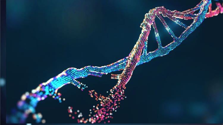
Liquid biopsy biomarkers are integral in modern cancer diagnostics and research. By offering minimally invasive access to tumor‑derived signals, they address the limitations of tissue biopsies and enable real‑time disease monitoring. This article explores the biology, workflows, applications and challenges of liquid biopsy, highlighting how these biomarkers advance precision oncology and translational research.
Key Takeaways
- Liquid biopsy advantages: Provides minimally invasive, repeatable access to tumor information compared to traditional tissue biopsies
- ctDNA and CTC roles: ctDNA is used in mutation detection and monitoring, while CTCs are employed in cellular profiling and metastasis research
- Expanded biomarker classes: cfRNA, exosomes and proteins broaden discovery by capturing transcriptional and functional signals
- Applications in oncology: Biomarker discovery, therapy response monitoring, patient stratification and companion diagnostic development
- Challenges and future needs: Low biomarker abundance, variability and lack of standardization demand robust tools and validated workflows
- Liquid biopsy advantages: Provides minimally invasive, repeatable access to tumor information compared to traditional tissue biopsies
- ctDNA and CTC roles: ctDNA is used in mutation detection and monitoring, while CTCs are employed in cellular profiling and metastasis research
- Expanded biomarker classes: cfRNA, exosomes and proteins broaden discovery by capturing transcriptional and functional signals
- Applications in oncology: Biomarker discovery, therapy response monitoring, patient stratification and companion diagnostic development
- Challenges and future needs: Low biomarker abundance, variability and lack of standardization demand robust tools and validated workflows
See how Danaher Life Sciences can help
What Is Liquid Biopsy?
Cancer diagnostics relies on robust technologies that not only detect malignancies early but also indicate disease stage and metastatic potential. Liquid biopsy biomarkers have become the cornerstone for rapid and reliable cancer detection by capturing circulating tumor DNA (ctDNA) and circulating tumor cells (CTCs) from body fluids.1
Liquid biopsy is a minimally invasive diagnostic approach that detects and analyzes these circulating tumor‑derived biomarkers, providing a dynamic snapshot of disease biology without the need for surgical sampling. It is frequently used in the clinic for its broad scope and minimal invasiveness.1
Tissue biopsy requires surgical or needle‑based sampling of tumor tissue, which can be painful, risky and limited to a single site. Liquid biopsy, by contrast, relies on accessible fluids such as blood, urine, cerebrospinal fluid or saliva, making it safer and more feasible for longitudinal studies.2
Different types of fluids are analyzed in liquid biopsy, with blood being the most widely used medium for ctDNA and CTC analysis2 and others, including:
- Urine: Urothelial and kidney cancers3
- Cerebrospinal fluid: CNS malignancies3
- Saliva: Head and neck cancers3
Together, these sample types expand the reach of cancer diagnostics, offering clinicians and researchers a versatile toolkit for precision oncology.
What Are Liquid Biopsy Biomarkers?
Liquid biopsy biomarkers reflect the molecular and cellular characteristics of a tumor. These biomarkers include nucleic acids, cells, vesicles and proteins that are shed into body fluids, each offering a different layer of information about cancer biology.4
Among the most widely studied are ctDNA and circulating tumor cells, which originate from malignant tissue and provide highly specific signals of disease presence and progression. ctDNA fragments carry tumor‑specific mutations, epigenetic changes and copy number alterations. At the same time, CTCs represent intact cancer cells that can reveal phenotypic traits such as morphology, protein expression and metastatic potential.5
The liquid biopsy biomarker landscape extends beyond ctDNA and CTCs, enabling a multi-layered analysis of tumor biology.
- Cell‑free RNA (cfRNA) can capture transcriptional activity and gene expression patterns6
- Exosomes, small extracellular vesicles secreted by tumor cells, transport nucleic acids and proteins that mirror the tumor microenvironment7
- Circulating proteins, including tumor‑associated antigens and signaling molecules, reflect functional changes in cancer metabolism and immune interactions8
An important caveat is that healthy individuals also release circulating markers, including DNA fragments, RNA, proteins and vesicles. The challenge lies in distinguishing cancer‑specific alterations from signals indicative of normal physiological processes.9
This is where biomarker sensitivity and specificity become essential. High sensitivity ensures that even low levels of tumor material can be detected, which is crucial for early diagnosis and monitoring minimal residual disease. Specificity, on the other hand, guarantees that detected signals truly reflect malignant processes rather than benign conditions.10
ctDNA in Liquid Biopsy
Circulating tumor DNA comprises small fragments of DNA released into the bloodstream by cancer cells. These fragments originate from processes such as apoptosis, necrosis and active secretion, which occur as tumors grow and interact with their microenvironment. Because ctDNA carries tumor‑specific genetic and epigenetic alterations, it provides a direct molecular fingerprint of the malignancy.11
ctDNA has become a cornerstone of liquid biopsy research due to its versatility. Use cases include:
- In targeted therapy, mutation detection allows clinicians to identify genetic changes that guide therapy selection12
- Longitudinal monitoring of ctDNA levels reveals treatment response, offering early indications of resistance or relapse13
- In minimal residual disease studies, ctDNA analysis can detect traces of solid tumors after therapy, even when imaging results appear clear14,15
The workflow for ctDNA analysis requires careful attention to technical details at every stage. Sample collection is typically performed using blood plasma, as serum can introduce background DNA from lysed cells. However, researchers must ensure stabilization of samples through specialized collection tubes, appropriate storage conditions and timely processing to prevent degradation and contamination.5
Extraction methods are equally important. Researchers should assess the efficiency of their method in isolating low-abundance ctDNA fragments with minimal loss. Successful extraction influences the accuracy of downstream PCR and next-generation sequencing, directly impacting the reproducibility of biomarker analyses that inform clinical decisions.5
Circulating Tumor Cell (CTC) in Liquid Biopsy
Circulating tumor cells are intact cancer cells that detach from a tumor and enter the bloodstream. Unlike ctDNA, which consists of fragmented genetic material, CTCs preserve cellular structure, protein expression and functional properties. This makes them extremely valuable for studying cancer biology at the cellular level. For example, their presence in circulation is closely linked to the metastatic process, offering direct insight into cancer progression.16
While both are indispensable in cancer diagnostics, CTCs and ctDNA detect distinct but complementary aspects of tumors. While ctDNA supports mutation detection and molecular monitoring, CTCs enable phenotypic profiling by uncovering morphology, protein expression and omics analysis. This level of insight makes them powerful biomarkers for studying tumor heterogeneity, metastasis mechanisms, prognosis and drug response, collectively informing personalized treatment strategies.16
Despite their promise, CTC workflows face challenges. CTCs are very rare, often just a few cells among billions, making detection difficult. Enrichment methods like immunomagnetic separation or microfluidic capture are needed, but each has trade-offs in sensitivity and specificity. Balancing purity and recovery complicates the collection of sufficient viable cells for analysis. Moreover, detection and analysis workflows, such as imaging, immunostaining, sequencing, or functional assays, vary.16
ctDNA vs. CTC Workflows: What is the difference between ctDNA and CTCs?
Here, we summarize how ctDNA workflows excel in sensitivity and molecular monitoring, while CTC workflows provide unique cellular insights despite technical challenges.5
Applications in Drug Discovery and Diagnostics
Liquid biopsy biomarkers are integrated not only into diagnostics but also into various other research and drug discovery applications.
One of their most essential roles is biomarker discovery, in which circulating signals are identified and validated as measurable endpoints during assay development. These biomarkers guide the development of new diagnostic assays that capture tumor‑specific alterations with high sensitivity and reproducibility.17
In therapy response monitoring, liquid biopsy enables clinicians to track patients' treatment response in real time. Shifts in ctDNA levels, changes in CTC phenotypes or variations in exosomal cargo can reveal whether a therapy is effective, whether resistance is emerging or whether residual disease persists. This capability is particularly valuable in oncology, where treatment decisions often hinge on early signs of progression or relapse.18
Patient stratification research is another critical application. By analyzing circulating biomarkers, researchers can identify patient subgroups more likely to benefit from specific therapies. This stratification supports precision medicine by ensuring that clinical trials enroll the right populations and that therapies are matched to individuals based on molecular and cellular profiles.19
Liquid biopsy is also instrumental in the development of companion diagnostics. These tests are designed to pair with targeted therapies, confirming the presence of mutations before treatment is prescribed. Companion diagnostics can be established with minimally invasive techniques, which is why they are suitable for personalized therapies and drug discovery pipelines with tight deadlines.20
Finally, liquid biopsy contributes to translational research and to the optimization of oncology workflows. They reduce reliance on invasive tissue sampling, shorten turnaround times and provide scalable tools for monitoring large patient cohorts. In this way, liquid biopsy not only advances scientific understanding but also enhances the efficiency and precision of cancer care.21
Selecting the Right Workflow Based on Research Objectives
The ideal choice between various circulating biomarkers depends on the specific goals of the study. Liquid biopsy biomarkers vary in the technologies used and in their suitability for research and clinical applications, each offering distinct advantages in efficiency and scientific relevance.9
Challenges in Liquid Biopsy Biomarker Analysis
Despite its advantages and clinical utility, liquid biopsy biomarker analysis faces several technical and biological hurdles that limit its widespread adoption.
One of the most persistent challenges is the low abundance of circulating biomarkers. ctDNA fragments, CTCs and other signals often occur at extremely low concentrations, requiring highly sensitive technologies to detect them reliably. This scarcity makes early diagnosis and monitoring of minimal residual disease particularly demanding.22
Sample variability further complicates analysis. Differences in collection methods, storage conditions and patient physiology can introduce inconsistencies that affect biomarker integrity. For example, plasma and serum may yield different ctDNA profiles, while delays in processing can lead to degradation or contamination. Such variability underscores the need for standardized pre‑analytical workflows to ensure reproducibility across studies and clinical settings.22
Achieving balance between sensitivity and specificity remains difficult. Although highly sensitive assays are essential for detecting rare biomarkers, they must also be specific to the biomarkers of interest and avoid false positives due to background noise from normal physiological processes. Balancing these two parameters is a constant challenge in assay design and validation.22
Another barrier is the lack of standardization and reproducibility across laboratories. Differences in protocols, analytical platforms and data interpretation can lead to inconsistent results, slowing the translation of liquid biopsy into routine practice. Programs involving multiple sites need to harmonize workflows and quality control systems to build confidence in large biomarker datasets.23
Finally, the field requires robust research tools and validated workflows to move beyond proof‑of‑concept studies. Advances in sequencing, imaging and computational analysis are helping address these needs. Still, widespread adoption will depend on rigorous validation and integration with other oncology workflows at the single-cell, population and tissue levels.23
Featured Product
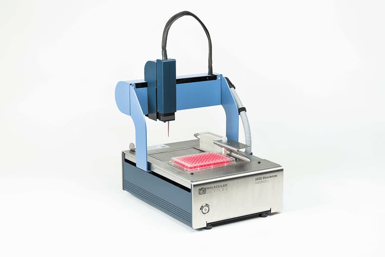
DispenCell™ Single-Cell Dispenser
DispenCell™ Single-Cell Dispenser - a compact, automated instrument for fast, gentle single-cell isolation with immediate, traceable proof of clonality, suited to rare-cell workflows.
See how Danaher Life Sciences can help
FAQ's
How does a liquid biopsy detect cancer?
It identifies tumor-derived signals such as ctDNA fragments or CTCs circulating in blood or other fluids, providing genetic and cellular evidence of malignancy.
Is a liquid biopsy better than a tissue biopsy?
It is less invasive and allows ongoing sampling, but tissue biopsy remains the gold standard for histological detail. The two approaches are complementary.
Can ctDNA and CTCs be analyzed from the same blood sample?
Yes. Plasma is processed for ctDNA, while whole blood is used for CTCs, offering molecular and cellular insights from a single draw.
What are the limitations of liquid biopsy for early cancer detection?
Low biomarker levels and background signals can reduce accuracy, making early-stage detection difficult.
What role does next-generation sequencing play in liquid biopsy?
NGS provides comprehensive profiling of ctDNA mutations and tumor heterogeneity, adding depth and precision to analysis.
Why are ctDNA and CTCs considered complementary biomarkers?
ctDNA supports molecular monitoring, while CTCs provide intact cells for functional and phenotypic studies, together giving a fuller view of cancer biology.
References
- Nikanjam M, Kato S, Kurzrock R. Liquid biopsy: current technology and clinical applications. J Hematol Oncol 2022;15(1):131.
- Armakolas A, Kotsari M, Koskinas J. Liquid biopsies, novel approaches and future directions. Cancers (Basel) 2023;15(5):1579.
- Bao Y, Zhang D, Guo H, Ma W. Beyond blood: Advancing the frontiers of liquid biopsy in oncology and personalized medicine. Cancer Sci 2024;115(4):1060‑1072.
- Wang W, Rong Z, Wang G, Hou Y, Yang F, Qiu M. Cancer metabolites: promising biomarkers for cancer liquid biopsy. Biomark Res 2023;11(1):66.
- Wang X, Wang L, Lin H, Zhu Y, Huang D, Lai M, et al. Research progress of CTC, ctDNA, and EVs in cancer liquid biopsy. Front Oncol 2024;14:1303335.
- Zhong P, Bai L, Hong M, Ouyang J, Wang R, Zhang X, et al. A comprehensive review on circulating cfRNA in plasma: implications for disease diagnosis and beyond. Diagnostics 2024;14(10):1045.
- Yu W, Hurley J, Roberts D, Chakrabortty S, Enderle D, Noerholm M, et al. Exosome‑based liquid biopsies in cancer: opportunities and challenges. Ann Oncol 2021;32(4):466‑477.
- Lapitz A, Azkargorta M, Milkiewicz P, Olaizola P, Zhuravleva E, Grimsrud MM, et al. Liquid biopsy‑based protein biomarkers for risk prediction, early diagnosis, and prognostication of cholangiocarcinoma. J Hepatol 2023;79(1):93‑108.
- Heidrich I, Ačkar L, Mossahebi Mohammadi P, Pantel K. Liquid biopsies: Potential and challenges. Int J Cancer 2021;148(3):528‑545.
- Garcia J, Kamps‑Hughes N, Geiguer F, Couraud S, Sarver B, Payen L, et al. Sensitivity, specificity, and accuracy of a liquid biopsy approach utilizing molecular amplification pools. Sci Rep 2021;11(1):10761.
- Yang W, Nguyen R, Safri F, Shiddiky MJ, Warkiani ME, George J, et al. Liquid biopsy in hepatocellular carcinoma: ctDNA as a potential biomarker for diagnosis and prognosis. Curr Oncol Rep 2025;27(6):791‑802.
- Alix‑Panabières C, Pantel K. Liquid biopsy: from discovery to clinical application. Cancer Discov 2021;11(4):858‑873.
- Lv J, Xu L‑X, Li Z‑X, Lin L, Wu C‑F, Quan T‑Q, et al. Longitudinal on‑treatment circulating tumor DNA as a biomarker for real‑time dynamic risk monitoring in cancer patients: the EP‑SEASON study. Cancer Cell 2024;42(8):1401‑1414.e4.
- Flach S, Howarth K, Hackinger S, Pipinikas C, Ellis P, McLay K, et al. Liquid BIOpsy for MiNimal RESidual DiSease Detection in Head and Neck Squamous Cell Carcinoma (LIONESS)—a personalised circulating tumour DNA analysis in head and neck squamous cell carcinoma. Br J Cancer 2022;126(8):1186‑1195.
- Aguilar H, López‑Roldán B, Vilalta‑Lacarra A, Alkorta‑Aranburu G, Claramunt R, López‑Guerrero JA, et al. Liquid biopsy for monitoring minimal residual disease in localized and locally‑advanced non‑small cell lung cancer after radical‑intent treatment. J Liquid Biopsy 2024;4:100145.
- Allen TA. The role of circulating tumor cells as a liquid biopsy for cancer: advances, biology, technical challenges, and clinical relevance. Cancers (Basel) 2024;16(7):1377.
- Diaz PM, Leehans A, Ravishankar P, Daily A. Multiomic approaches for cancer biomarker discovery in liquid biopsies: advances and challenges. Biomark Insights 2023;18:11772719231204508.
- Ghani MU, Du L, Moqbel AQ, Zhao E, Cui H, Yang L, et al. Exosomal ncRNAs in liquid biopsy: a new paradigm for early cancer diagnosis and monitoring. Front Oncol 2025;15:1615433.
- Haselmann V, Hedtke M, Neumaier M. Liquid profiling for cancer patient stratification in precision medicine–current status and challenges for successful implementation in standard care. J Lab Med 2022;46(4):225‑236.
- Sato Y. Clinical utility of liquid biopsy‑based companion diagnostics in the non‑small‑cell lung cancer treatment. Explor Target Antitumor Ther 2022;3(5):630.
- Bratulic S, Gatto F, Nielsen J. The translational status of cancer liquid biopsies. Regen Eng Transl Med 2021;7(3):312‑352.
- Batool SM, Yekula A, Khanna P, Hsia T, Gamblin AS, Ekanayake E, et al. The Liquid Biopsy Consortium: Challenges and opportunities for early cancer detection and monitoring. Cell Rep Med 2023;4(10).
- Ma L, Guo H, Zhao Y, Liu Z, Wang C, Bu J, et al. Liquid biopsy in cancer: current status, challenges and future prospects. Signal Transduct Target Ther 2024;9(1):336.