Adeno-associated Viruses (AAV)
Introduction
Viruses possess specialized mechanisms to enter into their hosts and integrate their genetic material with the host’s DNA. This unique capabiliy has been extensively employed in laboratories to transfer desired genes. Scientists modify viruses by removing their viral genes and then repurpose them as vectors for diverse drug research and development and medical applications, such as gene therapy.
Vectors derived from viruses, such as retroviruses, herpes simplex viruses (HSV), adenoviruses (AdVs) and adeno-associated viruses (AAVs), are studied and utilized extensively. Each viral vector type has its own specific applications. No single viral vector has yet to be established as a “fit for all” in research and development applications. The AAV vector plays a pivotal role in gene therapy applications. Although originally identified as a contaminant in adenovirus preparations, this viral vector has now become a key platform for gene delivery to treat various human diseases.
History and discovery of AAV
AAV was first discovered in the mid-1960s as a contaminant of adenovirus cultures. Over the following 15-20 years, extensive research explored into various aspect of AAV biology including its structure, genome configuration and composition, infectious latency, DNA replication and transcription, and virion assembly.
The potential of AAV as a vector was discovered after the wild-type AAV2 (Adeno-associated virus 2) vector was successfully cloned into plasmids, allowing for comprehensive genome studies. In 1984, the first recombinant adeno-associated viral vector for in vitro gene delivery was developed, followed by the application of recombinant AAV in humans in 1995. Further studies have provided more insight into the serotypes of AAV and their potential as an in vivo gene delivery system, which led to the introduction of the first European Medicines Agency (EMA) approved AAV-based gene therapy drug (Glybera) in 2012. The event was followed by the development of Luxturna—the first-ever AAV gene therapy approved by the U.S. Food and Drug Administration (FDA).¹
AAVs structure and properties
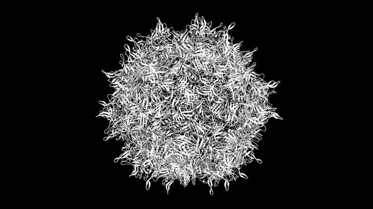
AAV, a member of the Dependoparvovirus (Parvoviridae family),² lacks the ability to replicate independently within a host and relies on assistance from other viruses like HSV or AdVs. Structurally, AAV can be categorized into two components: the capsid and the genome.
The AAV capsid is a non-enveloped and icosahedral protein shell with a diameter of 25 nm.³ The capsid of each AAV serotype is different, with only one VP3 common region, which consists of a small α-helix (αA) and eight-stranded β-barrel motif (βB-βI). The variable region on the capsid surface is essential to the virus’s life cycle, influencing its antigenic specificity, transduction efficiency, and receptor binding.⁴
AAV genome structure is composed of linear and single-stranded DNA.⁵ It contains two open reading frames (ORF), flanked at both ends by a 145-bp Inverted Terminal Repeat (ITR) sequence. The 5’ end ORF is involved in producing replication proteins (Rep 68, Rep 78, Rep 40, and Rep 52), while the 3’ end ORF encodes capsid proteins (VP1, VP2, and VP3)
AAV-mediated gene therapy vectors are designed by replacing the viral gene segment with a transgenic (or any desired) DNA sequence that is ideally less than 5kb. This segment is carefully inserted between the terminal inverted repeats of the viral genome.
See how Danaher Life Sciences can help
Classification of adeno-associated virus
Currently, there are 13 known AAV serotypes – found in both human and primates - all of which have been sequenced.⁶ Based on their antigenic creativity and sequence comparison, AAVs classification includes:
- Clades A to F - 6 genetic groups
- AAV4 & AAV5 - 2 clonal isolates
AAV serotypes harbor a sequence identity of about 65% to 99%, and a structural identity of around 95% to 99%.⁷ AAVs exhibit significant variations in their affinity for different target tissues, such as liver tissue, skeletal and cardiac muscle, lung tissue, and central nervous system cells. The distinctions between AAV serotypes can be leveraged in the development of vectors for gene therapy, allowing the targeted treatment of certain tissues.
For example, the AAV2 serotype has an affinity for cells with heparan sulfate proteoglycans (HSPG) receptors. On the other hand, adeno-associated virus serotype 8 (AAV8), AAV7, AAV6, and AAV1 have high affinity for muscle, liver, and lung cells.⁸ The specificity of different AAV serotypes depends on a suitable receptor for AAV on the surface of cells.
Natural and therapeutic applications of AAVs
Different AAV serotypes are currently under investigation for their efficacy as vectors in human gene therapy and the treatment of many critical diseases. This is mainly because AAVs can incorporate and express foreign genes in various tissues based on their binding affinity with cell surface receptors. AAV serotypes possess the unique capability to infect both dividing and non-dividing cells without causing any associated pathogenicity. Furthermore, recombinant AAV vectors (rAAVs) can be produced and purified at high concentrations, ensuring their availability for clinical applications.
Upon entering the target cell, AAV particles carrying the desired therapeutic gene travel to the nucleus. Once there, they unpack the cDNA and create extra-chromosomal episomes. Subsequently, the gene undergoes transcription and translation processes, resulting in the production of the therapeutic protein transgene.
Adeno-associated virus-mediated gene therapy is extensively used in clinical settings to treat many critical diseases. Some examples of successful clinical trials involving recombinant AAV vectors include the treatment of Leber’s congenital amaurosis (LCA) and hemophilia. B. Glybera, developed using the rAAV1 serotype, was the first vector approved by the European Commission.⁹ It was developed using the rAAV1 serotype to treat lipoprotein lipase (LPL) deficiency, a rare, inherited disorder.2012⁹ to treat lipoprotein lipase (LPL) deficiency, a rare, inherited disorder.
Advantages and challenges of using AAVs in gene therapy
AAV has emerged as the preferred vector in clinical trials for neurodegenerative diseases due to its safety and effectiveness in facilitating gene transfer to the central nervous system. Compared to adenovirus, AAV vectors exhibit lower immunogenicity and can infect a wide range of cells and hosts.
Other advantages of AAV include their ability to infect both dividing and non-dividing cells and sustain elevated levels of gene expression in vivo for extended periods, often lasting years.
However, one of the major limitations of AAV genomes or vectors is their small packaging capacity. You can package only 5 kb of gene segment in AAV vectors for various gene therapy applications. Other challenges include:
- Gene transfer to targeted cells
- Pre-existing immune responses to AAV vectors in host cells
- New transgene expression responses that have not yet been observed.
Additionally, a high amount of pre-existing neutralizing antibodies (NAbs) in individuals can hinder the delivery of AAV vectors for human gene therapy applications.
AAV vectors have also been found to cause off-target effects and liver toxicity. For instance, the AAV2 vector has shown liver toxicity in patients during clinical trials due to the generation of cytotoxic T lymphocyte response.¹⁰
Conclusion
Approximately 1900 gene therapy clinical trials are currently ongoing worldwide, with 99¹¹ of these trials utilizing AAV vectors for gene delivery applications. However, enhancing the effectiveness of AAV vectors demands overcoming challenges in the field.
For instance, one of the major challenges in using AAV vectors in human gene therapy is the presence of pre-existing immunity in patients. Thus, one active area of research in AAV vector applications is the isolation of AAV variants from nonhuman primates and the study of their efficacy in gene therapy, such as AAVrh32.33, derived from rhesus macaques.¹²
Researchers have also conducted peptide mapping of antigenic regions of novel AAVs. Moreover, scientists have successfully determined the structure of AAV capsids when bound to fragment antibodies (Fabs) derived from mouse monoclonal antibodies that neutralize AAV infections.
A pressing demand exists for the creation of advanced gene therapy vectors. However, to expand their applications, these vectors must possess the ability to target tissues that are typically resistant to transduction by naturally occurring AAV serotypes. Alternatively, limiting the virus's affinity to specific tissues is crucial.
Advancements in the field have introduced techniques like directed evolution and rational capsid engineering to overcome these challenges. These approaches not only address the obstacles in gene therapy but also aid in creating vectors capable of evading the pre-existing human immune response.
FAQs
What is an adeno-associated virus?
Adeno-associated virus (AAV) was organically discovered in 1965 as a contaminant in adenovirus cultures. It belongs to the genus Dependovirus of the Parvovirus family.
What is the difference between adenovirus (AdV) and adeno-associated virus (AAV)?
The major difference between AdV and AAV is in their genetic makeup. AAV is composed of a linear, single-stranded DNA, whereas AdV has a linear, double-stranded DNA (dsDNA). Additionally, the AAV vector has lower immunogenicity than AdV, and unlike AdV, AAV doesn't show any pathogenicity.
What does adeno-associated virus target?
Adeno-associated viruses selectively aim for distinct neuronal subtypes and non-neuronal cells within the nervous system.
What are the similarities between adenovirus and AAV?
The major similarity between AdV and AAV is that both of them are extensively utilized in gene therapy applications. This is primarily due to their ability to target a wide range of hosts and infect actively dividing and dormant cells.
References
- Wang D, Tai PW, Gao G. Adeno-associated virus vector as a platform for gene therapy delivery. Nature reviews Drug discovery. 2019 May;18(5):358-78.
- Ning K, Kuz CA, Cheng F, Feng Z, Yan Z, Qiu J. Adeno-associated virus monoinfection induces a DNA damage response and DNA repair that contributes to viral DNA replication. Mbio. 2023 Feb 28;14(1):e03528-22.
- Pupo A, Fernández A, Low SH, François A, Suárez-Amarán L, Samulski RJ. AAV vectors: The Rubik’s cube of human gene therapy. Molecular Therapy. 2022 Dec 7;30(12):3515-41.
- Halder S, Van Vliet K, Smith JK, Duong TT, McKenna R, Wilson JM, et al. Structure of neurotropic adeno-associated virus AAVrh.8. Journal of structural biology. 2015 Oct 1;192(1):21-36.
- Cervelli T, Backovic A, Galli A. Formation of AAV single stranded DNA genome from a circular plasmid in Saccharomyces cerevisiae. PloS one. 2011 Aug 10;6(8):e23474.
- Gadenstaetter AJ, Schmutzler L, Grimm D, Landegger LD. Intranasal application of adeno-associated viruses: a systematic review. Translational Research. 2022 Oct 1;248:87-110.
- Chu W, Shastry S, Barbieri E, Prodromou R, Greback‐Clarke P, Smith W, et al. Peptide ligands for the affinity purification of adeno‐associated viruses from HEK 293 cell lysates. Biotechnology and bioengineering. 2023 Aug;120(8):2283-300.
- Zhao L, Yang Z, Zheng M, Shi L, Gu M, Liu G, et al. Recombinant adeno-associated virus 8 vector in gene therapy: Opportunities and challenges. Genes & Diseases. 2024 Jan 1;11(1):283-93.
- Watanabe N, Yano K, Tsuyuki K, Okano T, Yamato M. Re-examination of regulatory opinions in Europe: possible contribution for the approval of the first gene therapy product Glybera. Molecular therapy Methods & clinical development. 2015 Jan 1;2.
- Li C, Hirsch M, Asokan A, Zeithaml B, Ma H, Kafri T, et al. Adeno-associated virus type 2 (AAV2) capsid-specific cytotoxic T lymphocytes eliminate only vector-transduced cells coexpressing the AAV2 capsid in vivo. Journal of virology. 2007 Jul 15;81(14):7540-7.
- Kaufmann KB, Büning H, Galy A, Schambach A, Grez M. Gene therapy on the move. EMBO molecular medicine. 2013 Nov 4;5(11):1642-61.
- Mikals K, Nam HJ, Van Vliet K, Vandenberghe LH, Mays LE, McKenna R, et al. The structure of AAVrh32. 33, a novel gene delivery vector. Journal of structural biology. 2014 May 1;186(2):308-17.