Automating 3D Cell Culture
Drug development can be like finding a needle in a haystack. The pressure to move drugs to clinical trials faster has required better drug screening models. This requires adjusting the technological capabilities to screen an increasing number of candidates during preclinical R&D and selecting promising molecules to advance to further development.
Drugs fail in clinical trials because data generated across pre-clinical R&D and CMC isn’t always human-relevant. 2D cell culture bioassays or animal models for drug screening and development are limited in the human-relevant insights they provide. Organoid systems are becoming more widely used as pre-clinical models because they:
- Provide relevant data on compound functionality, kinetics and toxicity
- Offer insights into the compounds' effects on human organs
- Present a more cost-effective and ethical solution than animal models
However, these 3D cell culture models face technological challenges in meeting the demand for consistent, large-scale throughput needed for drug screening.
The development of organoids is time- and resource-intensive. The workflow starts with a 2D pre-culture phase, progressing to 3D cell culture and organoid formation, which typically takes weeks. The process involves growing cultures on highly viscous media mimicking an extracellular matrix, which is challenging to manipulate. Manual handling increases inter-batch variability risk, potentially undermining the consistency and reliability of organoid data.
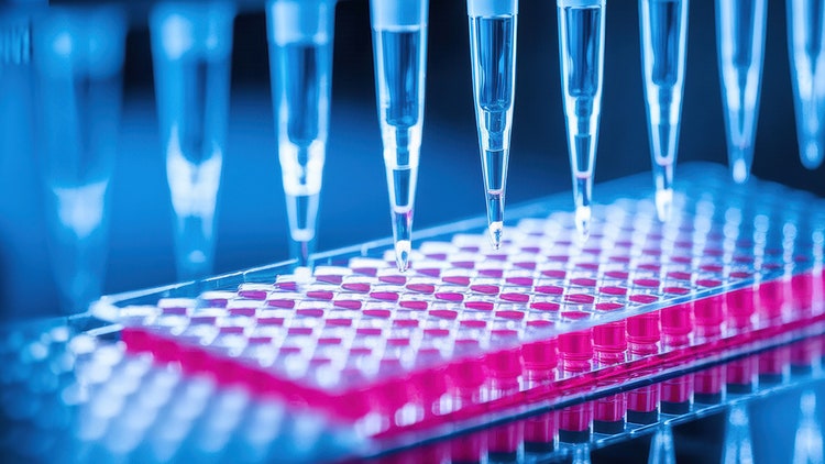
To address these challenges, the Life Sciences companies of Danaher offer automation solutions that enable standardized, hands-off production and culture of organoids with consistent methods and parameters. Leveraging tools such as the Beckman Coulter Life Sciences Biomek i-Series Automated Workstation, a liquid handler capable of custom protocols and integration with numerous devices, streamlines the workflow by handling tasks previously carried out manually.
Several steps can be automated, offering greater experimental repeatability and reproducibility, including:
- Cell seeding
- Media exchanges
- Organoid feeding
- Long-term incubation
Despite the desire to adopt organoids as model systems, there's still a lack of expertise and standardization in working with organoids and guiding these cell cultures to differentiate and organize sufficiently into 3D cell cultures. This leads to variability between screens and cultures and results that cannot be reproduced.
Automating these workflows can help decrease variability substantially.
For example, Molecular Devices’ CellXpress.ai offers a unique, automated solution that combines 3D workflows built with the extensive knowledge of in-house specialists, providing all-inclusive solutions for organoid culture. It offers:
- Cell cultures
- Incubation
- Sample processing
- Automated data analysis
This integrated solution ensures consistent, unbiased and relevant results from a single system while simplifying the use of organoids in preclinical testing.
Additional challenges in organoid development include:
-
Live imaging and quality control:
- Live imaging requires extended sample preparation
- Maintaining culture-like conditions to ensure that the preparation does not compromise the functionality and integrity
- Extracting meaningful data is challenging as the 3D nature of the samples can partially mask the sample details
-
High data volume and analysis burden:
- Screening multiple candidates generates a large amount of data
- Manual data collection is time-consuming and prone to errors
Introducing automation to organoid imaging can expedite data collection:
- The ImageXpress® Confocal HT.ai High Content Imaging System from Molecular Devices offers high-throughput analysis of 3D samples up to two times faster than manual preparation
- Integration with advanced analytical software, like IN Carta image analysis software, facilitates a deeper understanding of complex cellular interactions within 3D samples
- The Leica Microsystems Mica Microhub integrates a sample incubation chamber with an automated microscope to maintain conditions required for culture without compromising organoid health or integrity
The Life Sciences companies of Danaher provide automated microscopy solutions that enhance imaging, revealing greater detail in thick samples and delivering precise assay readouts for screening, ADME and toxicity studies. Integrating artificial intelligence and machine learning technologies simplifies advanced phenotypic classification and streamlines 3D image analysis. As a result, automated multi-scale imaging guarantees continuous oversight over organoid development and high-quality data collection from 3D cultures.
The Life Sciences companies of Danaher provide automation solutions to accelerate organoid research, optimize lab time and resource allocation and speed up the discovery and development of new drugs. To learn more, visit our integrated Human Relevant Model and Spatial Profiling solutions or contact an expert today.

Bridging the Gap: Advancing Human-Relevant Models for Real-World Impact Webinar