Immunocytochemistry/ Immunofluorescence - Anti-PRAME antibody [EPR20330] (ab219650)
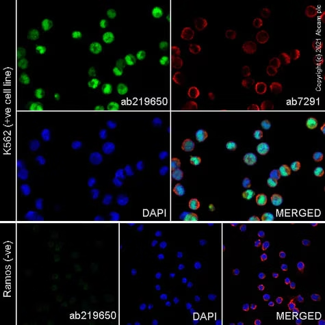
PRAME immunofluorescence with PRAME antibody ab219650 in K562 cells, with negative expression in Ramos cells. The cells were fixed with 4% formaldehyde (10 min), permeabilised with 0.1% Triton x-100 for 5 minutes and then blocked with 1% BSA/10% normal goat serum/0.3M glycine in 0.1% PBS-Tween for 1h. The cells were then incubated overnight at +4°C with ab219650 at 1 μg/ml and ab7291, Mouse monoclonal [DM1A] to alpha Tubulin at 0.5 μg/ml. Cells were then incubated with ab150081, Goat polyclonal Secondary Antibody to Rabbit IgG - H&L (Alexa Fluor® 488), pre-adsorbed at 1/1000 dilution (shown in green) and ab150119, Goat polyclonal Secondary Antibody to Mouse IgG - H&L (Alexa Fluor® 647), pre-adsorbed at 1/1000 dilution (shown in red). Nuclear DNA was labelled with DAPI (shown in blue).
Image was acquired with a confocal microscope (Leica-Microsystems TCS SP8) and a single confocal section is shown.
This product also work with 100% methanol (5 min) fixation under the same testing conditions.

Immunoprecipitation - Anti-PRAME antibody [EPR20330] (ab219650)
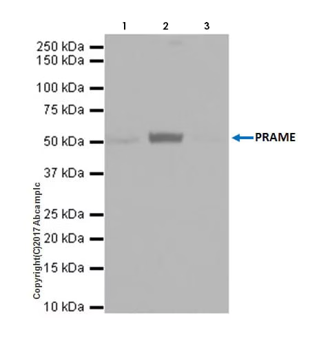
PRAME was immunoprecipitated from 0.35 mg of MeWo (Human malignant melanoma cell line) whole cell lysate with ab219650 at 1/30 dilution. Western blot was performed from the immunoprecipitate using ab219650 at 1/1000 dilution. VeriBlot for IP Detection Reagent (HRP) (ab131366), was used for detection at 1/10000 dilution.
Lane 1: MeWo whole cell lysate 10 μg (Input).
Lane 2: ab219650 IP in MeWo whole cell lysate.
Lane 3: Rabbit monoclonal IgG (ab172730) instead of ab219650 in MeWo whole cell lysate.
Blocking and dilution buffer and concentration: 5% NFDM/TBST.
Exposure time: 1 second.
All lanes: Immunoprecipitation - Anti-PRAME antibody [EPR20330] (ab219650)
Predicted band size: 57 kDa

Western blot - Anti-PRAME antibody [EPR20330] (ab219650)
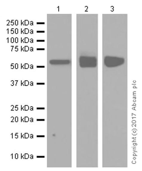
PRAME western blot with PRAME antibody ab219650.
Blocking/Dilution buffer: 5% NFDM/TBST.
Exposure time: Lane 1: 3 minutes; Lane 2: 5 seconds; Lane 3: 1 minute.
All lanes: Western blot - Anti-PRAME antibody [EPR20330] (ab219650) at 1/1000 dilution
Lane 1: Human ovary cancer lysate at 20 µg
Lane 2: A-375 (Human malignant melanoma cell line) whole cell lysate at 20 µg
Lane 3: Human testis lysate at 20 µg
Secondary
All lanes: Western blot - VeriBlot for IP Detection Reagent (HRP) (ab131366) at 1/1000 dilution
Predicted band size: 57 kDa
Observed band size: 57 kDa

Western blot - Anti-PRAME antibody [EPR20330] (ab219650)
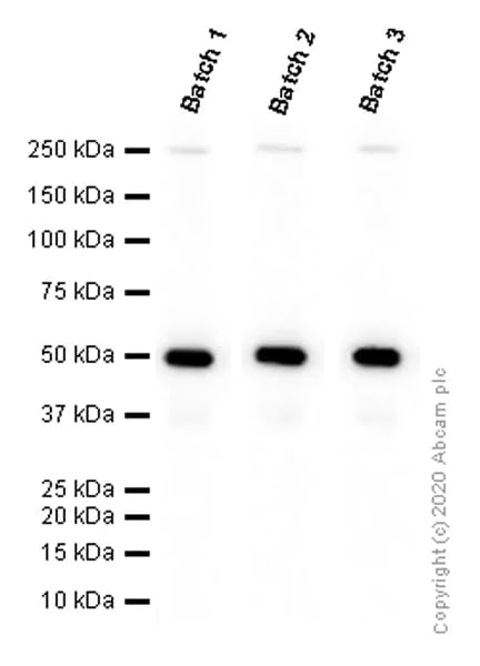
PRAME western blot with PRAME antibody ab219650.
Different batches of ab219650 were tested on MeWo (Human malignant melanoma fibroblast) whole cell lysate at 0.1 µg/ml. 15 µg of lysate was loaded in each lane. Bands observed at 50 kDa.
All lanes: Western blot - Anti-PRAME antibody [EPR20330] (ab219650)
Predicted band size: 57 kDa

Immunocytochemistry/ Immunofluorescence - Anti-PRAME antibody [EPR20330] (ab219650)
Immunofluorescent analysis of 4% paraformaldehyde-fixed, 0.1% Triton X-100 permeabilized MeWo (Human malignant melanoma cell line) cells labeling PRAME with ab219650 at 1/500 dilution, followed by Goat anti-rabbit IgG (Alexa Fluor® 488) (ab150077) secondary antibody at 1/1000 dilution (green). Confocal image showing mostly nuclear staining on MeWo cells.
The nuclear counter stain is DAPI (blue). Tubulin is detected with ab195889 (Anti-alpha Tubulin antibody [DM1A] - Microtubule Marker (Alexa Fluor® 594)) at 1/200 dilution (red).
Secondary antibody only control: Used PBS instead of primary antibody, secondary antibody is Goat anti-rabbit IgG (Alexa Fluor® 488) (ab150077) at 1/1000 dilution.
![Immunocytochemistry/ Immunofluorescence - Anti-PRAME antibody [EPR20330] (ab219650)](./media_1ad3cd1a3f9f0615316fc1d056da8836c5d404e10.avif?width=750&format=avif&optimize=medium)
Immunocytochemistry/ Immunofluorescence - Anti-PRAME antibody [EPR20330] (ab219650)
Immunofluorescent analysis of 4% paraformaldehyde-fixed, 0.1% Triton X-100 permeabilized A-375 (Human malignant melanoma cell line) cells labeling PRAME with ab219650 at 1/500 dilution, followed by Goat anti-rabbit IgG (Alexa Fluor® 488) (ab150077) secondary antibody at 1/1000 dilution (green). Confocal image showing mostly nuclear staining on A-375 cells.
The nuclear counter stain is DAPI (blue). Tubulin is detected with ab195889 (Anti-alpha Tubulin antibody [DM1A] - Microtubule Marker (Alexa Fluor® 594)) at 1/200 dilution (red).
Secondary antibody only control: Used PBS instead of primary antibody, secondary antibody is Goat anti-rabbit IgG (Alexa Fluor® 488) (ab150077) at 1/1000 dilution.
![Immunocytochemistry/ Immunofluorescence - Anti-PRAME antibody [EPR20330] (ab219650)](./media_1233fec0f0983e504796c65388c71de0b2fbe6f89.avif?width=750&format=avif&optimize=medium)
Western blot - Anti-PRAME antibody [EPR20330] (ab219650)
PRAME western blot with PRAME antibody ab219650.
Blocking/Dilution buffer: 5% NFDM/TBST.
All lanes: Western blot - Anti-PRAME antibody [EPR20330] (ab219650) at 1/1000 dilution
All lanes: MeWo (Human malignant melanoma cell line) whole cell lysate at 20 µg
Secondary
All lanes: Goat Anti-Rabbit IgG Peroxidase Conjugate, specific to the non-reduced form of IgG at 1/4000 dilution
Predicted band size: 57 kDa
Observed band size: 57 kDa
Exposure time: 15s
![Western blot - Anti-PRAME antibody [EPR20330] (ab219650)](./media_1275ec16e00b9c0e4a25d397dbb1d35e83e859563.avif?width=750&format=avif&optimize=medium)
Immunohistochemistry (Formalin/PFA-fixed paraffin-embedded sections) - Anti-PRAME antibody [EPR20330] (ab219650)
Tissue microarray (TMA) analysis using PRAME EPR20330 (ab219650).
Clickhereto view EPR20330 staining performance on human normal and cancer tissue microarray (TMA).
This table provides a detailed overview of positive PRAME staining (tick mark) and negative PRAME staining (cross mark) per sample type tested. PRAME is expressed in metastatic melanoma (PMID: 30045064). PRAME has low or no expression in normal tissues except in testis, ovary, placenta, adrenals and endometrium (PMID: 30045064).
The sections were pre-treated using Heat mediated antigen retrieval using ab93684 (Tris/EDTA buffer, pH 9.0). The sections were incubated with ab219650 at +4°C overnight followed by a ready to use Goat Anti-Rabbit IgG H&L (HRP polymer).
![Immunohistochemistry (Formalin/PFA-fixed paraffin-embedded sections) - Anti-PRAME antibody [EPR20330] (ab219650)](./media_1fcb492cf7b3fbab6920e86ee81c2eceaea874162.webp?width=750&format=webp&optimize=medium)
Immunohistochemistry (Formalin/PFA-fixed paraffin-embedded sections) - Anti-PRAME antibody [EPR20330] (ab219650)
Immunohistochemical analysis of paraffin-embedded human melanoma tissue labeling PRAME with ab219650 at 1/500 dilution, followed by LeicaDS9800 (Bond™ Polymer Refine Detection). Sections were counter stained with Hematoxylin. Antigen retrieval was heat mediated with Tris/EDTA buffer pH 9.0 before commencing with IHC staining protocol. Nuclear staining on human melanoma. The section was incubated with ab219650 for 30 mins at room temperature. The immunostaining was performed on a Leica Biosystems BOND® RX instrument. Secondary antibody only control: Used PBS instead of primary antibody.
![Immunohistochemistry (Formalin/PFA-fixed paraffin-embedded sections) - Anti-PRAME antibody [EPR20330] (ab219650)](./media_132aa7dbe5578e60fd0ea70a1902255f60f811bbc.jpeg?width=750&format=jpeg&optimize=medium)
Flow Cytometry (Intracellular) - Anti-PRAME antibody [EPR20330] (ab219650)
Flow cytometry overlay histogram showing left K562 positive cells and right negative HEK293 stained with ab219650 (red line). The cells were fixed with 4% formaldehyde (10 min) and then permeabilised with 0.1 % PBS-Triton X-100 for 15 min. The cells were then incubated in 1x PBS containing 10% normal goat serum to block non-specific protein-protein interaction followed by the antibody (ab219650) (1x 10⁶ in 100μl at 0.008μg/ml (1/267500)) for 30min at 22°C.
The secondary antibody Goat Anti-Rabbit IgG H&L (Alexa Fluor® 488) preadsorbed was incubated at 1/4000 for 30min at 22°C.
Isotype control antibody (black line) was Recombinant Rabbit IgG, monoclonal [EPR25A] - Isotype Control used at the same concentration and conditions as the primary antibody. Unlabelled sample (blue line) was also used as a control.
Acquisition of >5000 events were collected using a 50 mW Blue laser (488nm) and 525/40 bandpass filter.
This antibody gave a positive signal in K562 Fixed with 80% methanol (5 min) / permeabilised with 0.1 % PBS-Triton X-100 for 15 min under the same conditions.
![Flow Cytometry (Intracellular) - Anti-PRAME antibody [EPR20330] (ab219650)](./media_13453a409dca48aed1e044253c92395b8b9438970.avif?width=750&format=avif&optimize=medium)
Flow Cytometry (Intracellular) - Anti-PRAME antibody [EPR20330] (ab219650)
PRAME flow cytometry with PRAME antibody ab219650 of 4% paraformaldehyde-fixed and 90% Methanol-permeabilised MeWo (Human malignant melanoma cell line) cells labeling PRAME with ab219650 at 1/500 dilution (red) compared with a rabbit monoclonal IgG isotype control (ab172730; black) and an unlabeled control (cells without incubation with primary antibody and secondary antibody; blue). Goat anti rabbit IgG (Alexa Fluor® 488) (ab150077) at 1/2000 dilution was used as the secondary antibody.
![Flow Cytometry (Intracellular) - Anti-PRAME antibody [EPR20330] (ab219650)](./media_11eeebcb3d5b1d3f9fa735d1d9ebb95388ed25b16.avif?width=750&format=avif&optimize=medium)
Immunohistochemistry (Formalin/PFA-fixed paraffin-embedded sections) - Anti-PRAME antibody [EPR20330] (ab219650)
Immunohistochemical analysis of paraffin-embedded human stomach tissue labeling PRAME with ab219650 at 1/500 dilution, followed by LeicaDS9800 (Bond™ Polymer Refine Detection). Sections were counter stained with Hematoxylin. Antigen retrieval was heat mediated with Tris/EDTA buffer pH 9.0 before commencing with IHC staining protocol. Negative control: No staining on human stomach. The section was incubated with ab219650 for 30 mins at room temperature. The immunostaining was performed on a Leica Biosystems BOND® RX instrument.
![Immunohistochemistry (Formalin/PFA-fixed paraffin-embedded sections) - Anti-PRAME antibody [EPR20330] (ab219650)](./media_1e31455b521d25fa3e72449f7d464f2b58421d663.jpeg?width=750&format=jpeg&optimize=medium)
Immunohistochemistry (Formalin/PFA-fixed paraffin-embedded sections) - Anti-PRAME antibody [EPR20330] (ab219650)
Immunohistochemical analysis of paraffin-embedded human breast tissue labeling PRAME with ab219650 at 1/500 dilution, followed by LeicaDS9800 (Bond™ Polymer Refine Detection). Sections were counter stained with Hematoxylin. Antigen retrieval was heat mediated with Tris/EDTA buffer pH 9.0 before commencing with IHC staining protocol. Negative control: No staining on human breast. The section was incubated with ab219650 for 30 mins at room temperature. The immunostaining was performed on a Leica Biosystems BOND® RX instrument.
![Immunohistochemistry (Formalin/PFA-fixed paraffin-embedded sections) - Anti-PRAME antibody [EPR20330] (ab219650)](./media_1d835c802d04bf302a10a51e719a00f9b4a5ff66e.avif?width=750&format=avif&optimize=medium)
Immunohistochemistry (Formalin/PFA-fixed paraffin-embedded sections) - Anti-PRAME antibody [EPR20330] (ab219650)
Immunohistochemical analysis of paraffin-embedded human tonsil tissue labeling PRAME with ab219650 at 1/500 dilution, followed by LeicaDS9800 (Bond™ Polymer Refine Detection). Sections were counter stained with Hematoxylin. Antigen retrieval was heat mediated with Tris/EDTA buffer pH 9.0 before commencing with IHC staining protocol. Negative control: No staining on human tonsil. The section was incubated with ab219650 for 30 mins at room temperature. The immunostaining was performed on a Leica Biosystems BOND® RX instrument.
![Immunohistochemistry (Formalin/PFA-fixed paraffin-embedded sections) - Anti-PRAME antibody [EPR20330] (ab219650)](./media_1126f410ee59b033be6f35b687ef37736686ad126.jpeg?width=750&format=jpeg&optimize=medium)
Immunohistochemistry (Formalin/PFA-fixed paraffin-embedded sections) - Anti-PRAME antibody [EPR20330] (ab219650)
Immunohistochemical analysis of paraffin-embedded human testis tissue labeling PRAME with ab219650 at 1/500 dilution, followed by LeicaDS9800 (Bond™ Polymer Refine Detection). Sections were counter stained with Hematoxylin. Antigen retrieval was heat mediated with Tris/EDTA buffer pH 9.0 before commencing with IHC staining protocol. Nuclear staining on human testis. The section was incubated with ab219650 for 30 mins at room temperature. The immunostaining was performed on a Leica Biosystems BOND® RX instrument. Secondary antibody only control: Used PBS instead of primary antibody.
![Immunohistochemistry (Formalin/PFA-fixed paraffin-embedded sections) - Anti-PRAME antibody [EPR20330] (ab219650)](./media_1b21625ee6e26e6e10f4f57eb166581ebb22f167c.jpeg?width=750&format=jpeg&optimize=medium)
Immunohistochemistry (Formalin/PFA-fixed paraffin-embedded sections) - Anti-PRAME antibody [EPR20330] (ab219650)
Immunohistochemical analysis of paraffin-embedded human breast carcinoma tissue labeling PRAME with ab219650 at 1/500 dilution, followed by LeicaDS9800 (Bond™ Polymer Refine Detection). Sections were counter stained with Hematoxylin. Antigen retrieval was heat mediated with Tris/EDTA buffer pH 9.0 before commencing with IHC staining protocol. Nuclear staining on human breast carcinoma. The section was incubated with ab219650 for 30 mins at room temperature. The immunostaining was performed on a Leica Biosystems BOND® RX instrument. Secondary antibody only control: Used PBS instead of primary antibody.
![Immunohistochemistry (Formalin/PFA-fixed paraffin-embedded sections) - Anti-PRAME antibody [EPR20330] (ab219650)](./media_1cfa49d8e6150dcc668a748ad3a92f956ffaf145c.jpeg?width=750&format=jpeg&optimize=medium)