Mass Cytometry (CyTOF): A Complete Guide to Metal-Conjugated Antibodies

Key Takeaways
- Mass cytometry (CyTOF) is a single-cell analysis technique that uses metal isotope–tagged antibodies instead of fluorescent dyes to measure multiple cellular markers simultaneously
- It works by ionizing labeled cells in a plasma source and detecting metal tags according to their mass-to-charge ratio using time-of-flight mass spectrometry
- The technique provides highly multiplexed, quantitative data with minimal signal overlap, making it ideal for deep cellular profiling
- Mass cytometry is widely used in immunology, oncology and systems biology to study cellular heterogeneity and complex biological systems
What Is Mass Cytometry?
Mass cytometry is an advanced single-cell analysis technology that combines principles of flow cytometry with inductively coupled plasma time-of-flight mass spectrometry (ICP-TOF-MS). Instead of labeling antibodies with fluorescent dyes, mass cytometry uses stable metal isotopes as antibody tags. As individual cells pass through the instrument, they are atomized and ionized, enabling detection of the metal tags based on their unique atomic masses. This approach virtually eliminates spectral overlap, enabling measurement of more than 40 cellular markers within a single sample.1
Conventional flow cytometry has long been the standard for immunophenotyping and cellular analysis. It detects fluorescently labeled antibodies using lasers and optical filters, providing rapid, quantitative measurements of multiple proteins at the single-cell level. However, the number of measurable markers is constrained by overlapping fluorescence emission spectra, which require complex compensation and careful panel design.2
Mass cytometry was developed to overcome these limitations. By replacing fluorophores with rare earth metal isotopes, CyTOF technology dramatically expands multiplexing capacity while minimizing signal interference. This allows researchers to profile highly complex cellular populations with greater confidence and reduced challenges in panel optimization.2
Mass cytometry provides comprehensive immune profiling, detailed characterization of rare cell populations and deeper insights into cellular heterogeneity that may be difficult to achieve using conventional flow cytometry. These capabilities have made CyTOF an important platform in many areas of biomedical research, where understanding complex cellular interactions is critical.3
How Mass Cytometry Works
Mass cytometry combines antibody-based cell labeling with elemental mass spectrometry to generate high-dimensional single-cell data.
CyTOF or cytometry by time-of-flight integrates single-cell sample introduction with inductively coupled plasma time-of-flight mass spectrometry (ICP-TOF-MS). Cells stained with metal-conjugated antibodies are introduced individually into the instrument, ensuring that each cell is analyzed independently.4
Once inside the plasma, cellular material is converted into an ion cloud, and the attached metal isotopes are separated by the TOF analyzer, based on their mass-to-charge ratio. The resulting signals are translated into quantitative measurements for every marker expressed on each cell.4
A defining feature of mass cytometry is the use of stable rare-earth metal isotopes rather than fluorescent dyes. Each antibody is conjugated to a unique metal isotope, serving as an elemental barcode for a specific protein target. Because these isotopes possess discrete atomic masses, they can be distinguished with exceptional specificity. This strategy largely eliminates spectral overlap encountered in fluorescence-based assays, reducing the need for compensation methods for multiplexed antibody panels.5
Labeled cells are introduced into the mass cytometer, where they are nebulized into individual droplets, transported into an inductively coupled plasma and vaporized at high temperatures. The extreme heat vaporizes the cells, atomizes their components and ionizes the bound metal tags.6
The resulting ions enter a time-of-flight mass spectrometer, where they are accelerated through a flight tube. Since lighter ions travel faster than heavier ones, the instrument determines the identity and abundance of each metal isotope based on its flight time. The detected isotope signals are then reconstructed into a comprehensive expression profile for every analyzed cell.6
See how Danaher Life Sciences can help
Workflow of CyTOF Mass Cytometry
Step 1 – Antibody Labeling
Antibodies are conjugated to stable rare-earth metal isotopes, each acting as a unique tag for a specific cellular target.
Step 2 – Sample Preparation and Staining
Cells are prepared as single-cell suspensions and incubated with the metal-tagged antibody panel to label proteins of interest.
Step 3 – Atomization and Ionization
Stained cells are introduced into an inductively coupled plasma, where they are vaporized, atomized and converted into ion clouds.
Step 4 – Cytometry by Time of Flight (TOF) Analysis
Ions are accelerated through a TOF mass analyzer, which separates them based on their mass-to-charge ratios.
Step 5 – Detection and Quantification[ED1.1]
Metal ion signals are detected and converted into quantitative single-cell expression data for downstream computational analysis.
Advantages of Mass Cytometry
Mass cytometry offers several key benefits, making it a powerful tool for high-resolution single-cell analysis in biomedical research.7
- High-dimensional data from thousands of single cells: Mass cytometry enables simultaneous measurement of up to 50 markers across large cell populations, supporting deep phenotyping and systems-level analysis
- Minimal spectral overlap for cleaner results: Using metal isotope tags instead of fluorophores eliminates fluorescence spillover and reduces the need for signal compensation
- Quantitative and reproducible analysis: It provides highly stable digital signals, improving consistency across experiments
- Compatibility with rare-cell populations and complex samples: It supports accurate detection of low-frequency cell types in heterogeneous tissues, making it well-suited for immunology, oncology and clinical research applications
Applications of Mass Cytometry
Immunology and Cytokine Profiling
Mass cytometry is extensively applied in immunology to map immune cell diversity and function.
- Understanding immune cell heterogeneity: It supports detailed characterization of immune cell subsets, developmental states and activation profiles within complex immune systems8
- Detecting cytokine expression at the single-cell level: It allows simultaneous measurement of intracellular cytokines alongside surface markers, supporting functional immune profiling9
Cancer Biology and Tumor Microenvironment
Mass cytometry provides deep insights into tumor composition and immune interactions.
- Identifying immune infiltration patterns in tumors: It helps define immune cell types and their states within the tumor microenvironment10
- Biomarker discovery and therapy response tracking: It supports the identification of predictive biomarkers and monitoring of patient responses to immunotherapy or targeted treatments11
Drug Discovery and Bioprocessing
Mass cytometry technology is valuable for evaluating cellular responses to therapeutic compounds.
- Screening drug effects on immune or signaling pathways: A mass cytometer can be used to measure how drugs modulate phosphorylation states, signaling networks and immune activation at single-cell resolution12
- Cell-based assays for functional response measurements: It is a powerful method for assessing drug efficacy and toxicity in heterogeneous cell populations7
Systems Biology and Multi-Parameter Profiling
Mass cytometry is essential for systems biology and computational modeling of biological networks.
- Integrating mass cytometry data with transcriptomics and proteomics: It generates single-cell-level proteomics data that can be integrated with genomic and transcriptomic datasets for multi-omics insights13
- Building comprehensive cellular maps: The high-dimensional data obtained from mass cytometry can be used to construct detailed atlases of cellular states, differentiation pathways and tissue organization14
Limitations and Challenges
While mass cytometry offers powerful high-dimensional single-cell analysis, it also comes with several important limitations that can affect experimental design, accessibility and data interpretation.
One of the main barriers to widespread adoption is the cost of CyTOF instrumentation and the associated reagents. The mass cytometer itself is a sophisticated, expensive platform that typically requires a dedicated facility and trained personnel for operation and maintenance. In addition, metal-conjugated antibody panels are more costly and less universally available than conventional fluorophore-conjugated antibodies. These financial and logistical requirements can limit access to well-resourced research institutions or centralized core facilities.4
Mass cytometry generates highly complex, high-dimensional datasets that require advanced computational approaches for meaningful analysis. Each experiment can produce data for dozens of parameters across millions of cells, resulting in large file sizes that demand substantial storage and processing power. Interpreting these datasets often requires specialized bioinformatics expertise, including dimensionality reduction, clustering algorithms and visualization tools. As a result, the analytical workflow can be time-consuming and may present a steep learning curve for researchers new to the technique.4
Featured Product
Cytobank Premium Software
Chosen because it is a high-dimensional cytometry analytics platform, including CyTOF/mass cytometry data visualization and clustering, tying directly to this page's subject of mass cytometry cytof guide.

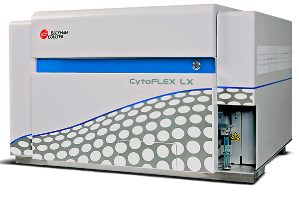
CytoFLEX LX Flow Cytometer
Chosen as a complementary second option because it is a multicolor flow cytometer for detailed immunophenotyping and multi-parameter cell analysis, also directly relevant to mass cytometry cytof guide.
See how Danaher Life Sciences can help
FAQ's
What is the difference between flow cytometry and mass cytometry?
Flow cytometry uses fluorescent dyes and lasers, while mass cytometry uses metal isotope–tagged antibodies detected by mass spectrometry, allowing higher multiplexing with minimal signal overlap.
What types of samples can be analyzed using CyTOF mass cytometry?
CyTOF can analyze blood, bone marrow, cultured cells and dissociated solid tissues, provided they are prepared as single-cell suspensions.
What kind of data does CyTOF provide?
It generates high-dimensional, quantitative single-cell data showing expression levels of dozens of markers per cell.
What is CyTOF antibody conjugation?
It is the process of attaching stable rare earth metal isotopes to antibodies so they can be detected by mass spectrometry.
References
- Xu S, Liu M, Bai Y, Liu H. Multi‐dimensional organic mass cytometry: simultaneous analysis of proteins and metabolites on single cells. Angew Chem Int Ed 2021;60(4):1806-1812.
- Jaimes MC, Leipold M, Kraker G, Amir Ea, Maecker H, Lannigan J. Full spectrum flow cytometry and mass cytometry: A 32‐marker panel comparison. Cytometry Part A 2022;101(11):942-959.
- Koladiya A, Davis KL. Advances in clinical mass cytometry. Clin Lab Med 2023;43(3):507-519.
- Iyer A, Hamers AA, Pillai AB. CyTOF® for the Masses. Front Immunol 2022;13:815828.
- Stevens CR, Atkuri K, Menard DL, King LE, Neubert H, Goihberg P. Mass cytometry for the multiplexed quantification and characterization of target expression on circulating cells in whole blood. Cytometry Part A 2023;103(8):631-645.
- Arnett LP, Rana R, Chung WW-Y, Li X, Abtahi M, Majonis D, et al. Reagents for mass cytometry. Chem Rev 2023;123(3):1166-1205.
- Tajik M, Baharfar M, Donald WA. Single-cell mass spectrometry. Trends Biotechnol 2022;40(11):1374-1392.
- Levine LS, Hiam-Galvez KJ, Marquez DM, Tenvooren I, Madden MZ, Contreras DC, et al. Single-cell analysis by mass cytometry reveals metabolic states of early-activated CD8+ T cells during the primary immune response. Immunity 2021;54(4):829-844. e5.
- Hoch T, Schulz D, Eling N, Gómez JM, Levesque MP, Bodenmiller B. Multiplexed imaging mass cytometry of the chemokine milieus in melanoma characterizes features of the response to immunotherapy. Sci Immunol 2022;7(70):eabk1692.
- Moldoveanu D, Ramsay L, Lajoie M, Anderson-Trocme L, Lingrand M, Berry D, et al. Spatially mapping the immune landscape of melanoma using imaging mass cytometry. Sci Immunol 2022;7(70):eabi5072.
- Yao H, Zhao H, Pan X, Zhao X, Feng J, Yang C, et al. Discriminating leukemia cellular heterogeneity and screening metabolite biomarker candidates using label-free mass cytometry. Anal Chem 2021;93(29):10282-10291.
- Zielinski JM, Luke JJ, Guglietta S, Krieg C. High throughput multi-omics approaches for clinical trial evaluation and drug discovery. Front Immunol 2021;12:590742.
- Nassar SF, Raddassi K, Wu T. Single-cell multiomics analysis for drug discovery. Metabolites 2021;11(11):729.
- Kuett L, Catena R, Özcan A, Plüss A, Schraml P, Moch H, et al. Three-dimensional imaging mass cytometry for highly multiplexed molecular and cellular mapping of tissues and the tumor microenvironment. Nat Cancer 2022;3(1):122-133.