Fluorescence-Activated Cell Sorting (FACS)

What is Fluorescence-Activated Cell Sorting (FACS)?
Fluorescence-activated cell sorting (FACS) is a specialized type of flow cytometry that involves the separation of cells in a sample based on their fluorescent characteristics. Researchers use FACS frequently for high-throughput, quantitative assessment of cell populations. They can also isolate specific cell subtypes for downstream experiments.1
In FACS, cells are tagged by fluorescently labelled antibodies or dyes that bind to specific cellular markers. These cells are transported through a fluid stream while a laser beam individually excites each cell. Simultaneously, a detector measures the fluorescence intensity and scatter patterns, which indicate cell size and granularity. Upon passage, cells are sorted based on user-defined parameters and sequestered into separate containers.1
Thanks to its ability to sort multiple cell populations simultaneously, FACS is widely used in immunology, oncology, stem cell research and drug discovery. Applications include:1
- Isolating rare cell populations
- Immune cell phenotypic analysis
- Assessment of protein expression
- Cell cycle dynamics
- Purifying cell subpopulations for genomic, proteomic or functional assays
Principle of Fluorescence-Activated Cell Sorting
Cell sorting in FACS is based on the ability to detect and quantify fluorescent signals emitted by cells as they pass individually through a laser beam. As each labelled cell flows through the interrogation point, the laser excites the bound fluorophores. The light emitted by the fluorophores at different wavelengths is captured by the detector, which measures fluorescence, forward-scattered light (FCS) and side-scattered light (SSC). The FACS cell sorter analyzes electrical signals from the detector to apply a charge to droplets, each of which contains a cell. The droplets are deflected into different tubes based on their charges, enabling cell separation based on size, granularity and the presence and intensity of biomarkers.2
Featured Product
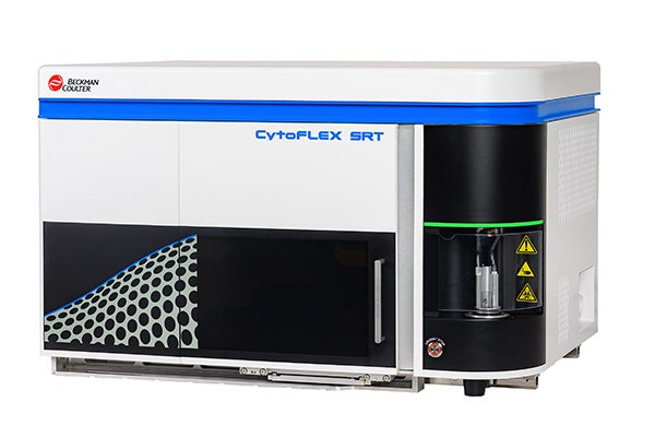
CytoFLEX SRT Benchtop Cell Sorters
CytoFLEX SRT Cell Sorter is a benchtop sorter. It is capable of meeting requirements for a wide range of sorting needs.
What are the Core Components in FACS?
- Fluidics System: The fluidics system transports cells in a narrow, single-file stream of sheath fluid so they pass one at a time through the laser interrogation point. Hydrodynamic focusing ensures that cells align in the center of the stream, allowing for accurate signal detection and precise sorting.2
- Optical System/ Lasers: The optical system consists of lasers, lenses, mirrors and detectors. Lasers provide excitation at specific wavelengths suitable for various fluorophores. As cells pass through the laser beams, emitted fluorescence and scattered light are collected by lenses and routed via dichroic mirrors to the optical detectors. These detectors convert the light signals into measurable electronic signals.1
- Electronics and Data Acquisition System: The electronics system processes the signals before cell sorting. The data processing software classifies cells based on user-defined gating strategies. The electronics ensure that the droplets, each containing one cell, are assigned the accurate charges.3
- Sorting Mechanism: Each droplet receives an electrostatic charge depending on the properties of the cell and its biomarkers. Charged deflection plates redirect droplets into separate collection containers, allowing precise isolation of specific cell populations.4
How Does Fluorescence-Activated Cell Sorting Work
Step 1 – Sample Preparation
Sample preparation in fluorescent-activated cell sorting involves isolating cells from tissue or culture as a single-cell suspension. Debris, clumps and dead cells are removed using filters or centrifugation to ensure clean flow through the cytometer. Furthermore, buffer conditions are optimized to maintain cell viability, prevent clumping and minimize background fluorescence.1
Step 2 – Fluorescent Labelling
Cells are incubated with fluorescently tagged antibodies or dyes that bind to specific markers. A key advantage of FACS is that multiple fluorophores can be used simultaneously to distinguish several cell types within the same sample. Unbound dye is washed away to reduce noise and maximize signal detection accuracy.5
Step 3 – Flow Cytometry Detection
The cells enter the fluidics system, where hydrodynamic focusing aligns cells in a single-file stream as they pass through laser beams. Lasers excite the fluorophores, while sensors detect emitted light and scatter signals. These signals are converted into electronic data, which serve as measurements for the characteristics of each cell, including size, granularity and marker expression.1
Step 4 – Sorting Process
Each cell is encapsulated into a droplet as it passes through the detection point. The droplet is then charged according to predefined sorting parameters. Electrostatic deflection plates direct charged droplets into specific collection tubes or wells, congregating cells with similar properties in the same container while separating them from the rest.1
Step 5 – Post-Sort Analysis
Sorted cells are transferred to appropriate culture media to maintain viability. Post-sort analysis is performed to validate purity, viability and population characteristics. If impurities (i.e., mixed cell populations) are detected in a particular culture, additional sorting methods may be required. The purified and isolated cells are used for performing microscopy or downstream assays, such as RNA sequencing.3
Applications of Fluorescence-Activated Cell Sorting
Drug Discovery and Development
FACS is widely used to isolate specific cell subpopulations for mechanism-of-action studies, compound screening, multi-omics analysis and target validation. Researchers can quantify biomarker expression and assess drug responses or potential toxicity, which helps improve the efficiency of early-stage drug discovery.6
Stem Cell and Regenerative Medicine Research
FACS is an essential tool for sorting stem cells based on markers specific to pluripotent, multipotent or lineage-committed populations. Thus, researchers can purify embryonic stem cells, induced pluripotent stem cells (iPSCs) and progenitor cells with high precision and accuracy. Isolated cells can be utilized for differentiation studies, tissue engineering and the development of cell-based therapies.7
Immunology and Disease Research
Immunologists use FACS to sort and analyze immune cell populations, including T cells, B cells, macrophages, dendritic cells and NK cells, enabling them to study their characteristics in various diseases, such as infectious diseases, cancer, inflammation and autoimmune diseases. Sorting immune cell subsets is critical for characterizing the tumor microenvironment, studying immune responses and developing patient-specific immunotherapy strategies.8,9
Biopharmaceutical Production and Diagnostics
FACS is used to isolate high-producing colonies, optimizing monoclonal antibody or recombinant protein production in biopharmaceutical manufacturing. Cells can be sorted with high precision based on the expression levels of productivity markers.10
In clinical diagnostics, FACS plays a crucial role in detecting and analyzing aberrant cell populations.11
Equipment, Technology and Innovations in FACS
Key Instruments and Components
FACS instruments combine flow cytometry detection with physical sorting capabilities. While the flow cytometer in FACS measures cell properties and markers, FACS systems also include sorting hardware, such as droplet generators, deflection plates and collection mechanisms, as well as software to streamline sorting based on user-defined criteria. Advanced FACS systems support multi-parameter detection, enabling the more sensitive sorting of heterogeneous cell populations and the isolation of rare cell types.1
The choice of fluorophores is critical, as it affects resolution, cell viability and overall sensitivity. Bright and photostable fluorophores should be selected to enhance the detection of cells with low-abundance markers. Additionally, spectral overlap must be minimized to improve the resolution of discrimination.1
Sorting buffers maintain cell health and integrity during the process by preventing cell clumping and providing optimum ionic conditions for cell survival.12
Advanced Sorting Technologies
Advanced sorting technologies improve throughput, purity and multiparametric separation in FACS.
Electrostatic droplet deflection is the key mechanism facilitating separation in FACS. Each characterized cell is encapsulated by a droplet, which gets assigned a specific charge. Deflection plates divert charged droplets into designated collection tubes.1
Microfluidic devices for high-precision sorting provide a more compact alternative to droplet-based sorters. They comprise microchannel networks, optical traps or dielectrophoretic fields to steer cells with lower shear stress. Therefore, they may be ideal for sensitive rare cell subtypes, stem cells or clinical samples.11,13
Other advanced technologies include:
- Spectral flow cytometry for multiparametric sorting14
- High-speed sorters with real-time digital processing and adaptive optics1
- Machine learning-assisted gating15
- Automated sample handling16
Advantages, Limitations and Considerations of FACS
Key Advantages of FACS
Fluorescence-activated cell sorting offers significant advantages for life sciences applications requiring rapid and precise cell sorting.
- High precision, purity and throughput: A FACS instrument can sort thousands of cells per second, allowing researchers to generate well-defined cell subsets.1
- Multi-parameter analysis capability: FACS systems can simultaneously measure dozens of biomarkers using multiple fluorophores, allowing researchers to segregate heterogeneous populations with high resolution.17
- Sorting of rare and heterogeneous cell populations: FACS is a powerful tool for isolating rare cell types, such as stem cells, from heterogeneous samples. Therefore, it is ideal for the detailed investigation of complex tissue samples and tumor microenvironments.18
Limitations and Practical Considerations
Despite their cutting-edge separation mechanism, FACS systems require special investment and care to maximize their utility.
- High instrument cost and maintenance: High-end lasers, detectors, fluidics components and software can increase the cost of purchase and maintenance in FACS instruments.2
- Sample preparation complexity: Successful sorting relies on high-quality single-cell suspensions free from debris and aggregates. Rigorous filtration, staining, washing and viability assessment are required to prevent false signals and inaccurate separation.2
- Potential stress or loss of cells during sorting: The sorting process exposes cells to shear forces, changes in pressure, droplet formation, lasers and extended handling times, which may reduce the viability and alter the properties of sensitive cells. Post-sort recovery, microfluidic systems and post-FACS analysis are critical in ensuring that cells remain functional and intact.19
FAQs
What does fluorescence-activated cell sorting (FACS) do?
FACS separates and analyzes cells based on fluorescent markers, allowing researchers to isolate specific subpopulations with high precision and purity.
What’s the difference between Flow Cytometry and FACS?
Flow cytometry measures physical and fluorescent characteristics of cells, while FACS is a specialized form of flow cytometry that also physically sorts cells into separate containers.
What is the standard fluorescence-activated cell sorting (FACS) protocol?
A typical FACS protocol involves preparing a single-cell suspension, fluorescent labelling, washing to remove unbound dyes, running the sample through a flow cytometer, gating target populations and collecting sorted cells for downstream analysis.
What is the difference between FACS and MACS?
FACS uses fluorescent markers and laser-based detection to sort cells individually. MACS uses magnetic beads and magnetic fields, offering simpler but less precise bulk separation.
What types of cells can be sorted using FACS?
FACS can sort immune cells, stem cells, cancer cells, primary cells and genetically modified reporter cells.
References
- Garg G, Patel P, Gupta GD, Kurmi BD. A Review on Working Principle and Advanced Applications of Fluorescence activated Cell Sorting Machine (FACS). Curr Pharm Anal 2024;20(2):85-97.
- Agarwal A, Khushalani D, Harkare A, Agrawal R. A review of FACS: fluorescence activated cell sorting system. Helix 2020;10(04):204-7.
- Telford WG. Flow cytometry and cell sorting. Front Med 2023;10:1287884.
- Cai B, Ding J, Yalikun Y, Ren D. Progress of Cell Sorting in Flow Cytometry. iLABMED 2025;3(1):96-105.
- Gautier A. Fluorescence-activating and absorption-shifting tags for advanced imaging and biosensing. Acc Chem Res 2022;55(21):3125-3135.
- Vitelli M, Budman H, Pritzker M, Tamer M. Applications of flow cytometry sorting in the pharmaceutical industry: A review. Biotechnol Prog 2021;37(4):e3146.
- Cai Y, Wang J, Zou K. The progresses of spermatogonial stem cells sorting using fluorescence-activated cell sorting. Stem Cell Rev Rep 2020;16(1):94-102.
- Fei C, Nie L, Zhang J, Chen J. Potential applications of fluorescence-activated cell sorting (facs) and droplet-based microfluidics in promoting the discoveryof specific antibodies for characterizations of fish immune cells. Front Immunol 2021;12:771231.
- Miwa H, Dimatteo R, de Rutte J, Ghosh R, Di Carlo D. Single-cell sorting based on secreted products for functionally defined cell therapies. Microsyst Nanoeng 2022;8(1):84.
- Kol S, Ley D, Wulff T, Decker M, Arnsdorf J, Schoffelen S, et al. Multiplex secretome engineering enhances recombinant protein production and purity. Nat Commun 2020;11(1):1908.
- Sivaramakrishnan M, Kothandan R, Govindarajan DK, Meganathan Y, Kandaswamy K. Active microfluidic systems for cell sorting and separation. Curr Opin Biomed Eng 2020;13:60-68.
- Pan J, Wan J. Methodological comparison of FACS and MACS isolation of enriched microglia and astrocytes from mouse brain. J Immunol Methods 2020;486:112834.
- Cha H, Fallahi H, Dai Y, Yuan D, An H, Nguyen N-T, et al. Multiphysics microfluidics for cell manipulation and separation: A review. LAB CHIP 2022;22(3):423-444.
- Bonilla DL, Reinin G, Chua E. Full spectrum flow cytometry as a powerful technology for cancer immunotherapy research. Front Mol Biosci 2021;7:612801.
- Lee E, Torres R, Schulz W, Durant T. Automated gating and interpretation of clinical flow cytometry data: A computational approach using artificial intelligence and deep learning. Am J Clin Pathol 2022;158:S7.
- Wiener DM, Huynh E, Jeyakumar I, Bax S, Sama S, Cabrera JP, et al. An open-source FACS automation system for high-throughput cell biology. PLoS One 2024;19(3):e0299402.
- Ofir N, Rozenberg E, Sharabi O, Zektser M, Rouvio O, Gazit R. Advanced Multicolor Flow Cytometry Method for Multiple Myeloma. Clin Lymphoma Myeloma Leuk2025.
- Maes E, Cools N, Willems H, Baggerman G. FACS-based proteomics enables profiling of proteins in rare cell populations. Int JMol Sci 2020;21(18):6557.
- Box A, DeLay M, Tighe S, Chittur SV, Bergeron A, Cochran M, et al. Evaluating the effects of cell sorting on gene expression. JBT 2020.