Human-relevant model discussion with Leica Microsystems
Dr. Andrew Barazia shares his expertise on characterizing human-relevant models, particularly organoids, using Leica Microsystems’ advanced imaging platforms and AI-based image analysis software. These tools can help effectively and efficiently scale human-relevant model programs. Andrew will further discuss how these techniques will enable a deeper understanding of some of the most complex biological processes.
Q&A
- What are the main challenges with existing microscopy techniques when imaging 3D human-relevant models?
- Why is light sheet microscopy well-suited for long-term live imaging of 3D human-relevant models?
- How is Leica Microsystems addressing these challenges of phototoxicity and photobleaching?
1. What is the main challenge with existing microscopy techniques when imaging 3D human-relevant models?
When we think about wanting to image 3D human-relevant models, we want to image that sample for days, if not weeks, which has not been as easy or as common in many imaging platforms. We also want to image that sample at high resolution, at relatively high speed, and get enough temporal resolution to see the cell divisions or the cellular movements gently enough that we're not causing phototoxicity or photobleaching on these samples. When looking at a sample with a wide field or confocal microscope, you often image from above or below, where we're focusing light onto the focal plane and then detecting everything in that focal plane that we can. But in wide field microscopy, collecting everything above and below that plane, or in the case of a confocal microscope, throwing away everything above and below that focal plane with a pinhole so that we can get nice optical sectioning and increase the signal to noise, that puts a lot of light on the sample in order to obtain high resolution.
2. Why is light sheet microscopy well-suited for long-term live imaging of 3D human-relevant models?
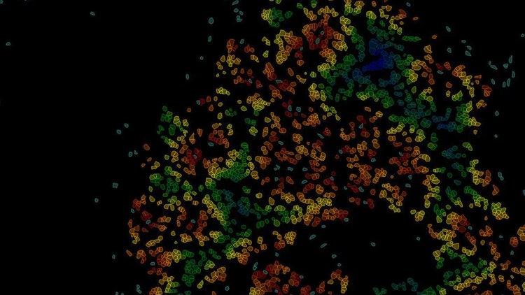
Light sheet microscopy is a newer technique allowing us to image organoids or even some larger developmental biology-esque samples like Drosophila embryos and zebrafish embryos. With light sheet microscopy, the idea is to actually excite the sample parallel to the focal plane and perpendicular to the detection objective and get that optical sectioning by just having that thin excitation plane. This gives us some benefits when it comes to phototoxicity and photobleaching but also gives us a really nice imaging speed that we can then take advantage of with some high-speed (complementary metal-oxide-semiconductor) CMOS cameras. When we're talking about imaging these human-relevant models for long periods, we want to ensure that we minimize phototoxicity so as not to cause excessive ROS generation or excessive thermal damage, just by having too much laser on the sample at any given moment. Light sheet microscopy offers this advantage over some of the other existing microscopy techniques. For example, in a brain organoid, you can see both the maximum intensity projection and the 3D rendering of hundreds of cells migrating and interacting with each other over time, with minimal phototoxicity and bleaching.
38 hour time-lapse of brain organoid
3. How is Leica Microsystems addressing these challenges of phototoxicity and photobleaching?
Regarding light-sheet microscopy, Leica Microsystems asked, "What if we could get both multiple views and positions in one system while decreasing phototoxicity and photobleaching?" With Leica Microsystems’ Viventis Deep system, large multicellular sample imaging is improved due to the dual-view detection. First, the excitation portion of the system is tilted a little bit below these wells, which we can illuminate along that vertical light sheet axis. And because we have two illumination objectives, we can evenly illuminate and not cause an intensity gradient as we go from left to right across the samples or top to bottom, which is common in many single-objective systems. Secondly, there are two objectives that give us significant benefits from imaging both sides of the sample at the same time. Suppose we have an organoid that is a few hundred microns across. In that case, we can image that in its entirety. We're not limited by the single photon imaging depth of 100 to 150 microns, and we can see both sides of some samples and fuse them back together using our AIVIA software. The Viventis Deep system allows us to effectively go twice as deep, combined with the dual illumination objectives and maintain low phototoxicity and photobleaching.
67 hour multi-position time-lapse of intestinal organoids
Listen to the full webinar here to learn more about standardizing human-relevant model culture and deep characterization with cutting-edge imaging and AI-based image analysis.

Bridging the Gap: Advancing Human-Relevant Models for Real-World Impact Webinar