Obtaining high-quality tissue sample images is essential during drug screening and development. For example, high-quality spatial imaging can facilitate cancer research, such as evaluating the tumor microenvironment and analyzing human organoids. However, the difficulty in obtaining clear and representative sample data due to heterogeneity within tissue samples and the need to maintain sample culture and incubation conditions can make tissue imaging complicated. Sample preparation can also be challenging. Many stains can be involved in the process, and antibodies often need to penetrate specific tissues to reach antigens.
Complexities associated with tissue imaging do not end there. Capturing images and analyzing them manually is time-consuming, inefficient and subjective. These bottlenecks are exacerbated when using techniques such as confocal microscopy and when using multiple antibody stains and incubations, which require more precise and involved sample preparation. Imaging instruments that automate image capture and high-content image analysis with guided workflows and software for image segmentation and classification like Molecular Devices ImageXPress Confocal HT.ai High-Content Imaging System, can enable laboratories to go beyond one or two analysis parameters and generate more valuable data from high-content image capture.
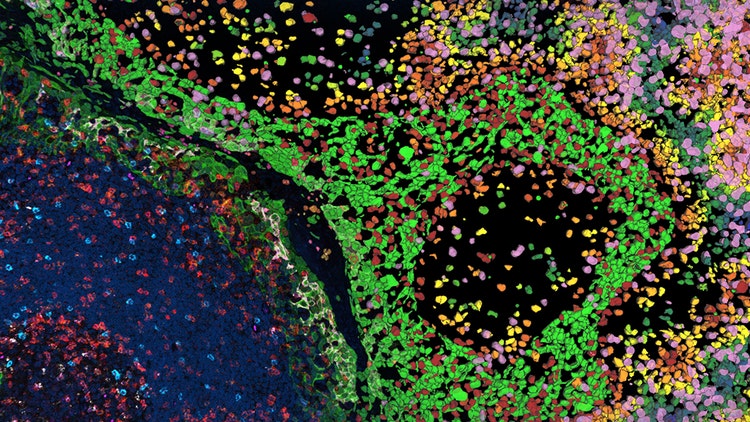
If a picture is worth one thousand words, multiplexed images may be worth a few publications. Maximizing data yield from samples and ensuring every appropriate insight is being gleaned from tissues can seem like an endless task. Without appropriate technology, it can become extremely difficult to capture all the required data from a single biopsy or tissue sample. Precise, open multiplexing solutions, such as Leica Microsystems Cell DIVE, can help researchers transform tissue research with multiple validated antibodies — from a choice of over 350 — to be applied simultaneously, facilitating high-quality automated image capture from multiple conditions in parallel. Automated image processing and feature extraction across acquired images can enable visualization, identification and quantification of biomarkers and other research areas for maximizing insights.
Unlocking productivity, removing bottlenecks in workflows and improving quality by reducing subjectivity resulting from human error calls for scalable automation. At the Life Sciences companies of Danaher we help pioneer more effective treatments and develop more efficient workflows. To learn how our offerings can help you achieve your imaging needs or to explore ways of partnering with us, contact an expert at the Life Sciences companies of Danaher today.
Improving the efficiency and quality of advanced imaging and analysis