JavaScript is disabled in your browser. Please enable JavaScript to view this website.

Make label-free screening decision-grade. Turn transmitted-light images into accurate nuclei and cell readouts without making fluorescent staining the limiting step

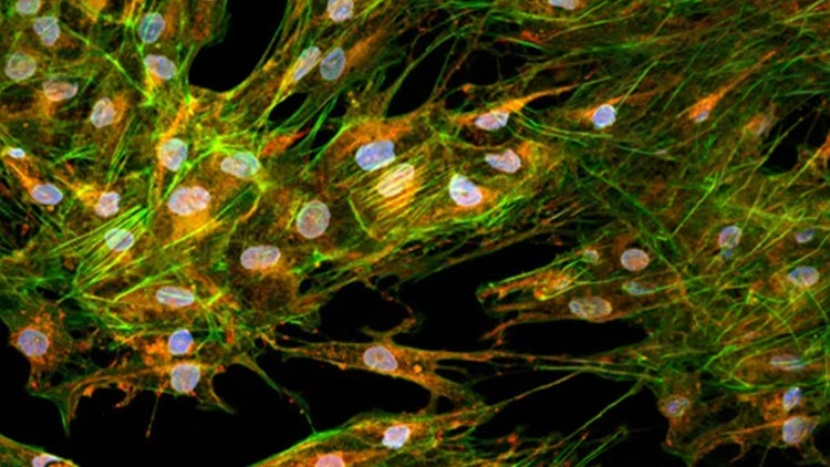
Pharma screening organizations are measured on how confidently they can move from a completed plate to a defensible go/no-go decision, not simply on how many images they can acquire. Traditional high-content workflows often depend on fluorescent or chemical labels to segment nuclei and cells, adding reagent burden, staining steps, endpoint constraints, and potential disruption to live-cell biology. The label-free analysis materials show a different path: customizable AI models in IN Carta® Image Analysis Software can use transmitted-light (TL) images alone to segment nuclei and cells after being trained with fluorescence-derived masks. This enables label-free nuclear detection validated against Hoechst/DAPI-based counts, supports live-cell and non-invasive monitoring, reduces reliance on fluorescent labels for the readout, and produces dose-response behavior comparable to traditional fluorescence-based methods. For screening leaders, the strategic value is not “AI segmentation” as a feature; it is the ability to simplify assay execution while preserving confidence in the biological decision.
Molecular devices

Fewer labels. Less perturbation. Decision-grade screening confidence

With ImageXpress HCS.ai + IN Carta, transmitted-light images can become AI-derived nuclei and cell counts. SINAP cross-channel supervision uses fluorescence masks to train TL models, then applies those models directly to label-free TL images. The result is a simplified workflow that reduces fluorescent-label dependency, preserves live-cell observation, supports high-throughput phenotypic screening, and maintains confidence through documented accuracy and dose-response agreement with fluorescence-based approaches.

AI-Enabled Label-Free Segmentation, Validated Against Fluorescence-Based Ground Truth

The label-free evidence base supports quality, time, cost, and capacity narrative for HCS.ai + IN Carta. In the AI-enabled label-free detection materials, Molecular Devices used customizable AI models within IN Carta to develop deep learning-based segmentation models for accurate cell and nuclear detection using transmitted-light images alone. A TL-based nuclear segmentation model was trained by first segmenting nuclei in Hoechst/DAPI-stained images, pairing those segmentation masks with corresponding TL images, and retraining a model capable of segmenting nuclei from TL images only. A parallel approach trained a TL cell segmentation model using Calcein AM-derived masks. The U2OS model achieved over 97% label-free nuclear detection accuracy validated against Hoechst-based nuclear counts and a 97% whole-cell segmentation performance compared to nuclear counts. Across compound-screening experiments, TL-based cell counts showed extremely strong correlation and comparable IC50 values versus traditional nuclear staining methods. We also document faster processing, workflow simplicity, reduced reagent cost, real-time/non-invasive monitoring, and the ability to preserve cell viability by eliminating fluorescent or chemical labels. ImageXpress HCS.ai + IN Carta lets screening teams move label-free analysis from a convenience feature to a decision-grade AI workflow, reducing staining dependency while maintaining the accuracy required for screening decisions.

A transmitted-light AI workflow that simplifies screening without disconnecting from fluorescence-validated confidence

The workflow is grounded in cross-channel supervision, not unsupported black-box inference. During model development, fluorescence channels generate masks for nuclei or cells; those masks are paired with corresponding TL images to train a model that can later segment directly from TL images. IN Carta’s SINAP module supports this customizable AI workflow, and user-defined targets can compare TL-derived nuclei against DAPI/Hoechst ground truth to calculate detection and false-positive performance. This allows screening teams to validate TL-only readouts against familiar fluorescence-based reference methods before applying label-free segmentation at scale. In the documented studies, TL-derived dose-response curves showed consistent trends with DAPI/Hoechst-derived curves and comparable IC50 values, supporting confidence that fewer staining dependencies do not mean weaker screening decisions.

Quality increase: TL-derived nuclei/cell counts were validated against fluorescence-based ground truth, with over 97% nuclear detection accuracy, 97% whole-cell segmentation performance, and strong correlation/comparable IC50 values versus traditional staining methods. Time reduction: the source materials state label-free imaging provides faster processing and workflow simplicity, and the TL workflow reduces dependence on staining as the final readout. Cost reduction: label-free imaging reduces fluorescent reagent burden by eliminating the need for fluorescent or chemical labels for the label-free readout. Capacity increase: multi-cell-line training using SINAP export/import and customizable AI models supports scalability across diverse cell lines and makes the workflow suitable for high-throughput and phenotypic screening applications.

Speak to one of our leading life sciences experts

TTAE

TTAE

546006278

TTAE

add

Talk to an Expert

https://lifesciences.danaher.com/

https://lifesciences.danaher.com/us/en/solutions/gene-therapy/crispr-gene-mediated.html