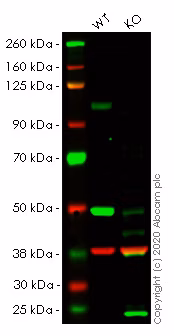
Western blot - Anti-GAPDH antibody [6C5] - Loading Control (ab8245)
Lanes 1- 2: Merged signal (red and green). Green - ab16640 observed at 100 kDa. Red - Anti-GAPDH antibody [6C5] - Loading Control (ab8245) observed at 37 kDa.
ab16640 was shown to react with Sortilin/NT3 in wild-type HeLa cells in western blot. Loss of signal was observed when knockout cell line ab264772 (knockout cell lysate ab257696) was used. Wild-type HeLa and SORT1 knockout HeLa cell lysates were subjected to SDS-PAGE. Membrane was blocked for 1 hour at room temperature in 0.1% TBST with 3% non-fat dried milk. ab16640 and Anti-GAPDH antibody [6C5] - Loading Control (ab8245) were incubated overnight at 4°C at a 1 μg/ml and a 1 in 20000 dilution respectively. Blots were developed with Goat anti-Rabbit IgG H&L (IRDye®800CW) preadsorbed (ab216773) and Goat anti-Mouse IgG H&L (IRDye®680RD) preadsorbed (ab216776) secondary antibodies at 1 in 20000 dilution for 1 hour at room temperature before imaging.
All lanes:
Western blot - Anti-Sortilin/NT3 antibody (ab16640) at 1 µg/mL
Lane 1:
Wild-type HeLa cell lysate at 20 µg
Lane 2:
Western blot - Human SORT1 (Sortilin/NT3) knockout HeLa cell lysate (ab257696) at 20 µg
Secondary
Lanes 1 - 2:
Western blot - Goat anti-Rabbit IgG H&L (IRDye® 800CW) preadsorbed (ab216773) at 1/20000 dilution
Lanes 1 - 2:
Western blot - Goat anti-Mouse IgG H&L (IRDye® 680RD) preadsorbed (ab216776) at 1/20000 dilution
Performed under reducing conditions.
Predicted band size: 92 kDa
Observed band size: 100 kDa

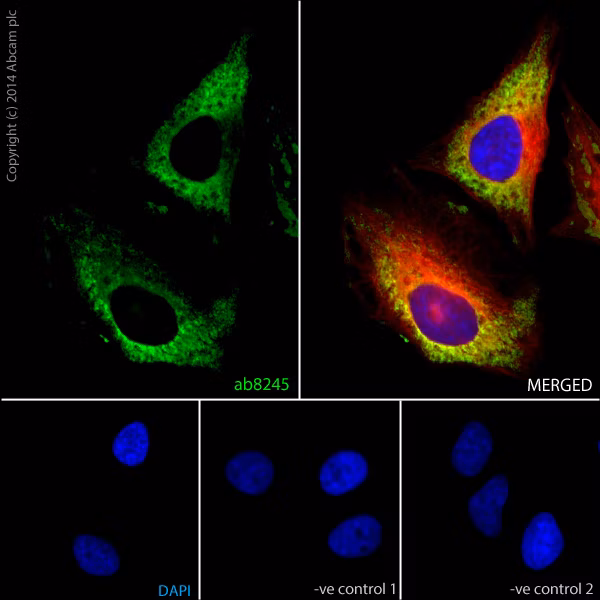
Immunocytochemistry/ Immunofluorescence - Anti-GAPDH antibody [6C5] - Loading Control (ab8245)
ab8245 staining GAPDH in HeLa (Human epithelial cell line from cervix adenocarcinoma) cells.
The cells were fixed with 100% methanol (5 minutes) and then blocked in 1% BSA/10% normal goat serum/0.3M glycine in 0.1%PBS-Tween for 1 hour. The cells were then incubated with ab8245 at 5 μg/ml and ab6046 at 1 μg/ml overnight at +4°C, followed by a further incubation at room temperature for 1 hour with Goat Anti-Mouse IgG H&L (Alexa Fluor® 488) preadsorbed (ab150117) at 2 μg/ml (shown in green) and Goat Anti-Rabbit IgG H&L (Alexa Fluor® 594) preadsorbed (ab150088) at 2 μg/ml (shown in pseudo color red). Nuclear DNA was labeled in blue with DAPI.
Negative controls: 1– Rabbit primary antibody and anti-mouse secondary antibody; 2 – Mouse primary antibody and anti-rabbit secondary antibody. Controls 1 and 2 indicate that there is no unspecific reaction between primary and secondary antibodies used.
Western blot - Anti-GAPDH antibody [6C5] - Loading Control (ab8245)
This blot was produced using a 4-12% Bis-tris gel under the MES buffer system. The gel was run at 200V for 50 minutes before being transferred onto a Nitrocellulose membrane at 30V for 70 minutes. The membrane was then blocked for an hour before being incubated with ab140751 overnight at 4°C. Antibody binding was detected using the Donkey Anti-Rabbit IgG H&L (IRDye® 680RD) preadsorbed ab216779 at a 1:10,000 dilution for 1hr at room temperature and then imaged using the Licor Odyssey CLx.
All lanes:
Western blot - Anti-GAPDH antibody [6C5] - Loading Control (ab8245) at 1/2000 dilution
Lane 1:
Wild-type HAP1 cell lysate (20 µg)
Lane 2:
NF-κB p60 knockout HAP1 cell lysate (20 µg)
Lane 3:
HeLa cell lysate (20 µg)
Lane 4:
A431 cell lysate (20 µg)
Secondary
All lanes:
Western blot - Donkey Anti-Rabbit IgG H&L (IRDye® 680RD) preadsorbed (ab216779) at 1/10000 dilution
Predicted band size: 36 kDa

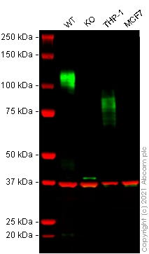
Western blot - Anti-GAPDH antibody [6C5] - Loading Control (ab8245)
False colour image of Western blot: Anti-SIRPA antibody staining at 1/1000 dilution, shown in green; Mouse anti-GAPDH antibody [6C5] (ab8245) loading control staining at 1/20000 dilution, shown in red. In Western blot, the antibody was shown to bind specifically to SIRPA. A band was observed at 100-140 kDa (mouse SIRPA, isoform 1), in wild-type RAW 264.7 cell lysates (band observed at 70-100 kDa in THP-1 is Human SIRPA) with no signal observed at this size in SIRPA knockout cell line ab281618 (knockout cell lysate ab282969). To generate this image, wild-type and SIRPA knockout RAW 264.7 cell lysates were analysed. First, samples were run on an SDS-PAGE gel then transferred onto a nitrocellulose membrane. Membranes were blocked in 3 % milk in TBS-0.1 % Tween® 20 (TBS-T) before incubation with primary antibodies overnight at 4 °C. Blots were washed four times in TBS-T, incubated with secondary antibodies for 1 h at room temperature, washed again four times then imaged. Secondary antibodies used were Goat anti-Rabbit IgG H&L (IRDye® 800CW) preabsorbed (ab216773) and Goat anti-Mouse IgG H&L (IRDye® 680RD) preabsorbed (ab216776) at 1/20000 dilution.
All lanes:
Western blot - Anti-GAPDH antibody [6C5] - Loading Control (ab8245) at 1/20000 dilution
Lane 1:
Wild-type RAW 264.7 cell lysate at 20 µg
Lane 2:
RAW 264.7 cell lysate at 20 µg
Lane 3:
THP-1 cell lysate at 20 µg
Lane 4:
MCF7 cell lysate at 20 µg
Predicted band size: 55 kDa
Western blot - Anti-GAPDH antibody [6C5] - Loading Control (ab8245)
Lane 1: Wild-type HeLa cell lysate (20µg)
Lane 2: SORT1 knockout HeLa cell lysate (20µg)
Lanes 1- 2: Merged signal (red and green). Green - ab188586 observed at 100 kDa. Red - loading control ab8245 observed at 37 kDa.
ab188586 Anti-Sortilin/NT3 antibody [EPR15010] was shown to specifically react with Sortilin/NT3 in wild-type HeLa cells in western blot. Loss of signal was observed when knockout cell line ab264772 (knockout cell lysate ab257696) was used. Wild-type and Sortilin/NT3 knockout samples were subjected to SDS-PAGE. Membrane was blocked for 1 hour at room temperature in 0.1% TBST with 3% non-fat dried milk. ab188586 and Anti-GAPDH antibody [6C5] - Loading Control (ab8245) were incubated overnight at 4°C at 1 in 1000 and 1 in 20000 dilution respectively. Blots were developed with Goat anti-Rabbit IgG H&L (IRDye® 800CW) preadsorbed (ab216773) and Goat anti-Mouse IgG H&L (IRDye® 680RD) preadsorbed (ab216776) secondary antibodies at 1 in 20000 dilution for 1 hour at room temperature before imaging.
All lanes:
Western blot - Anti-Sortilin/NT3 antibody [EPR15010] (ab188586) at 1/1000 dilution
Lane 1:
Wild-type HeLa cell lysate at 20 µg
Lane 2:
Western blot - Human SORT1 (Sortilin/NT3) knockout HeLa cell lysate (ab257696) at 20 µg
Secondary
Lanes 1 - 2:
Western blot - Goat anti-Rabbit IgG H&L (IRDye® 800CW) preadsorbed (ab216773) at 1/20000 dilution
Lanes 1 - 2:
Western blot - Goat anti-Mouse IgG H&L (IRDye® 680RD) preadsorbed (ab216776) at 1/20000 dilution
Performed under reducing conditions.
Predicted band size: 92 kDa
Observed band size: 100 kDa