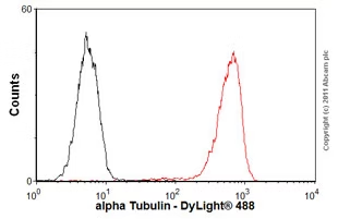
Flow Cytometry (Intracellular) - Anti-alpha Tubulin antibody [DM1A] - Loading Control (ab7291)
Overlay histogram showing HeLa cells stained with ab7291 (red line). The cells were fixed with 80% methanol (5 min) and then permeabilized with 0.1% PBS-Tween for 20 min. The cells were then incubated in 1x PBS / 10% normal goat serum / 0.3M glycine to block non-specific protein-protein interactions followed by the antibody (ab7291, 1μg/1x106 cells) for 30 min at 22°C. The secondary antibody used was an anti-mouse DyLight® 488 (ab96879) at 1/500 dilution for 30 min at 22°C. Isotype control antibody (black line) was mouse IgG1 [ICIGG1] (ab91353, 2μg/1x106 cells) used under the same conditions. Acquisition of >5,000 events was performed.
Western blot - Anti-alpha Tubulin antibody [DM1A] - Loading Control (ab7291)
This blot was produced using a 4-12% Bis-tris gel under the MOPS buffer system. The gel was run at 200V for 50 minutes before being transferred onto a Nitrocellulose membrane at 30V for 70 minutes. The membrane was then blocked for an hour using 5% Milk before being incubated with ab7291 overnight at 4°C. Antibody binding was detected using ab175738 at a 1:10,000 dilution for 1hr at room temperature and then imaged using the Licor Odyssey CLx.
All lanes:
Western blot - Anti-alpha Tubulin antibody [DM1A] - Loading Control (ab7291) at 1 µg/mL
All lanes:
HeLa (Human epithelial carcinoma cell line) Whole Cell Lysate at 10 µg
Secondary
All lanes:
Western blot - Donkey Anti-Mouse IgG H&L (Alexa Fluor® 750) (ab175738) at 1/10000 dilution
Predicted band size: 33 kDa, 36 kDa, 42 kDa, 47 kDa, 61 kDa, 80 kDa
Observed band size: 100 kDa, 50 kDa, 60 kDa, 74 kDa
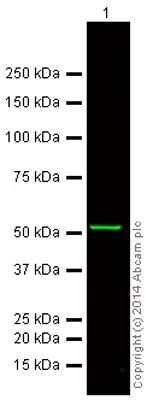
Western blot - Anti-alpha Tubulin antibody [DM1A] - Loading Control (ab7291)
This blot was produced using a 4-12% Bis-tris gel under the MOPS buffer system. The gel was run at 200V for 50 minutes before being transferred onto a Nitrocellulose membrane at 30V for 70 minutes. The membrane was then blocked for an hour using 5% Milk before being incubated with ab7291 overnight at 4°C. Antibody binding was detected using ab175739 at a 1:10,000 dilution for 1hr at room temperature and then imaged using the Licor Odyssey CLx.
All lanes:
Western blot - Anti-alpha Tubulin antibody [DM1A] - Loading Control (ab7291) at 1 µg/mL
All lanes:
HeLa (Human epithelial carcinoma cell line) Whole Cell Lysate at 10 µg
Secondary
All lanes:
Western blot - Donkey Anti-Mouse IgG H&L (Alexa Fluor® 750) preadsorbed (ab175739) at 1/10000 dilution
Predicted band size: 36 kDa
Observed band size: 50 kDa

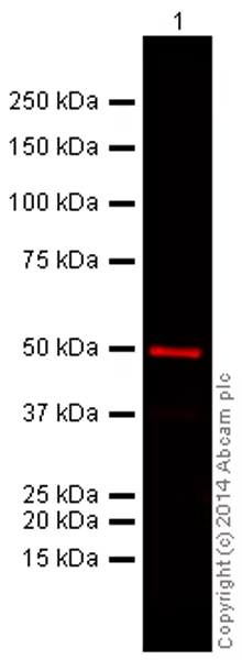
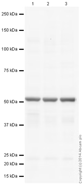
Western blot - Anti-alpha Tubulin antibody [DM1A] - Loading Control (ab7291)
This blot was produced using a 4-12% Bis-tris gel under the MOPS buffer system. The gel was run at 200V for 50 minutes before being transferred onto a Nitrocellulose membrane at 30V for 70 minutes. The membrane was then blocked for an hour using 2% Bovine Serum Albumin before being incubated with ab7291 overnight at 4°C. Antibody binding was detected using an anti-mouse HRP (ab97040), and visualised using ECL development solution ab133406.
All lanes:
Western blot - Anti-alpha Tubulin antibody [DM1A] - Loading Control (ab7291) at 1 µg/mL
Lane 1:
HeLa (Human epithelial cell line from cervix adenocarcinoma cell line) whole cell lysate at 10 µg
Lane 2:
NIH/3T3 (Mouse embryonic fibroblast cell line) whole cell lysate at 10 µg
Lane 3:
PC12 (Rat adrenal gland pheochromocytoma cell line) whole cell lysate at 10 µg
Secondary
All lanes:
Western blot - Goat Anti-Mouse IgG H&L (HRP) preadsorbed (ab97040) at 1/50000 dilution
Developed using the ECL technique.
Performed under reducing conditions.
Predicted band size: 36 kDa, 50 kDa
Observed band size: 50 kDa
Exposure time: 150s
Western blot - Anti-alpha Tubulin antibody [DM1A] - Loading Control (ab7291)
This blot was produced using a 4-12% Bis-tris gel under the MOPS buffer system. The gel was run at 200V for 50 minutes before being transferred onto a Nitrocellulose membrane at 30V for 70 minutes. The membrane was then blocked for an hour using 5% Milk before being incubated with ab7291 overnight at 4°C. Antibody binding was detected using ab186694 at a 1:10,000 dilution for 1hr at room temperature and then imaged using the Licor Odyssey CLx.
All lanes:
Western blot - Anti-alpha Tubulin antibody [DM1A] - Loading Control (ab7291) at 1 µg/mL
All lanes:
HeLa (Human epithelial carcinoma cell line) Whole Cell Lysate at 10 µg
Secondary
All lanes:
Western blot - Goat Anti-Mouse IgG H&L (Alexa Fluor® 680) preadsorbed (ab186694) at 1/10000 dilution
Predicted band size: 36 kDa
Observed band size: 50 kDa