M844 Ophthalmic microscope for advanced eye surgery
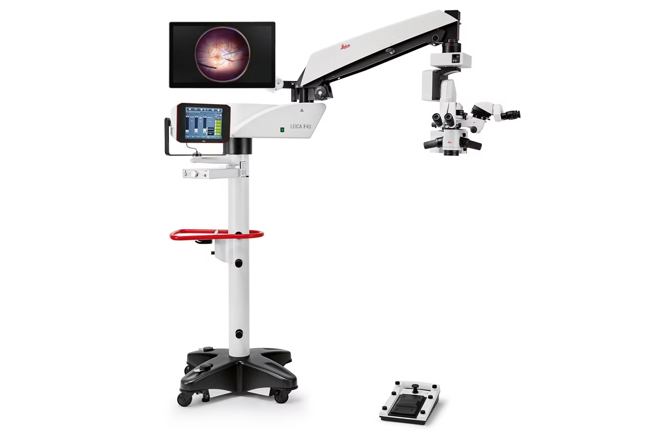
The M844 belongs to the premium class of ophthalmic surgical microscopes and provides an optimal solution particularly for advanced posterior and anterior segment surgery.
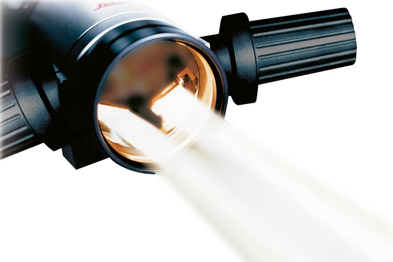
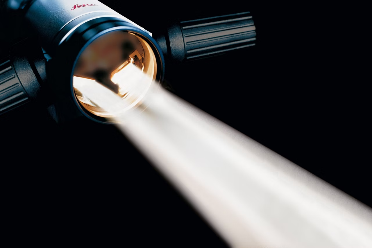
Combining APO OptiChrome optics and direct halogen illumination, the M844 provides crisp, bright, true-color images, even when operating with low light levels.
Innovative features such as the QuadZoom technology, double wing assistant bridge, and HD recording make it an ideal microscope for teaching institutes and university hospitals.
This item has been phased out and is no longer available, except in China. Please contact us to enquire about recent alternative products that may suit your needs.
See more with less light

Leica Optics have superior light gathering abilities. Sharp, brilliant, true anatomical color images with excellent depth of field can be obtained even with very low or no microscope illumination. This helps protect the eyes of both the surgeon and patient, but still enables clear visualization of small details..
Intuitive user interface

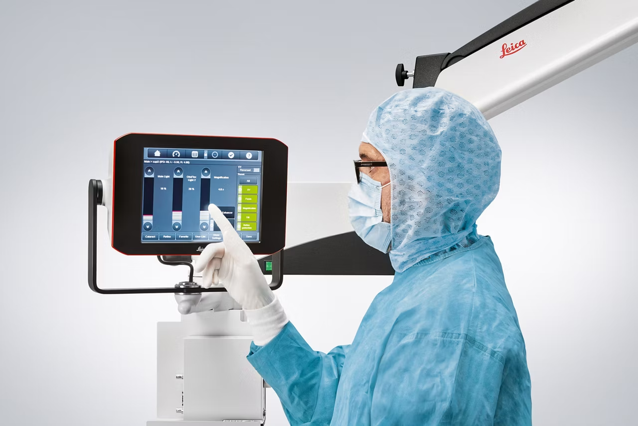
Select, modify, and load the surgeon preferences using the intuitive touch screen user interface of the M844.
Double beam stereo illumination

With two bulbs and two prisms, the M844 delivers homogenous illumination for a true three-dimensional vision with high depth perception.
Identical views for all observers

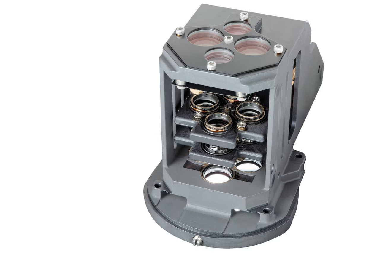
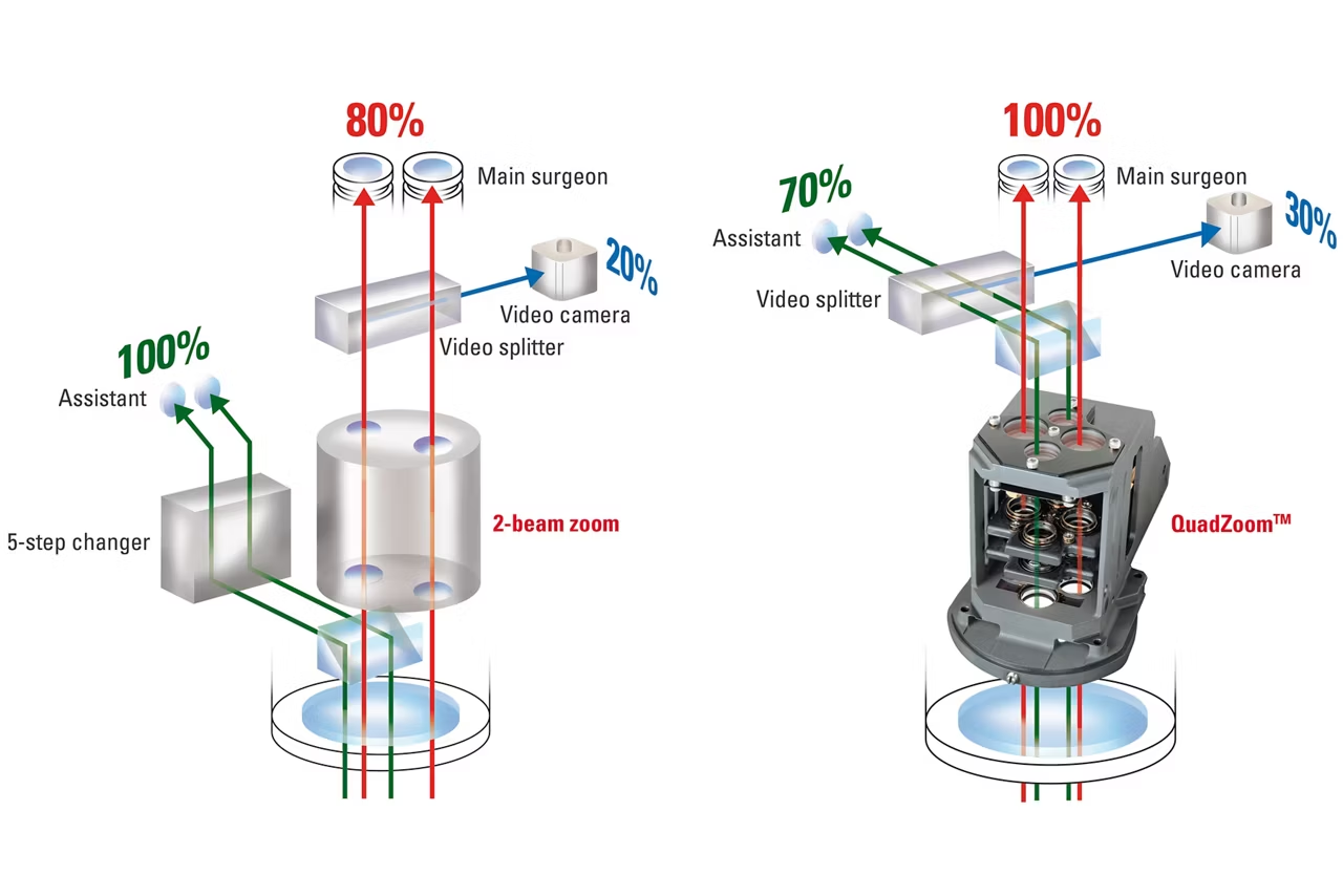
QuadZoom technology with four separate beam paths in the zoom system of the M844 provides an identically bright stereo view for both the main surgeon and the assistants. No manual adjustment by the teacher is necessary. Furthermore, the optional double wing assistant bridge can give a full stereo view with the same magnification to two assistants.
QuadZoom technology

Conventional microscope (left image): The main surgeon loses up to 20% of the light to the video camera. M844 (right image): the main surgeon benefits from 100% light, even if a video camera is added. QuadZoom features four separate beam paths - two for the surgeon/teacher and two for the assistant/student. The result: identical magnification and stereo view for all observers.
Work in comfort

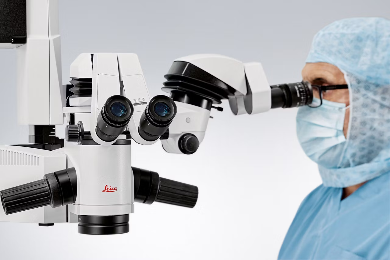
The M844 ophthalmic microscope offers various features to enhance the comfort of both the surgeon and assistant, as well as to support an ergonomic workflow. For example: individually adjustable binocular tubes such as the UltraLow III binocular tube, wireless footswitch, StepCycle, and Auto Reset function.
Wide-angle observation systems and image inverters

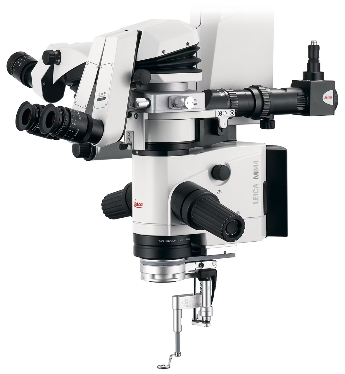
M844 fully integrates the most used wide-angle observation systems or stereo image inverters such as the BIOM 5 and SDI 4 from OCULUS. The surgeon can operate these vitreoretinal accessories via footswitch.
Visualize and document in HD quality

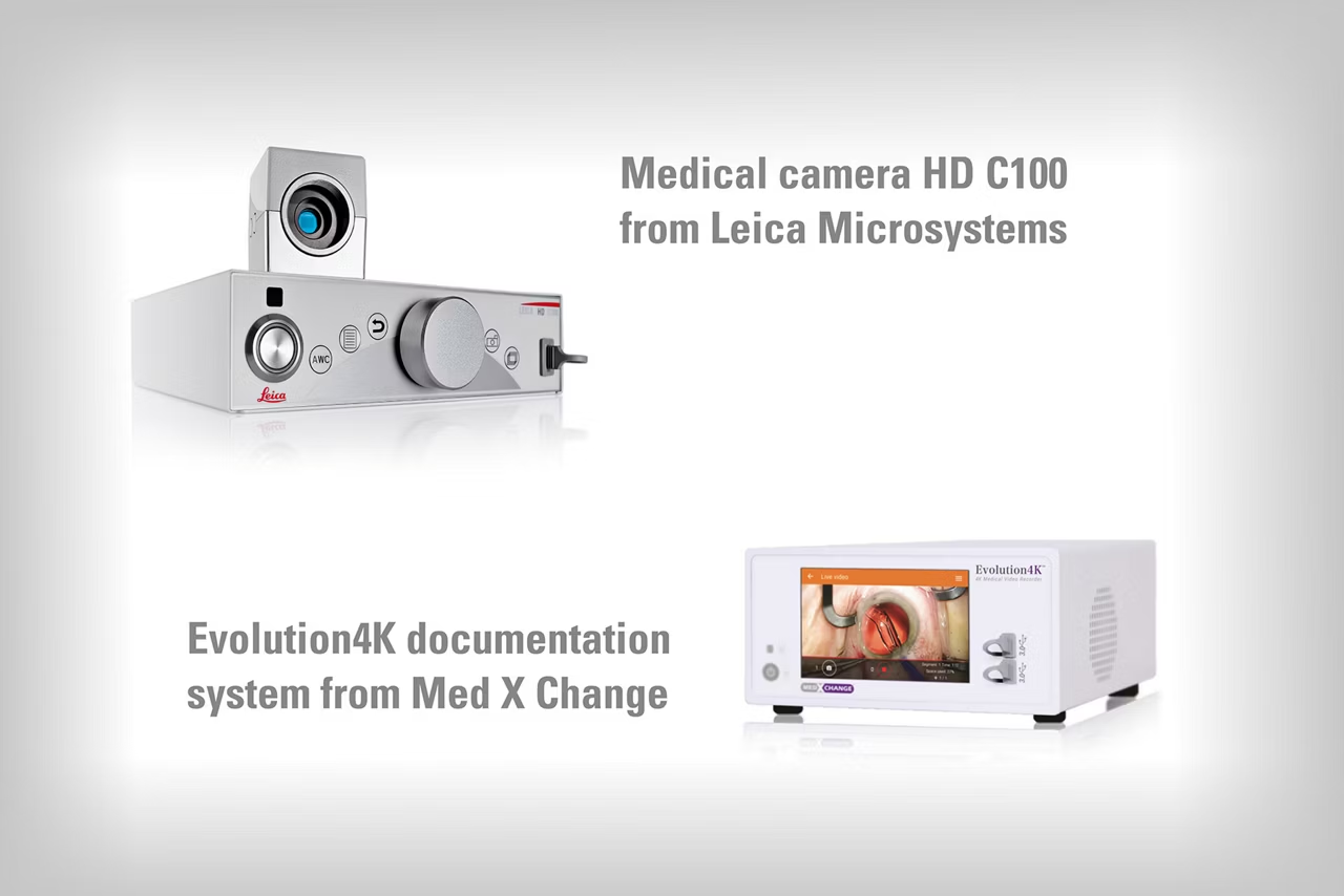
Combine the M844 ophthalmic microscope with the HD C100 medical video camera from Leica Microsystems to record and easily share procedures in HD quality. For more comprehensive documentation, the M844 is also compatible with the EVO 4K recording and documentation system from Med X Change.
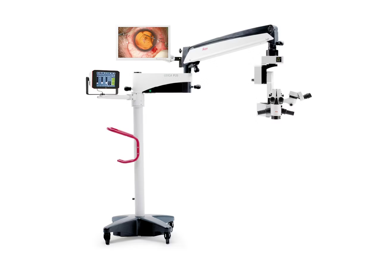
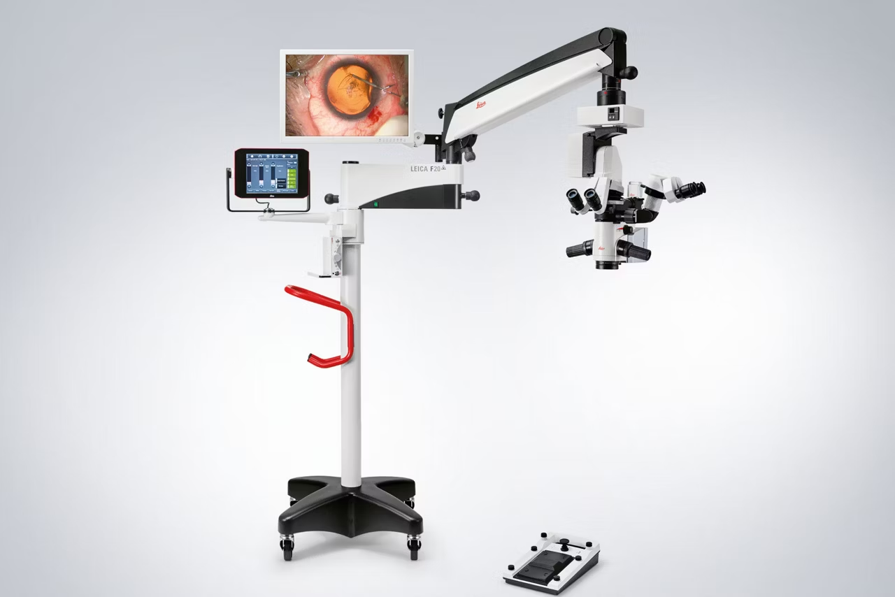
M844 F20

Floor stand microscope featuring mechanical friction brakes and 24-inch microscope-mounted monitor.
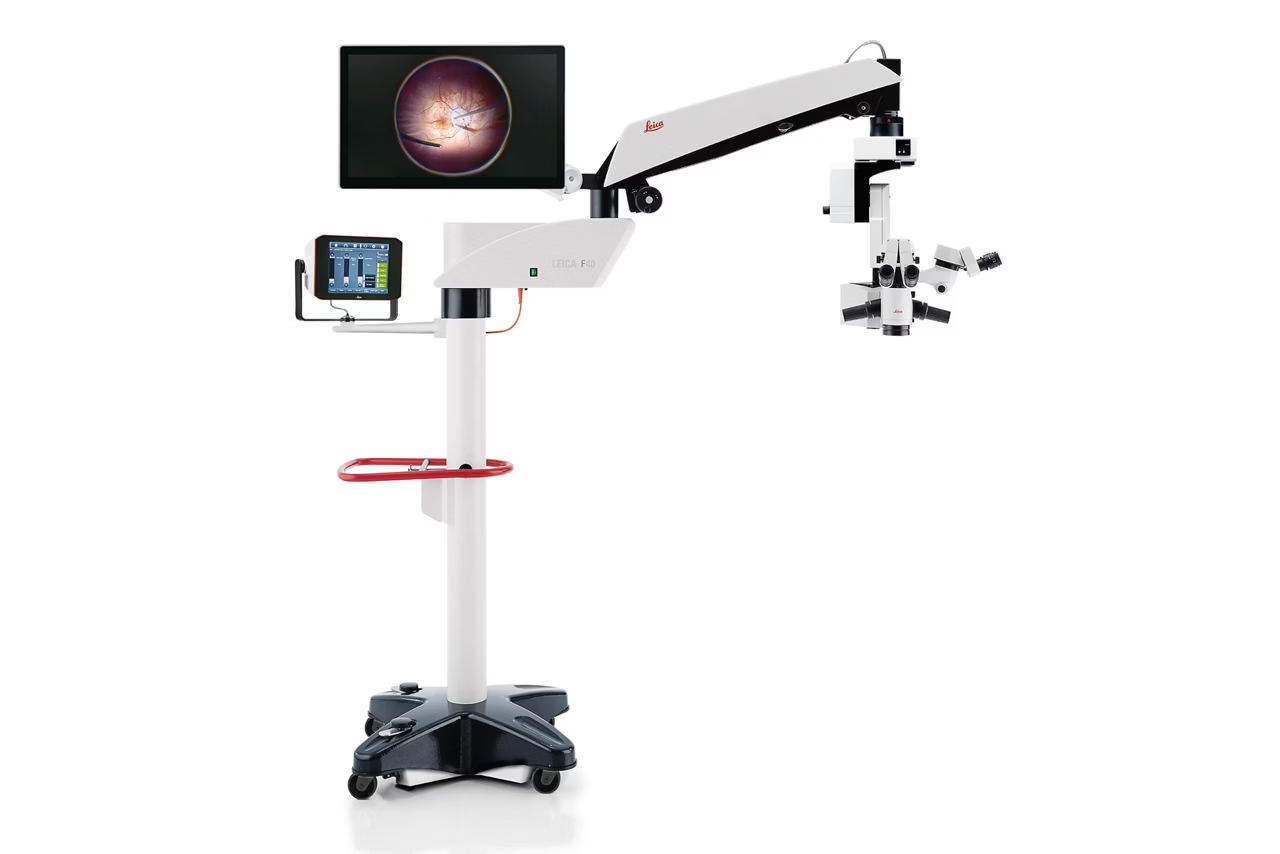
M844 F40

Floor stand microscope featuring electromagnetic breaks and 27-inch microscope-mounted monitor.
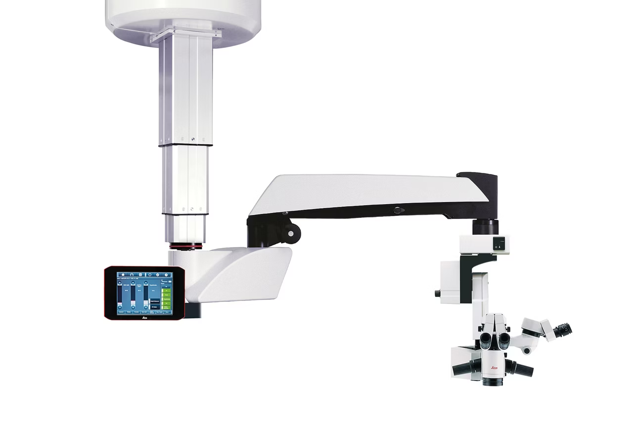
M844 CT40

Space-saving telescope ceiling mount version frees up floor space in a small or multifunctional OR.
RUV800 Panoramic Viewing System for Retinal Surgery
Wide angle retinal viewing system with integrated inverter for upright views. Compatible with anterior and posterior use. Request a quote!
M822 Ophthalmic surgical microscope for anterior and posterior segment surgery
The M822 ophthalmic surgical microscope supports anterior and posterior segment procedures with high quality optics and dual direct illumination. Get a quote!
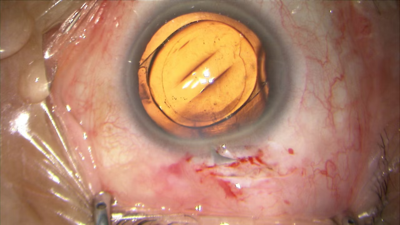
ToricEyePiece The Perfect Aid for Placement of Premium IOL's
Rotatable angle scale eyepiece for accurate toric IOL alignment and astigmatism correction during cataract surgery. Request a quote today.