M530 OHX Surgical Microscope
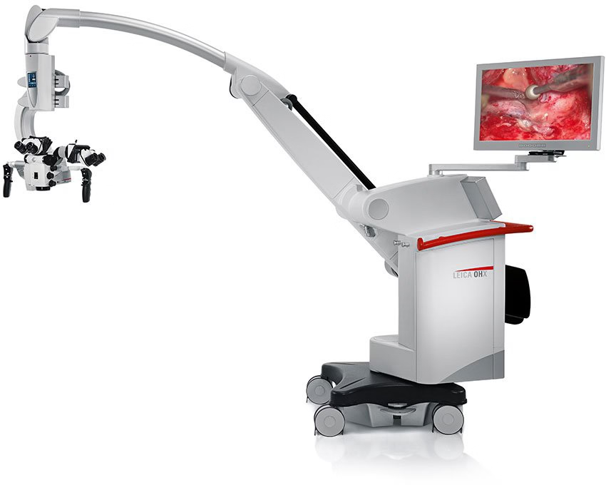
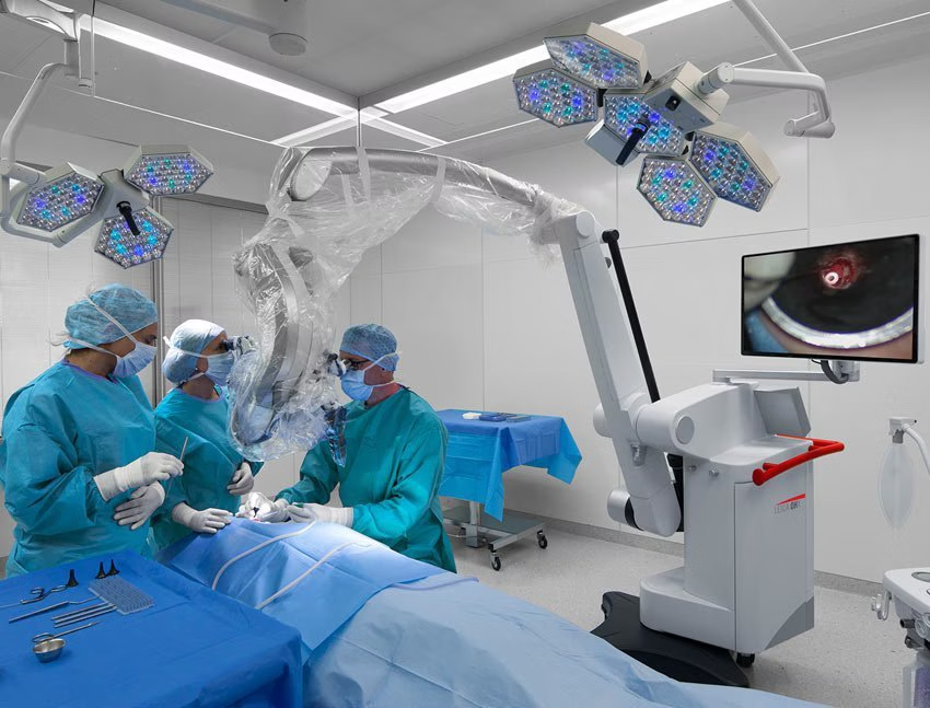
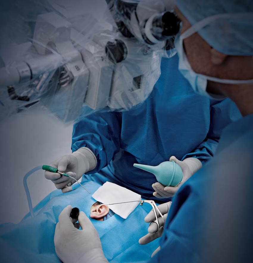

The M530 OHX surgical microscope for neurosurgery, spine, and plastic reconstructive procedures integrates FusionOptics and xenon illumination, flexible positioning, long working distance and fluorescence modes. These enable effortless work in deep and narrow cavities and visualization of vascular and tumoral structures.
Not all products or services are approved or offered in every market and approved labeling and instructions may vary between countries. Please contact your local Leica representative for details.
High resolution & enhanced depth of field

FusionOptics technology from Leica Microsystems combines enhanced depth of field with high resolution for an optimal view of the surgical field. This results in a larger area in full focus, reducing the need for frequent refocusing.
- Two separate optical beam paths
- One path provides great depth of field
- The other path provides high resolution
- The brain effortlessly merges the images into a single, optimal spatial view.
High quality optics

For brilliant images and enhanced depth perception, the M530 OHX surgical microscope combines FusionOptics, apochromatic optics, and 400-Watt xenon illumination.
- FusionOptics and xenon light deliver a bright, sharply focused, true-color image
- Small Angle Illumination (SAI) reduces shadows in deep cavities
- Optional Magnification Multiplier boosts magnification by 40%
Beyond the visible: advanced fluorescence imaging

The M530 OHX surgical microscope offers three fluorescence filters*:
- FL560 yellow fluorescence
- FL400 tumor fluorescence
- FL800 vascular fluorescence
Switch between TriFluoro fluorescence modes with just a few button clicks to compare techniques efficiently and maintain surgical flow without interruption.
*Please check with your local Leica Microsystems representative for product registration status.
Workflow efficiency in spine surgery

Designed to streamline spine procedures, M530 OHX offers features that support fast setup, precise positioning, and efficient visualization—even in minimally invasive environments.
- Fast and effortless positioning to reduce potential strain
- 600 mm working distance for complete flexibility
- Motorized XY microscope tilt and selective brake settings for optimal microscope positioning
- SAI for minimally invasive spine surgery allows shadow-free visualization of deep cavities
- Quick focusing via SpeedSpot laser focus
Effortless positioning for your OR setup

Compact and adaptable design
The M530 OHX surgical microscope's compact footprint, extended arm reach, and generous overhead clearance support flexible positioning—even in crowded operating rooms.
Smooth maneuverability
- Electromagnetic brakes for precise control
- Internally routed cables for a clean setup
- Large range of motion of the optical carrier for high ergonomic comfort
Ideal for cross-table spine setups, cranial procedures, and reconstructive surgery.
Enhanced ergonomics for surgical comfort
Designed for you and your team
M530 OHX adapts to individual needs, helping both surgeon and assistant maintain a comfortable posture during cranial and spine procedures.
Ergonomic features
- Compact optics carrier for close access to the operative field
- A selection of binoculars to suit your preferences, all featuring 360° rotation
- Independent fine focus for optimal visualization by the rear assistant
Fast and effortless positioning reduces the potential strain of harsh movements and enhances efficiency.
You can see much better. You have a much wider field of view with the M530 OHX microscope, which allows you to have an overall view. This is very important, since with a conventional microscope you have to move the camera, which can be quite awkward when you have a flap that is quite large.
Dr. Harold Chatel, Plastic and reconstructive surgeon in Paris
Share and document your work

Visualize your surgery in brilliant 4K resolution on a 27" monitor. The flexible monitor arm allows easy positioning, so your team can follow the surgical workflow in real-time.
For teaching and digital patient documentation, a recording system is available with image streaming to mobile devices as well as DICOM and PACS connectivity. Additionally, the M530 OHX can be connected to an external monitor in your operating room.
Easily connect to compatible surgical devices

Combining preoperative images with intraoperative imaging is crucial during surgical procedures. Image-guided surgery (IGS) systems allow you to enhance your microscope view by overlaying anatomical and functional data onto the white light image and displaying it on the IGS monitor.
The M530 OHX surgical microscope is compatible with neuro-navigation systems from leading manufacturers, supporting seamless integration for advanced surgical guidance.
Precision Comes Standard
Precision optics
FusionOptics technology and xenon illumination provide precise while Small Angle Illumination minimizes shadows in deep cavities, enhancing clarity during cranial, spinal, and reconstructive procedures.
Integrated fluorescence visualization

Access to a range of fluorescence modes during vascular and tumoral visualization.
Flexible setup for surgical efficiency
Compact design, extended arm reach, and motorized positioning enable smooth integration into various OR setups, enhancing workflow efficiency and comfort.
FL560 Fluorescence Module
The FL560 module enables simultaneous real-time observation of fluorescent and non-fluorescent tissue with clear contrast during neurosurgery. Get a quote.
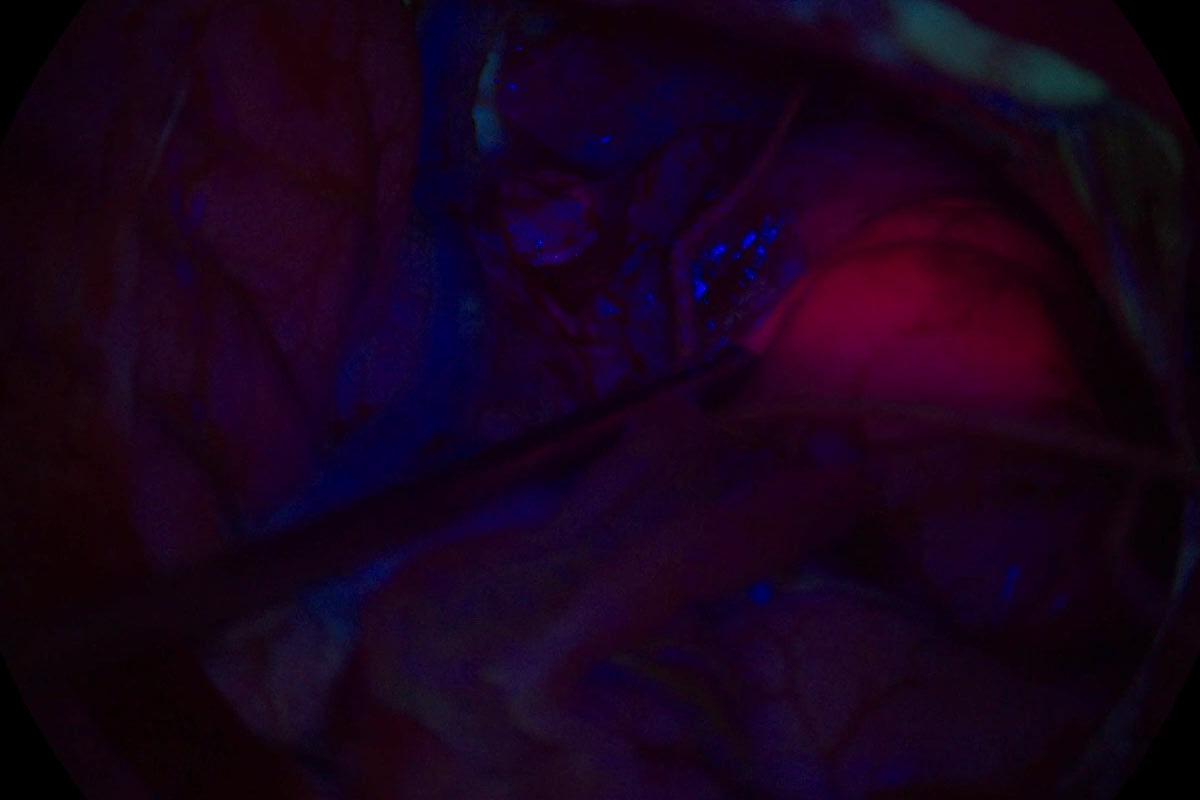
FL400 Blue Light Fluorescence Module
FL400 fluorescence module with 5-ALA helps differentiate tumor tissue from healthy brain tissue during glioma resection. Request a quote today.
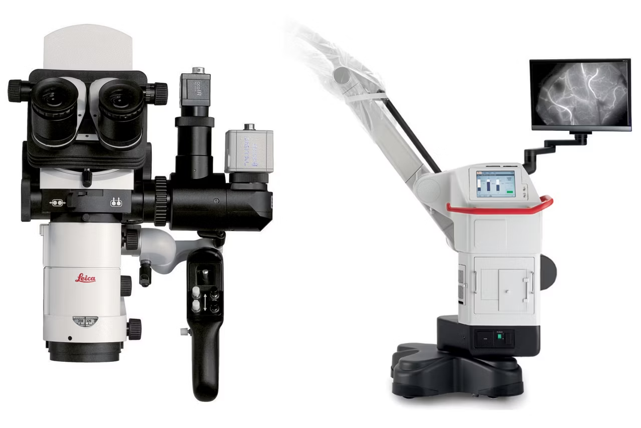
FL800
FL800 uses ICG dye and near infrared camera to visualize vessel patency during neurosurgery. Supports eyepiece and video monitor viewing. Request a quote.