JavaScript is disabled in your browser. Please enable JavaScript to view this website.

ENVISU OCT Imaging for Preclinical Researchers
Buehnenbild Clinical Kid21755949

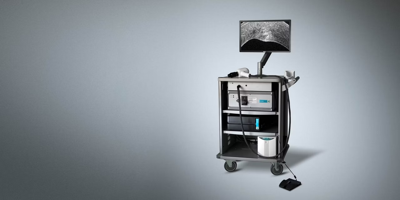
See more and don’t miss pathology with high-volume, high-density, and high-resolution images. The Envisu Image Guided SDOCT systems allow you to:

Explore tissue structure with stunning full-volume detail

Experience superb visualization with high signal-to-noise-ratio provided by Bioptigen-engineered VHR and UHR spectrometers

Conduct reproducible longitudinal studies with an organized framework for customized studies, reporting features, and analysis station.

Acquire great images quickly with a broad array of precision imaging and alignment accessories for small and large animals alike

For research use only. R-Class SDOCT Systems, Diver & Envisu Application Software are Contraindicated for use with humans.

What is Optical Coherence Tomography?
Optical Coherence Tomography, abbreviated OCT, is a non-invasive, contact-free imaging technique to detect and monitor morphological changes of ocular tissue.
What is SDOCT?
Spectral Domain Optical Coherence Tomography (SDOCT) is a particular implementation of OCT that collects all of the wavelengths of light at the same time using a specially designed spectrometer. Through judicious design of the spectrometer, selection of the light source, and implementation of mathematical techniques, SDOCT systems can be tailored to provide exceptional images for a wide range of research and clinical applications.
How does OCT work?
OCT employs interferometry, in which waves are superimposed in order to extract information, to create cross-sectional images. Using low-power, near-infrared light, the OCT system generates high-resolution, volumetric images of the internal tissue microstructures of cornea and retina. OCT measures the reflectivity of tissue microstructures as a function of depth. A single vertical scan through the depth of tissue is called an A-scan. A 2D tomographic slice (or a series of A-scans), is called a B-scan, as in ultrasound. Multiple adjacent B-scans create a 3D volume. B-scans provide in vivo observation of ocular tissues, including all retinal layers. Longitudinal studies quantify retinal layer thickening and thinning.