Envisu C-Class
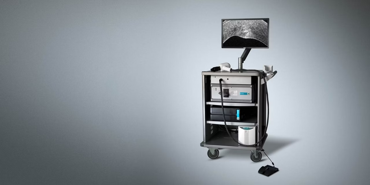
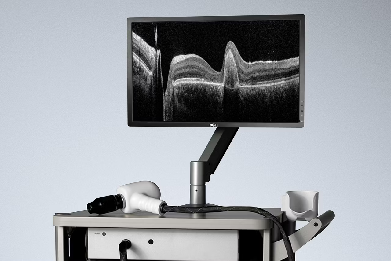
Quickly capture and observe brilliant, high-resolution, and high-density optical coherence tomography (OCT) images where you need it. The gentle, non-contact Envisu C2300 system is suitable for even premature infants.
Envisu, is a hand-held OCT solution with an intended use to aid in the diagnosis of physiological and pathologic conditions of the eye.
- ake OCT to your patient: The handheld OCT scan head and mobile cart provides flexibility to image from the ICU, operating room, and clinic.
- Focus with confidence: The Envisu C2300 can be mounted on a surgical microscope for steady, high-quality live imaging.
- Get brilliant detail when you need it: High-resolution, real-time images can be seen with detail down to 3 μm.
- Explore with full volume: Featuring full 3D volumetric data capturing capability with fully customizable high density data scans up to 1000 x 1000.
The intended use of this product is to acquire, process, display and save depth-resolved images of ocular tissue. Please refer to your country specific regulatory organization for clearance/approval status.
How does OCT work?
OCT employs interferometry, in which waves are superimposed in order to extract information, to create cross-sectional images. Using low-power, near-infrared light, the OCT system generates high-resolution, volumetric images of the internal tissue microstructures of cornea and retina.
OCT measures the reflectivity of tissue microstructures as a function of depth. A single vertical scan through the depth of tissue is called an A-scan. A 2D tomographic slice (or a series of A-scans), is called a B-scan, as in ultrasound. Multiple adjacent B-scans create a 3D volume. B-scans provide in vivo observation of ocular tissues, including all retinal layers. Longitudinal studies quantify retinal layer thickening and thinning.
What is SDOCT?
Spectral Domain Optical Coherence Tomography (SDOCT) is a particular implementation of OCT that collects all of the wavelengths of light at the same time using a specially designed spectrometer. Through judicious design of the spectrometer, selection of the light source, and implementation of mathematical techniques, SDOCT systems can be tailored to provide exceptional images for a wide range of research and clinical applications.
What is Optical Coherence Tomography?
Optical Coherence Tomography, abbreviated OCT, is a non-invasive, contact-free imaging technique to detect and monitor morphological changes of ocular tissue.