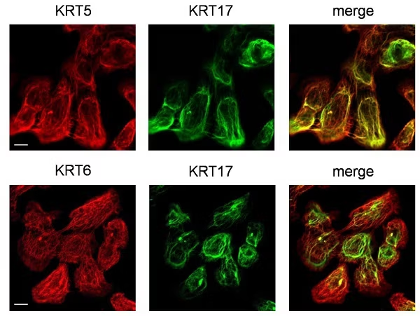
Immunocytochemistry/ Immunofluorescence - Anti-Cytokeratin 5 antibody [EP1601Y] - Cytoskeleton Marker (ab52635)
Colocalization of KRT5, KRT6 and KRT17 in HSC3 cells
Immunocytochemistry in HSC3 (human oral squamous carcinoma cell line) cells. Scale bar, 10 μm.
(Taken from Figure S3 of Khanom et al)
Image from Khanom R. et al PLoS One. 2016 Aug 11;11(8):e0161163. doi: 10.1371/journal.pone.0161163. eCollection 2016.
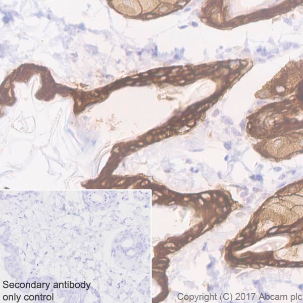
Immunohistochemistry (Formalin/PFA-fixed paraffin-embedded sections) - Anti-Cytokeratin 5 antibody [EP1601Y] - Cytoskeleton Marker (ab52635)
Immunohistochemistry (Formalin/PFA-fixed paraffin-embedded sections) analysis of rat skin tissue sections labeling Cytokeratin 5 with Purified ab52635 at 1:200 dilution. Heat mediated antigen retrieval was performed using ab93684 (Tris/EDTA buffer, pH 9.0). Tissue was counterstained with Hematoxylin. ImmunoHistoProbe one step HRP Polymer (ready to use) secondary antibody was used. PBS instead of the primary antibody was used as the negative control.
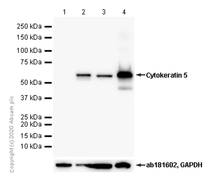
Western blot - Anti-Cytokeratin 5 antibody [EP1601Y] - Cytoskeleton Marker (ab52635)
The lysates were prepared in 1%SDS Hot lysis method.
Observed MW: 62kDa
Blocking/diluting buffer and concentration: 5% NFDM/TBST
All lanes:
Western blot - Anti-Cytokeratin 5 antibody [EP1601Y] - Cytoskeleton Marker (ab52635) at 1/1000 dilution
Lane 1:
Human skin lysates prepared in RIPA lysis method at 20 µg
Lane 2:
Human skin lysates prepared in 1%SDS Hot lysis method at 20 µg
Lane 3:
Mouse skin lysates prepared in RIPA lysis method at 20 µg
Lane 4:
Mouse skin lysates prepared in 1%SDS Hot lysis method at 20 µg
Secondary
All lanes:
Goat Anti-Rabbit IgG (HRP) with minimal cross-reactivity with human IgG at 1/2000 dilution
Predicted band size: 62 kDa
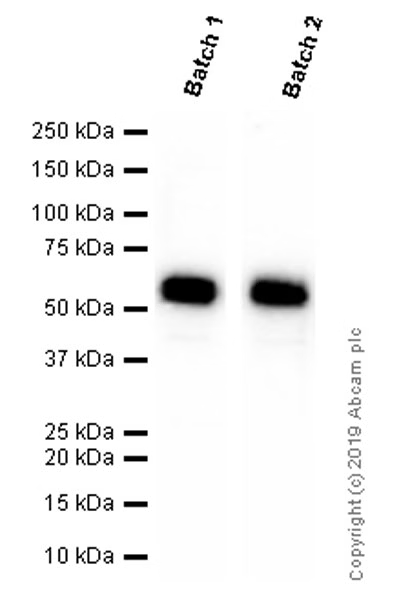
Western blot - Anti-Cytokeratin 5 antibody [EP1601Y] - Cytoskeleton Marker (ab52635)
Different batches of ab52635 were tested on Rat skin lysate at 1.0 µg/ml. 15 µg of lysate was loaded in each lane. Bands observed at 62 kDa.
All lanes:
Western blot - Anti-Cytokeratin 5 antibody [EP1601Y] - Cytoskeleton Marker (ab52635)
Predicted band size: 62 kDa
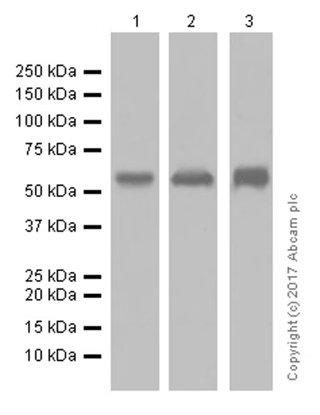
Western blot - Anti-Cytokeratin 5 antibody [EP1601Y] - Cytoskeleton Marker (ab52635)
Blocking and diluting buffer: 5% NFDM/TBST.
The lysates were prepared in 1%SDS Hot lysis method.
All lanes:
Western blot - Anti-Cytokeratin 5 antibody [EP1601Y] - Cytoskeleton Marker (ab52635) at 1/10000 dilution
Lane 1:
Human fetal skin lysates at 20 µg
Lane 2:
Rat skin lysates at 20 µg
Lane 3:
Mouse skin lysates at 20 µg
Secondary
All lanes:
Goat Anti-Rabbit IgG (HRP) with minimal cross-reactivity with human IgG at 1/2000 dilution
Predicted band size: 62 kDa
Observed band size: 62 kDa
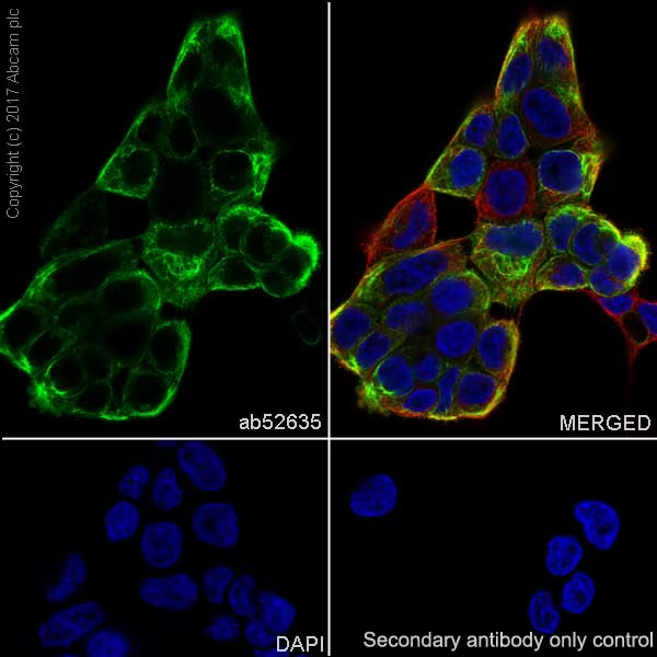
Immunocytochemistry/ Immunofluorescence - Anti-Cytokeratin 5 antibody [EP1601Y] - Cytoskeleton Marker (ab52635)
Immunocytochemistry/ Immunofluorescence analysis of A431 (Human epidermoid carcinoma epithelial cell) cells labeling Cytokeratin 6 with Purified ab52635 at 1/100 dilution. Cells were fixed in 4% Paraformaldehyde and permeabilized with 0.1% tritonX-100. Cells were counterstained with ab195889 Anti-alpha Tubulin antibody [DM1A] - Microtubule Marker (Alexa Fluor® 594) 1/200 (2.5 μg/ml). ab150077 Goat anti rabbit IgG(Alexa Fluor® 488) was used as the secondary antibody at 1/1000 dilution. DAPI nuclear counterstain. PBS instead of the primary antibody was used as the secondary antibody only control.
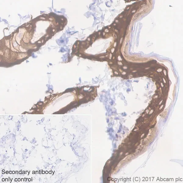
Immunohistochemistry (Formalin/PFA-fixed paraffin-embedded sections) - Anti-Cytokeratin 5 antibody [EP1601Y] - Cytoskeleton Marker (ab52635)
Immunohistochemistry (Formalin/PFA-fixed paraffin-embedded sections) analysis of mouse skin tissue sections labeling Cytokeratin 5 with Purified ab52635 at 1:200 dilution. Heat mediated antigen retrieval was performed using ab93684 (Tris/EDTA buffer, pH 9.0). Tissue was counterstained with Hematoxylin. ImmunoHistoProbe one step HRP Polymer (ready to use) secondary antibody was used. PBS instead of the primary antibody was used as the negative control.
