Anti-Collagen I antibody [RM1131](ab316222)
$undefined


Anti-Collagen I antibody [RM1131] (ab316222) is a rabbit recombinant multiclonal antibody detecting Collagen I in Western Blot, Flow Cytometry (Intra), IP, IHC-P, IHC-Fr, ICC/IF. Suitable for Human, Mouse, Rat.
- Biophysical QC for unrivalled batch-batch consistency
Product details - Anti-Collagen I antibody [RM1131] (ab316222)
Product Specifications
Anti-Collagen I antibody [RM1131] (ab316222) is a recombinant rabbit multiclonal antibody which is comprised of multiple monoclonal antibodies targeting different epitopes and recapitulating the performance of a polyclonal. Each component clone was developed by Abcam using patented rabbit monoclonal antibody technology.
Anti-Collagen I antibody [RM1131] (ab316222) is validated for use in Flow Cyt (Intra), ICC/IF, IHC-Fr, IHC-P, IP, WB in human, mouse, rat samples.
Anti-Collagen I antibody [RM1131] (ab316222) specifically detects Collagen I (UniProt ID: P02452; Molecular weight: 95kDa) and is sold in a convenient trial size to enable initial testing (20 µL) and larger sizes for subsequent scaling up experiments (100 µL and 1 mL).
Quality and Validation
Abcams high quality manufacturing and validation processes ensure Anti-Collagen I antibody [RM1131] (ab316222) has high sensitivity and specificity alongside high lot-to-lot consistency and reproducibility.
Target Information
Collagen I, the most abundant collagen type in the human body, plays a significant role in oncology. It is a major component of the extracellular matrix (ECM) and contributes to the structural integrity of tissues. In cancer, Collagen I can influence tumor progression by promoting tumor cell proliferation, invasion, and metastasis through interactions with various cell surface receptors and signaling pathways.
What are recombinant multiclonals?
Recombinant multiclonals are a mixture of recombinant antibodies co-expressed from a library of heavy and light chains. They offer several advantages including:
- The sensitivity of polyclonal antibodies by recognising multiple epitopes
- High batch-to-batch consistency and reproducibility
- Improved sensitivity and specificity
- Long-term security of supply
- Animal-free batch production
Patented technology
Our RabMAb® technology is a patented hybridoma-based technology for making rabbit monoclonal antibodies.
Supplementary information
This supplementary information is collated from multiple sources and compiled automatically.
Collagen type I also called collagen I is a structural protein expressed mainly in connective tissues such as skin tendon bone and ligaments. It serves as an important component in providing mechanical strength and integrity to these tissues. Collagen I is a fibrillar collagen known for its triple-helix structure composed of two alpha-1 chains and one alpha-2 chain and has a molecular mass of approximately 300 kDa. Researchers often employ collagen western blot and collagen ELISA techniques for its detection. Collagen suppliers offer various collagen antibodies used in these assays to study its distribution and function.
Biological function summary
Collagen type I plays a central role in maintaining the extracellular matrix and supporting cellular environments. It interacts with other matrix proteins and cells forming complexes that help in tissue development and repair. Type I collagen is especially important in bone matrix working alongside minerals like hydroxyapatite to provide rigidity and support. Anti-collagen antibodies aid in studying its biological functions and interactions which are critical to understanding tissue dynamics.
Pathways
Collagen type I interacts with multiple signaling cascades involved in tissue remodeling and repair. It is a significant player in the TGF-β pathway which regulates fibrosis and wound healing processes. In these pathways proteins such as fibronectin and integrins work in concert with collagen type I to orchestrate cellular responses to damage. Researchers often examine its role in these pathways to uncover therapeutic possibilities for disease interventions.
Collagen type I has strong connections to conditions like osteogenesis imperfecta and fibrosis. Mutations or irregularities in collagen I production can lead to osteogenesis imperfecta a genetic disorder characterized by brittle bones. In fibrosis excessive collagen deposition disrupts normal tissue architecture contributing to organ dysfunction. In both conditions type I collagen interacts with other proteins like matrix metalloproteinases which modulate its breakdown and remodeling highlighting its importance in disease pathology.
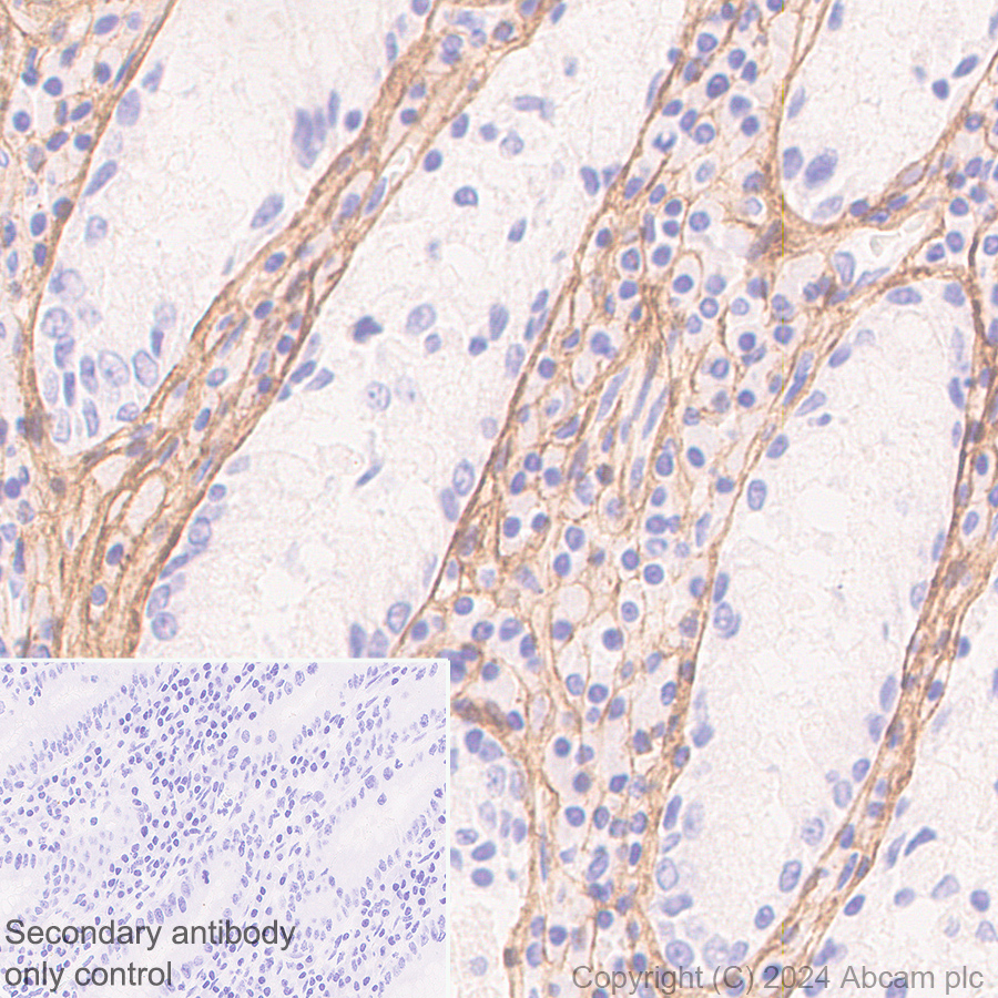
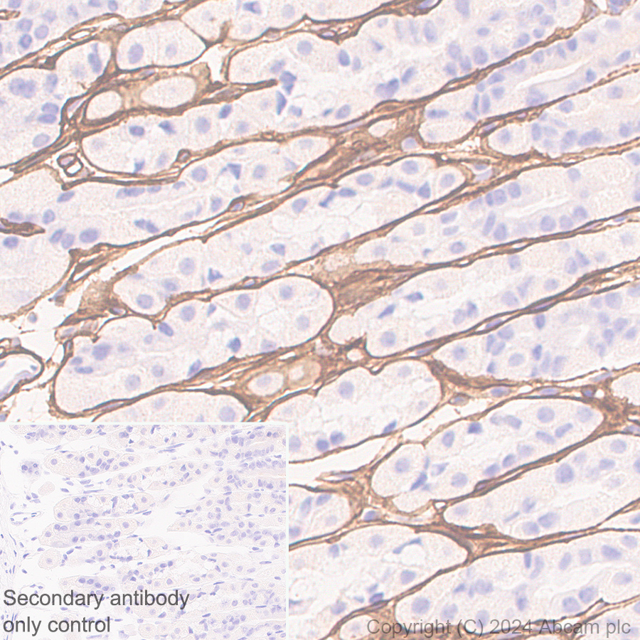
Immunohistochemistry (Formalin/PFA-fixed paraffin-embedded sections) - Anti-Collagen I antibody [RM1131] (AB316222)
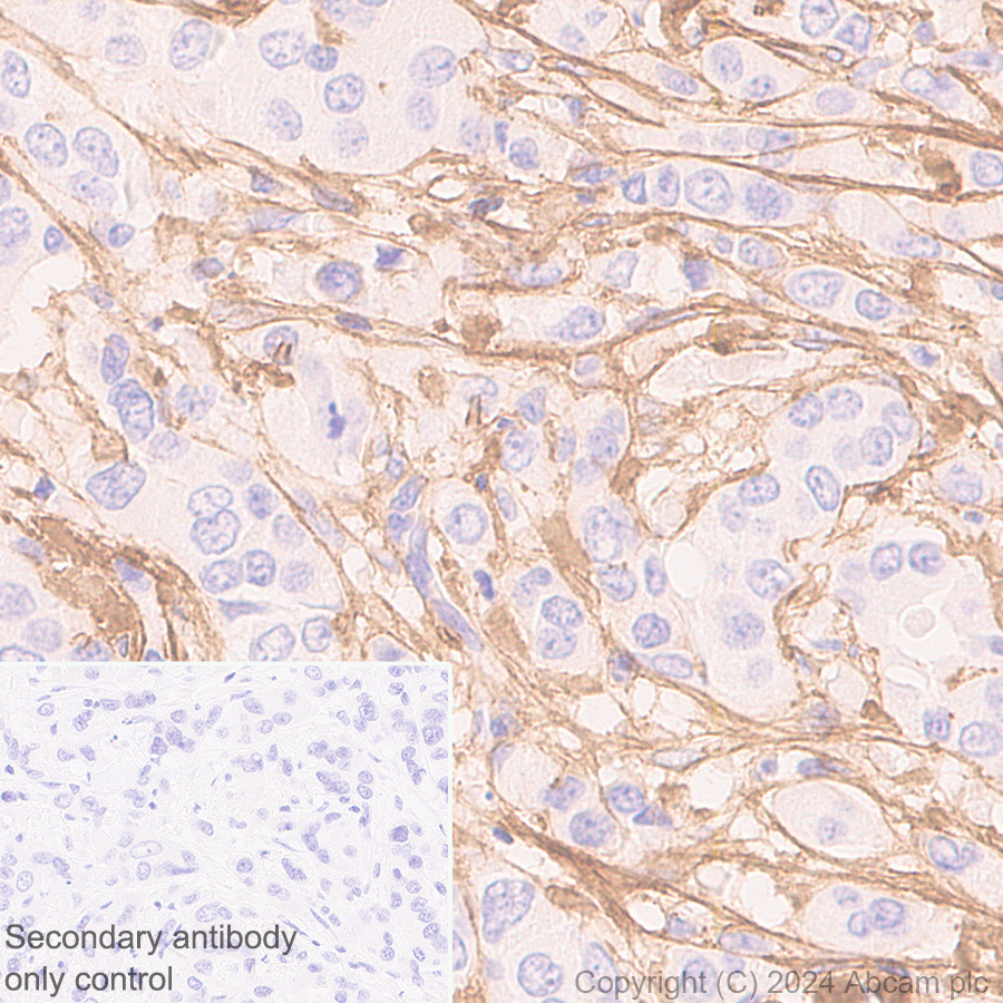
Immunohistochemical analysis of paraffin-embedded Human breast cancer tissue labeling Collagen I with ab316222 at 1/500 (1.012 ug/ml) dilution, followed by a ready to use LeicaDS9800 (Bond™ Polymer Refine Detection).
Positive staining on stroma of human breast cancer.
The section was incubated with ab316222 for 30 mins at room temperature.
The immunostaining was performed on a Leica Biosystems BOND® RX instrument
Counterstained with Hematoxylin.
Secondary antibody only control : Secondary antibody is a ready to use LeicaDS9800 (Bond™ Polymer Refine Detection).
Heat mediated antigen retrieval was performed with Tris-EDTA buffer (pH 9.0, Epitope Retrieval Solution2) for 20 mins
Immunohistochemistry (Formalin/PFA-fixed paraffin-embedded sections) - Anti-Collagen I antibody [RM1131] (AB316222)
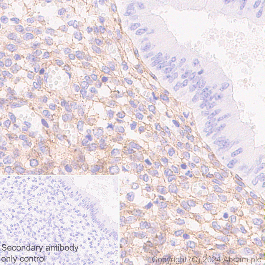
Immunohistochemical analysis of paraffin-embedded Human endometrium tissue labeling Collagen I with ab316222 at 1/500 (1.012 ug/ml) dilution, followed by a ready to use LeicaDS9800 (Bond™ Polymer Refine Detection).
Positive staining on stroma of human endometrium.
The section was incubated with ab316222 for 30 mins at room temperature.
The immunostaining was performed on a Leica Biosystems BOND® RX instrument
Counterstained with Hematoxylin.
Secondary antibody only control : Secondary antibody is a ready to use LeicaDS9800 (Bond™ Polymer Refine Detection).
Heat mediated antigen retrieval was performed with Tris-EDTA buffer (pH 9.0, Epitope Retrieval Solution2) for 20 mins
Immunohistochemistry (Formalin/PFA-fixed paraffin-embedded sections) - Anti-Collagen I antibody [RM1131] (AB316222)
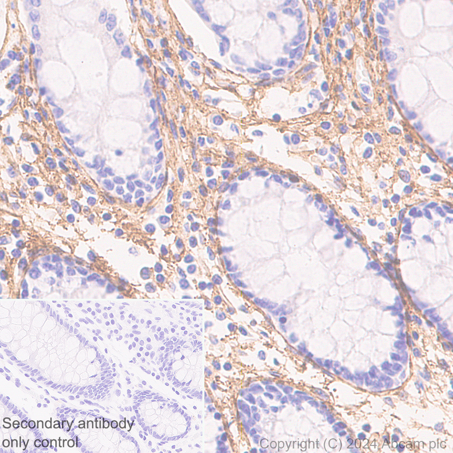
Immunohistochemical analysis of paraffin-embedded Human colon tissue labeling Collagen I with ab316222 at 1/500 (1.012 ug/ml) dilution, followed by a ready to use LeicaDS9800 (Bond™ Polymer Refine Detection).
Positive staining on stroma of human colon.
The section was incubated with ab316222 for 30 mins at room temperature.
The immunostaining was performed on a Leica Biosystems BOND® RX instrument
Counterstained with Hematoxylin.
Secondary antibody only control : Secondary antibody is a ready to use LeicaDS9800 (Bond™ Polymer Refine Detection).
Heat mediated antigen retrieval was performed with Tris-EDTA buffer (pH 9.0, Epitope Retrieval Solution2) for 20 mins
Immunocytochemistry/ Immunofluorescence - Anti-Collagen I antibody [RM1131] (AB316222)
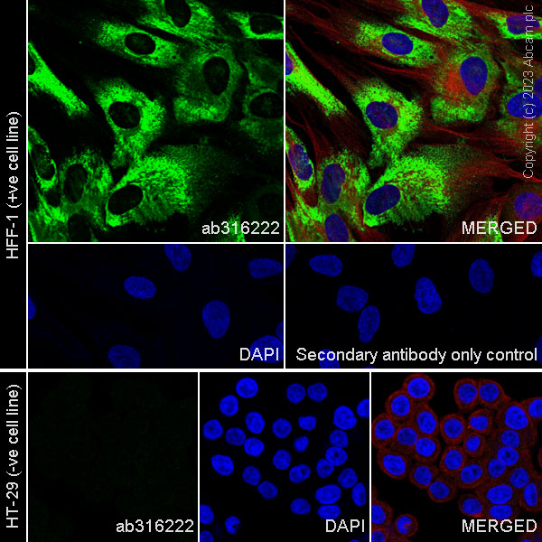
Immunofluorescent analysis of 4% Paraformaldehyde-fixed, 0.1% TritonX-100 permeabilized HFF-1 (human skin fibroblast) cells labelling Collagen I with ab316222 at 1/100 (5.06 ug/ml) dilution, followed by ab150081 Goat Anti-Rabbit IgG H&L (Alexa Fluor® 488) preadsorbed antibody at 1/1000 (2 ug/ml) dilution (Green).
Confocal image showing cytoplasmic staining in HFF-1 cell line.
Negative control : HT-29.
Image was taken with a confocal microscope (Leica-Microsystems, TCS SP8).
ab195889 Anti-alpha Tubulin mouse monoclonal antibody - Microtubule Marker (Alexa Fluor® 594) was used to counterstain tubulin at 1/500 (1ug/ml) dilution (Red). The Nuclear counterstain was DAPI (Blue).
Secondary antibody only control : Secondary antibody is ab150081 Goat Anti-Rabbit IgG H&L (Alexa Fluor® 488) preadsorbed at 1/1000 (2 ug/ml) dilution.
Immunohistochemistry (Formalin/PFA-fixed paraffin-embedded sections) - Anti-Collagen I antibody [RM1131] (AB316222)
Immunohistochemical analysis of paraffin-embedded Human stomach tissue labeling Collagen I with ab316222 at 1/500 (1.012 ug/ml) dilution, followed by a ready to use LeicaDS9800 (Bond™ Polymer Refine Detection).
Positive staining on stroma of human stomach.
The section was incubated with ab316222 for 30 mins at room temperature.
The immunostaining was performed on a Leica Biosystems BOND® RX instrument
Counterstained with Hematoxylin.
Secondary antibody only control : Secondary antibody is a ready to use LeicaDS9800 (Bond™ Polymer Refine Detection).
Heat mediated antigen retrieval was performed with Tris-EDTA buffer (pH 9.0, Epitope Retrieval Solution2) for 20 mins
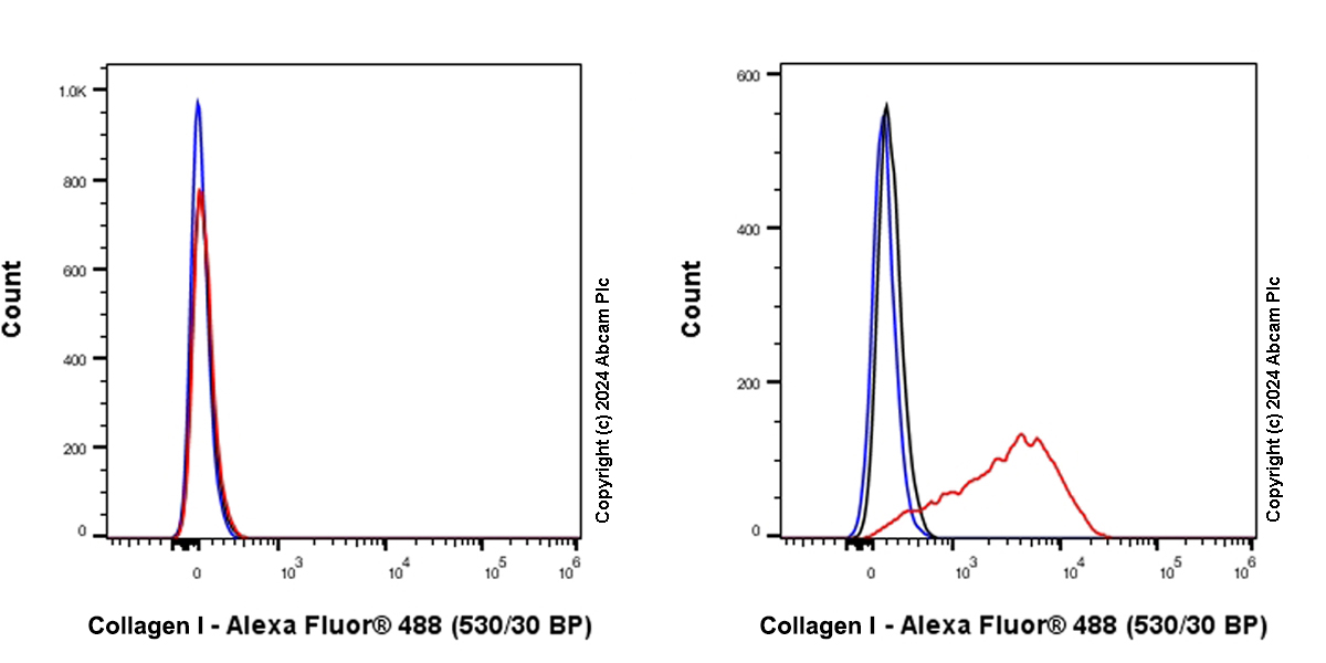
Flow Cytometry (Intracellular) - Anti-Collagen I antibody [RM1131] (AB316222)
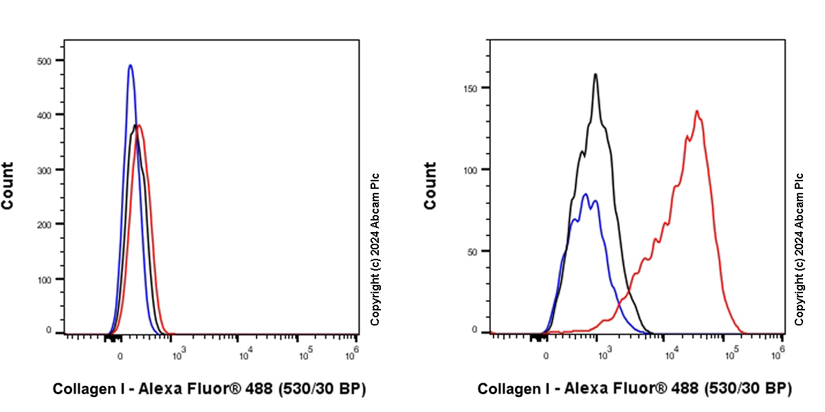
Flow cytometric analysis of 4% paraformaldehyde fixed 90% methanol permeabilized HT-29 (human colorectal adenocarcinoma epithelial cell, Left) / HFF-1 (human skin fibroblast, Right) cells labelling Collagen I with ab316222 at 1/5000 dilution (0.01 ug)/Red compared with a Rabbit monoclonal IgG (ab172730) (Black) isotype control and an unlabelled control (cells without incubation with primary antibody and secondary antibody) (Blue).
Goat Anti-Rabbit IgG (Alexa Fluor® 488, ab150081) at 1/5000 dilution was used as the secondary antibody.
Negative control : HT-29.
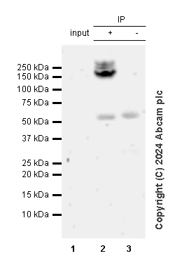
Immunoprecipitation - Anti-Collagen I antibody [RM1131] (AB316222)
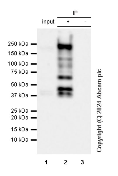
Collagen I was immunoprecipitated from 0.35 mg HFF-1 (human skin fibroblast) whole cell lysate with ab316222 at 1/30 dilution (2ug in 0.35mg lysates). Western blot was performed on the immunoprecipitate using ab316222 at 1/1000 dilution. VeriBlot for IP secondary antibody(HRP)(ab131366) was used at 1/5000 dilution.
Lane 1 : HFF-1 (human skin fibroblast) whole cell lysate
Lane 2 : ab316222 IP in HFF-1 (human skin fibroblast) whole cell lysate
Lane 3 : Rabbit monoclonal IgG (ab172730) instead of ab316222 in HFF-1 whole cell lysate
All lanes:
Immunoprecipitation - Anti-Collagen I antibody [RM1131] (ab316222) at 1/30 dilution
All lanes:
HFF-1 (human skin fibroblast) whole cell lysate
Secondary
All lanes:
Immunoprecipitation - VeriBlot for IP Detection Reagent (HRP) (ab131366) at 1/5000 dilution
Exposure time: 10s
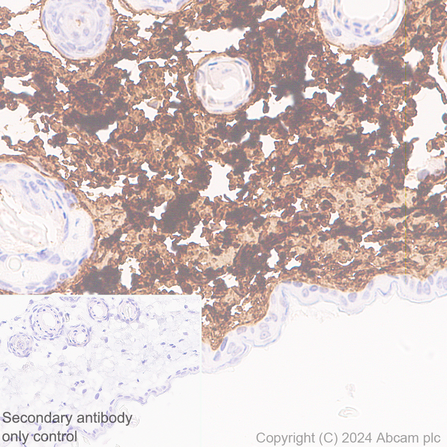
Immunohistochemistry (Frozen sections) - Anti-Collagen I antibody [RM1131] (AB316222)
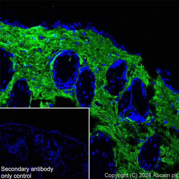
Immunohistochemical analysis of 4% PFA-fixed, 0.2% Triton X-100 permeabilized frozen Mouse skin (fresh frozen) tissue labeling Collagen I with ab316222 at 1/500 (1.012 ug/ml) dilution followed by ab150081 Goat Anti-Rabbit IgG H&L (Alexa Fluor® 488) preadsorbed at 1/1000 (2 ug/mL) dilution (Green).
Confocal image showing positive staining on mouse skin. The nuclear counterstain was DAPI (Blue). The section was incubated with ab316222 for 60 mins at room temperature. The section was then mounted using Fluoromount®.The immunostaining was performed on a Leica Biosystems BOND® RX instrument. Image was taken with a confocal microscope (Leica-Microsystems, TCS SP8).
Secondary antibody control : Secondary antibody is ab150081 Goat Anti-Rabbit IgG H&L (Alexa Fluor® 488) preadsorbedat 1/1000 (2 ug/mL) dilution.
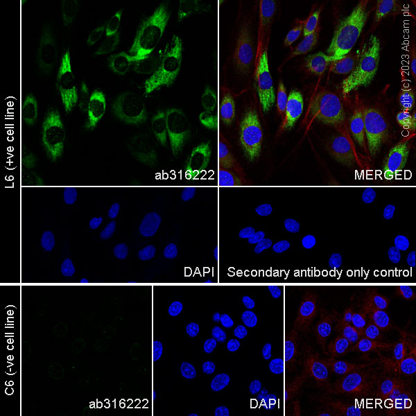
Flow Cytometry (Intracellular) - Anti-Collagen I antibody [RM1131] (AB316222)
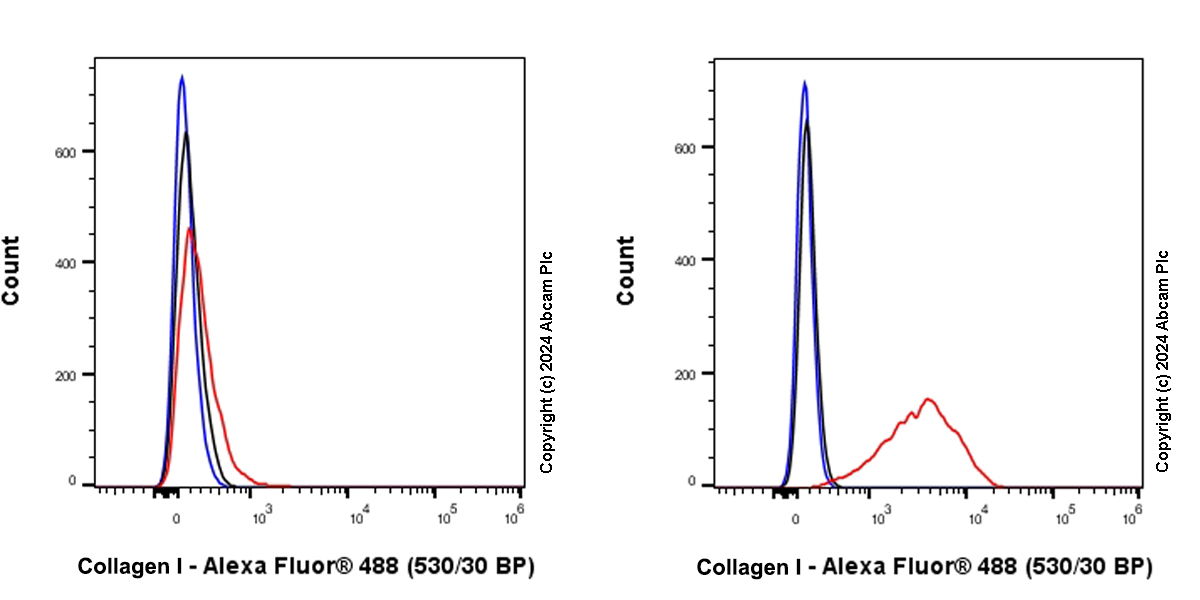
Flow cytometric analysis of 4% paraformaldehyde fixed 90% methanol permeabilized C6 (rat glial tumor glial cell, Left) / L6 (rat skeletal muscle myoblast, Right) cells labelling Collagen I with ab316222 at 1/5000 dilution (0.01 ug)/Red compared with a Rabbit monoclonal IgG (ab172730) (Black) isotype control and an unlabelled control (cells without incubation with primary antibody and secondary antibody) (Blue).
Goat Anti-Rabbit IgG (Alexa Fluor® 488, ab150081) at 1/5000 dilution was used as the secondary antibody.
Negative control : C6.
Immunohistochemistry (Formalin/PFA-fixed paraffin-embedded sections) - Anti-Collagen I antibody [RM1131] (AB316222)
Immunohistochemical analysis of paraffin-embedded Rat stomach tissue labeling Collagen I with ab316222 at 1/500 (1.012 ug/ml) dilution, followed by a ready to use LeicaDS9800 (Bond™ Polymer Refine Detection).
Positive staining on stroma of rat stomach.
The section was incubated with ab316222 for 30 mins at room temperature.
The immunostaining was performed on a Leica Biosystems BOND® RX instrument
Counterstained with Hematoxylin.
Secondary antibody only control : Secondary antibody is a ready to use LeicaDS9800 (Bond™ Polymer Refine Detection).
Heat mediated antigen retrieval was performed with Tris-EDTA buffer (pH 9.0, Epitope Retrieval Solution2) for 20 mins
Immunohistochemistry (Formalin/PFA-fixed paraffin-embedded sections) - Anti-Collagen I antibody [RM1131] (AB316222)
Immunohistochemical analysis of paraffin-embedded Mouse skin tissue labeling Collagen I with ab316222 at 1/500 (1.012 ug/ml) dilution, followed by a ready to use LeicaDS9800 (Bond™ Polymer Refine Detection).
Positive staining on stroma of mouse skin.
The section was incubated with ab316222 for 30 mins at room temperature.
The immunostaining was performed on a Leica Biosystems BOND® RX instrument
Counterstained with Hematoxylin.
Secondary antibody only control : Secondary antibody is a ready to use LeicaDS9800 (Bond™ Polymer Refine Detection).
Heat mediated antigen retrieval was performed with Tris-EDTA buffer (pH 9.0, Epitope Retrieval Solution2) for 20 mins
Immunohistochemistry (Frozen sections) - Anti-Collagen I antibody [RM1131] (AB316222)
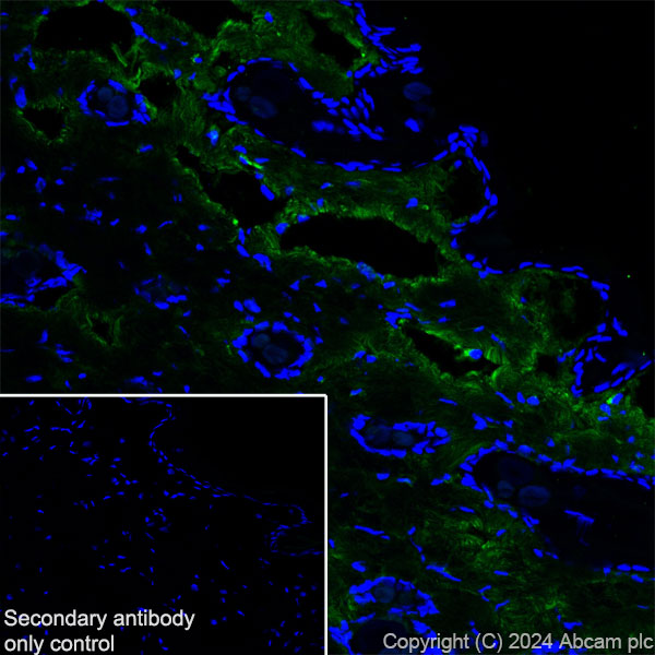
Immunohistochemical analysis of 4% PFA-fixed, 0.2% Triton X-100 permeabilized frozen Rat skin (fresh frozen) tissue labeling Collagen I with ab316222 at 1/500 (1.012 ug/ml) dilution followed by ab150081 Goat Anti-Rabbit IgG H&L (Alexa Fluor® 488) preadsorbed at 1/1000 (2 ug/mL) dilution (Green).
Confocal image showing positive staining on rat skin. The nuclear counterstain was DAPI (Blue). The section was incubated with ab316222 for 60 mins at room temperature. The section was then mounted using Fluoromount®.The immunostaining was performed on a Leica Biosystems BOND® RX instrument. Image was taken with a confocal microscope (Leica-Microsystems, TCS SP8).
Secondary antibody control : Secondary antibody is ab150081 Goat Anti-Rabbit IgG H&L (Alexa Fluor® 488) preadsorbedat 1/1000 (2 ug/mL) dilution.
Immunocytochemistry/ Immunofluorescence - Anti-Collagen I antibody [RM1131] (AB316222)
Immunofluorescent analysis of 100% methanol-fixed, 0.1% TritonX-100 permeabilized L6 (rat skeletal muscle myoblast) cells labelling Collagen I with ab316222 at 1/100 (5.06 ug/ml) dilution, followed by ab150081 Goat Anti-Rabbit IgG H&L (Alexa Fluor® 488) preadsorbed antibody at 1/1000 (2 ug/ml) dilution (Green).
Confocal image showing cytoplasmic staining in L6 cell line.
Negative control : C6.
Image was taken with a confocal microscope(Leica-Microsystems, TCS SP8).
ab195889 Anti-alpha Tubulin mouse monoclonal antibody - Microtubule Marker (Alexa Fluor® 594) was used to counterstain tubulin at 1/500 (1ug/ml) dilution (Red). The Nuclear counterstain was DAPI (Blue).
Secondary antibody only control : Secondary antibody is ab150081 Goat Anti-Rabbit IgG H&L (Alexa Fluor® 488) preadsorbed at 1/1000 (2 ug/ml) dilution.
Immunocytochemistry/ Immunofluorescence - Anti-Collagen I antibody [RM1131] (AB316222)
Immunofluorescent analysis of 100% methanol-fixed, 0.1% TritonX-100 permeabilized NIH/3T3 (mouse embryonic fibroblast) cells labelling Collagen I with ab316222 at 1/100 (5.06 ug/ml) dilution, followed by ab150081 Goat Anti-Rabbit IgG H&L (Alexa Fluor® 488) preadsorbed antibody at 1/1000 (2 ug/ml) dilution (Green).
Confocal image showing cytoplasmic staining in NIH/3T3 cell line.
Negative control : RAW 264.7.
Image was taken with a confocal microscope(Leica-Microsystems, TCS SP8).
ab195889 Anti-alpha Tubulin mouse monoclonal antibody - Microtubule Marker (Alexa Fluor® 594) was used to counterstain tubulin at 1/500 (1ug/ml) dilution (Red). The Nuclear counterstain was DAPI (Blue).
Secondary antibody only control : Secondary antibody is ab150081 Goat Anti-Rabbit IgG H&L (Alexa Fluor® 488) preadsorbed at 1/1000 (2 ug/ml) dilution.
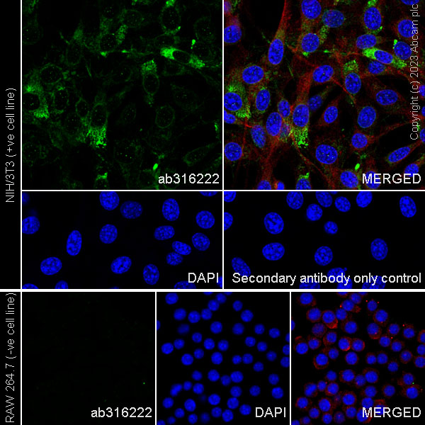
Flow Cytometry (Intracellular) - Anti-Collagen I antibody [RM1131] (AB316222)
Flow cytometric analysis of 4% paraformaldehyde fixed 90% methanol permeabilized Raw 264.7 (mouse Abelson murine leukemia virus-induced tumor macrophage, Left) / NIH/3T3 (mouse embryonic fibroblast, Right) cells labelling Collagen I with ab316222 at 1/5000 dilution (0.01 ug)/Red compared with a Rabbit monoclonal IgG (ab172730) (Black) isotype control and an unlabelled control (cells without incubation with primary antibody and secondary antibody) (Blue).
Goat Anti-Rabbit IgG (Alexa Fluor® 488, ab150081) at 1/5000 dilution was used as the secondary antibody.
Negative control : Raw 264.7.
Immunohistochemistry (Formalin/PFA-fixed paraffin-embedded sections) - Anti-Collagen I antibody [RM1131] (AB316222)
Immunohistochemical analysis of paraffin-embedded Mouse stomach tissue labeling Collagen I with ab316222 at 1/500 (1.012 ug/ml) dilution, followed by a ready to use LeicaDS9800 (Bond™ Polymer Refine Detection).
Positive staining on stroma of mouse stomach.
The section was incubated with ab316222 for 30 mins at room temperature.
The immunostaining was performed on a Leica Biosystems BOND® RX instrument
Counterstained with Hematoxylin.
Secondary antibody only control : Secondary antibody is a ready to use LeicaDS9800 (Bond™ Polymer Refine Detection).
Heat mediated antigen retrieval was performed with Tris-EDTA buffer (pH 9.0, Epitope Retrieval Solution2) for 20 mins
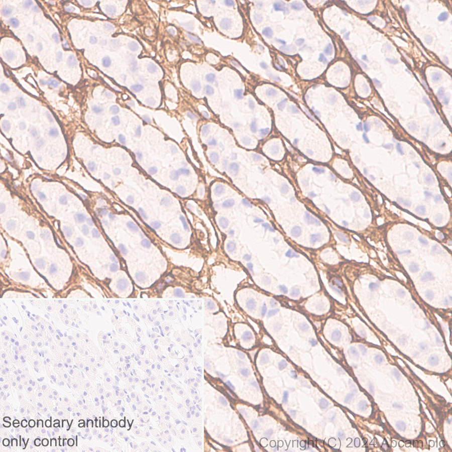
Immunohistochemistry (Formalin/PFA-fixed paraffin-embedded sections) - Anti-Collagen I antibody [RM1131] (AB316222)
Immunohistochemical analysis of paraffin-embedded Rat skin tissue labeling Collagen I with ab316222 at 1/500 (1.012 ug/ml) dilution, followed by a ready to use LeicaDS9800 (Bond™ Polymer Refine Detection).
Positive staining on stroma of rat skin.
The section was incubated with ab316222 for 30 mins at room temperature.
The immunostaining was performed on a Leica Biosystems BOND® RX instrument
Counterstained with Hematoxylin.
Secondary antibody only control : Secondary antibody is a ready to use LeicaDS9800 (Bond™ Polymer Refine Detection).
Heat mediated antigen retrieval was performed with Tris-EDTA buffer (pH 9.0, Epitope Retrieval Solution2) for 20 mins
Immunoprecipitation - Anti-Collagen I antibody [RM1131] (AB316222)
Collagen I was immunoprecipitated from 0.35 mg Mouse skin tissue lysate with ab316222 at 1/30 dilution (2ug in 0.35mg lysates). Western blot was performed on the immunoprecipitate using ab316222 at 1/1000 dilution. VeriBlot for IP secondary antibody(HRP)(ab131366) was used at 1/5000 dilution.
Lane 1 : Mouse skin tissue lysate
Lane 2 : ab316222 IP in Mouse skin tissue lysate
Lane 3 : Rabbit monoclonal IgG (ab172730) instead of ab316222 in mouse skin tissue lysate
All lanes:
Immunoprecipitation - Anti-Collagen I antibody [RM1131] (ab316222) at 1/30 dilution
All lanes:
Mouse skin tissue lysate
Secondary
All lanes:
Immunoprecipitation - VeriBlot for IP Detection Reagent (HRP) (ab131366) at 1/5000 dilution
Exposure time: 180s
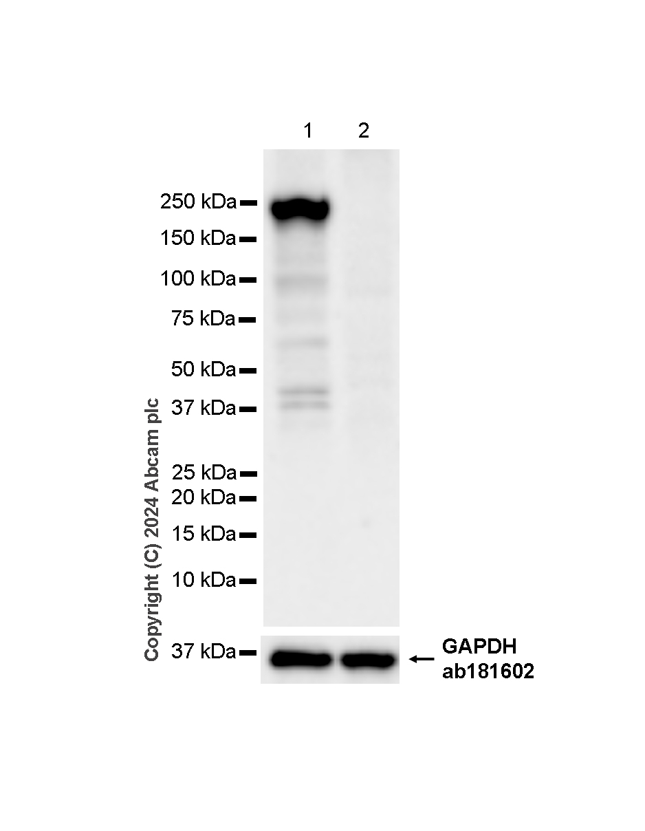
Western blot - Anti-Collagen I antibody [RM1131] (AB316222)
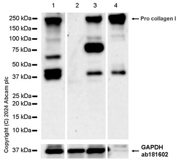
Blocking and diluting buffer and concentration : 5% NFDM/TBST.
Negative control : HT-29.
We are unsure how to define these extra bands below 100kDa.
All lanes:
Western blot - Anti-Collagen I antibody [RM1131] (ab316222) at 1/1000 dilution
Lane 1:
HFF-1 (human skin fibroblast) whole cell lysate at 20 µg
Lane 2:
HT-29 (human colorectal adenocarcinoma epithelial cell) whole cell lysate at 20 µg
Lane 3:
NIH/3T3 (mouse embryonic fibroblast) whole cell lysate at 20 µg
Lane 4:
NIH/3T3 (mouse embryonic fibroblast) culture supernatant at 10 µL
Secondary
All lanes:
Goat Anti-Rabbit IgG (HRP) with minimal cross-reactivity with human IgG at 1/2000 dilution
Observed band size: 220 kDa
Exposure time: 180s
Western blot - Anti-Collagen I antibody [RM1131] (AB316222)
The molecular weight observed is consistent with what has been described in the literature (PMID : 23940311)
In Western blot, Anti-GAPDH antibody [EPR16891] - Loading Control (ab181602) staining at 1/200000 dilution.
Exposure time : Lane 1 : 180 seconds, Lane 2 : 8 seconds, Lane 3 : 81 seconds
All lanes:
Western blot - Anti-Collagen I antibody [RM1131] (ab316222) at 1/5000 dilution
Lane 1:
Mouse skin tissue lysate at 20 µg
Lane 2:
Rat skin tissue lysate at 20 µg
Lane 3:
Human uterus tissue lysate at 20 µg
Secondary
All lanes:
Goat Anti-Rabbit IgG (HRP) with minimal cross-reactivity with human IgG at 1/2000 dilution
Observed band size: 139 kDa,220 kDa,36 kDa
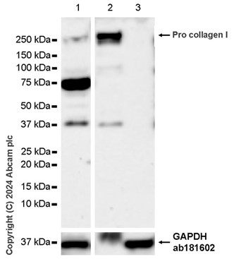
Western blot - Anti-Collagen I antibody [RM1131] (AB316222)
Collagen I might be easily degraded. Lysates were freshly made and used for Western blotting immediately to minimize protein degradation.
In Western blot, Anti-GAPDH antibody [EPR16891] - Loading Control (ab181602) staining at 1/200000 dilution.
All lanes:
Western blot - Anti-Collagen I antibody [RM1131] (ab316222) at 1/1000 dilution
Lane 1:
HFF-1 (human skin fibroblast) whole cell lysate at 20 µg
Lane 2:
HT-29 (human colorectal adenocarcinoma epithelial cell) whole cell lysate at 20 µg
Secondary
All lanes:
Goat Anti-Rabbit IgG (HRP) with minimal cross-reactivity with human IgG at 1/2000 dilution
Observed band size: 220 kDa,36 kDa
Exposure time: 6s
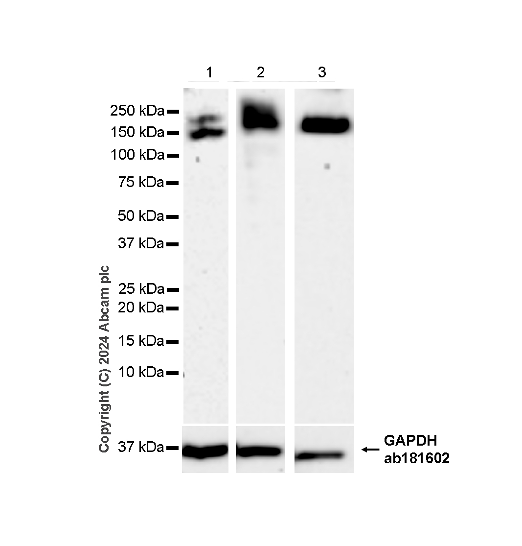
Western blot - Anti-Collagen I antibody [RM1131] (AB316222)
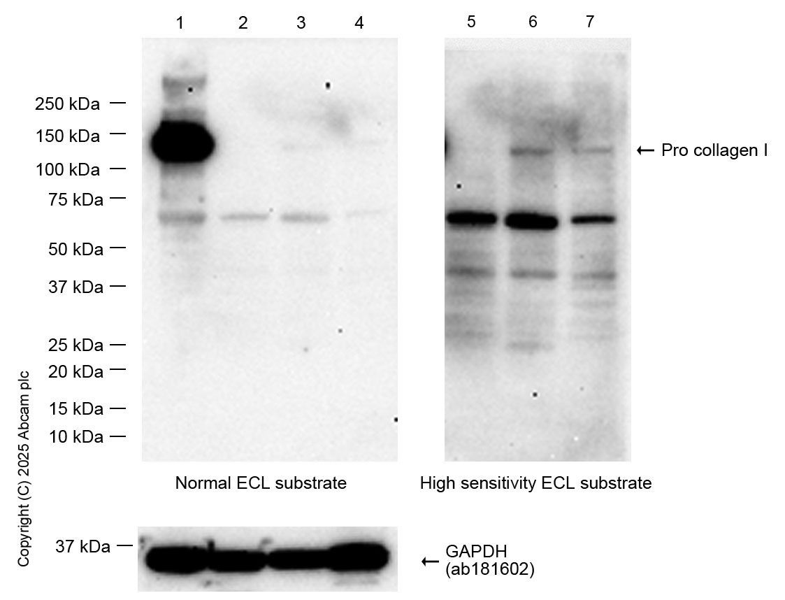
Blocking and diluting buffer and concentration : 5% NFDM/TBST.
This blot was developed using a high sensitivity ECL substrate.
Low expression tissue : normal mouse kidney (PMID : 33758176, PMID : 29102563)
All lanes:
Western blot - Anti-Collagen I antibody [RM1131] (ab316222) at 1/1000 dilution
Lane 1:
Mouse skin tissue lysate at 20 µg
Lanes 2 and 5:
Mouse kidney tissue lysate at 20 µg
Lanes 3 and 6:
Mouse lung tissue lysate at 20 µg
Lanes 4 and 8:
Mouse heart tissue lysate at 20 µg
Secondary
All lanes:
Western blot - Goat Anti-Rabbit IgG H&L (HRP) (ab97051) at 1/20000 dilution
Predicted band size: 139 kDa
Observed band size: 139 kDa
Exposure time: 180s
Western blot - Anti-Collagen I antibody [RM1131] (AB316222)
Blocking and diluting buffer and concentration : 5% NFDM/TBST.
Negative control : RAW 264.7.
We are unsure how to define these extra bands below 100kDa.
All lanes:
Western blot - Anti-Collagen I antibody [RM1131] (ab316222) at 1/5000 dilution
Lane 1:
NIH/3T3 (mouse embryonic fibroblast) whole cell lysate at 20 µg
Lane 2:
NIH/3T3 (mouse embryonic fibroblast) culture supernatant at 10 µL
Lane 3:
RAW 264.7 (mouse Abelson murine leukemia virus-induced tumor macrophage) whole cell lysate at 20 µg
Secondary
All lanes:
Goat Anti-Rabbit IgG (HRP) with minimal cross-reactivity with human IgG at 1/2000 dilution
Observed band size: 220 kDa
Exposure time: 81s
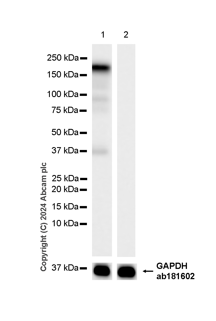
Western blot - Anti-Collagen I antibody [RM1131] (AB316222)
Negative control : C6.
In Western blot, Anti-GAPDH antibody [EPR16891] - Loading Control (ab181602) staining at 1/200000 dilution.
All lanes:
Western blot - Anti-Collagen I antibody [RM1131] (ab316222) at 1/1000 dilution
Lane 1:
L6 (rat skeletal muscle myoblast) whole cell lysate at 20 µg
Lane 2:
C6 (rat glial tumor glial cell) whole cell lysate at 20 µg
Secondary
All lanes:
Goat Anti-Rabbit IgG (HRP) with minimal cross-reactivity with human IgG at 1/2000 dilution
Observed band size: 220 kDa,36 kDa
Exposure time: 10s
| Shipped At Conditions | Blue Ice |
| Appropriate Long term Storage Conditions | -20°C |
| Clonality | Multiclonal |
| Applications | Flow Cyt (Intra), ICC/IF, IHC-Fr, IHC-P, IP, WB |
| Species Reactivity | Human, Mouse, Rat |
| Isotype | IgG |
| Appropriate short-term storage conditions | +4°C |
| Storage information | Avoid freeze / thaw cycle |
| Form | Liquid |
| Purification technique | Affinity purification Protein A |
| Storage Buffer | pH: 7.2 – 7.4; Preservative: 0.01% Sodium azide; Constituents: PBS, 40% Glycerol (glycerin, glycerine), 0.05% BSA |
| Appropriate Short-Term Storage Duration | 1–2 weeks |
| Aliquoting Information | Upon delivery aliquot |