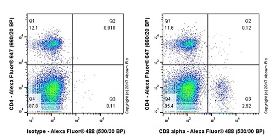
Flow Cytometry - Anti-CD8 alpha antibody [EPR21769] (ab217344)
Flow cytometric analysis of mouse primary splenocytes labeling CD8 alpha with ab217344 at 1/500 dilution (right panel) compared with a Rabbit IgG, monoclonal [EPR25A] - Isotype Control (ab172730) (left panel). Goat Anti-Rabbit IgG H&L (Alexa Fluor® 488) (ab150077) at 1/2000 dilution was used as the secondary antibody.
Cells were surface stained with CD4-Alexa Fluor® 647, then stained with rabbit IgG (Left) / ab217344 (Right) separately. CD4 and CD8 alpha are mutually exclusive expressed in mouse spleen. Gated on total viable cells.
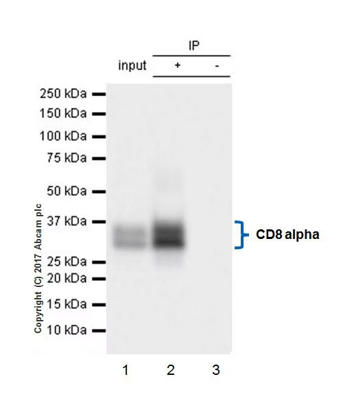
Immunoprecipitation - Anti-CD8 alpha antibody [EPR21769] (ab217344)
CD8 alpha was immunoprecipitated from 0.35 mg of mouse thymus lysate with ab217344 at 1/30 dilution. Western blot was performed from the immunoprecipitate using ab217344 at 1/2000 dilution. VeriBlot for IP Detection Reagent (HRP) (ab131366), was used for detection at 1/10000 dilution.
Lane 1: Mouse thymus lysate 10 μg (Input).
Lane 2: ab217344 IP in mouse thymus lysate.
Lane 3: Rabbit monoclonal IgG (ab172730) instead of ab214344 in mouse thymus lysate.
Exposure time: 5 seconds.
Blocking and dilution buffer and concentration: 5% NFDM/TBST.
The two bands are different isoforms that are consistent with the literature (PMID 3085089).
All lanes:
Immunoprecipitation - Anti-CD8 alpha antibody [EPR21769] (ab217344)
Predicted band size: 26 kDa
Observed band size: 34-38 kDa
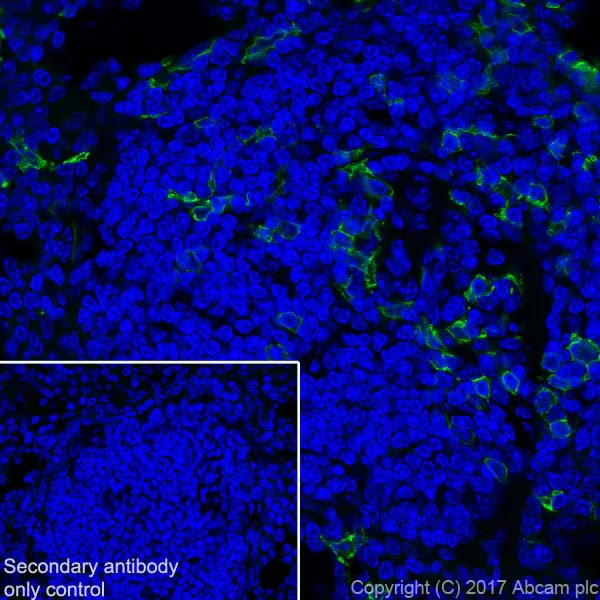
Immunohistochemistry (Frozen sections) - Anti-CD8 alpha antibody [EPR21769] (ab217344)
Immunohistochemical analysis of 4% paraformaldehyde-fixed, 0.2% Triton X-100 permeabilized frozen mouse spleen tissue labeling CD8 alpha with ab217344 at 1/500 dilution, followed by Goat Anti-Rabbit IgG H&L (Alexa Fluor® 488) (ab150077) secondary antibody at 1/1000 dilution (green). Positive membrane staining on mouse spleen (PMID: 25616911).
The nuclear counter stain is DAPI (blue).
Secondary antibody only control: Used PBS instead of primary antibody, secondary antibody is Goat Anti-Rabbit IgG H&L (Alexa Fluor® 488) (ab150077) at 1/1000 dilution.
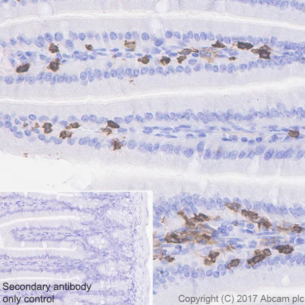
Immunohistochemistry (Formalin/PFA-fixed paraffin-embedded sections) - Anti-CD8 alpha antibody [EPR21769] (ab217344)
Immunohistochemical analysis of paraffin-embedded mouse colon tissue labeling CD8 alpha with ab217344 at 1/2000 dilution, followed by Goat Anti-Rabbit IgG H&L (HRP) Ready to use. Positive staining on stromal cells of mouse colon. Counter stained with Hematoxylin. Heat mediated antigen retrieval was performed using Tris/EDTA buffer pH 9.0 before commencing with IHC staining protocol.
Secondary antibody only control: Used PBS instead of primary antibody, secondary antibody is Goat Anti-Rabbit IgG H&L (HRP) Ready to use.
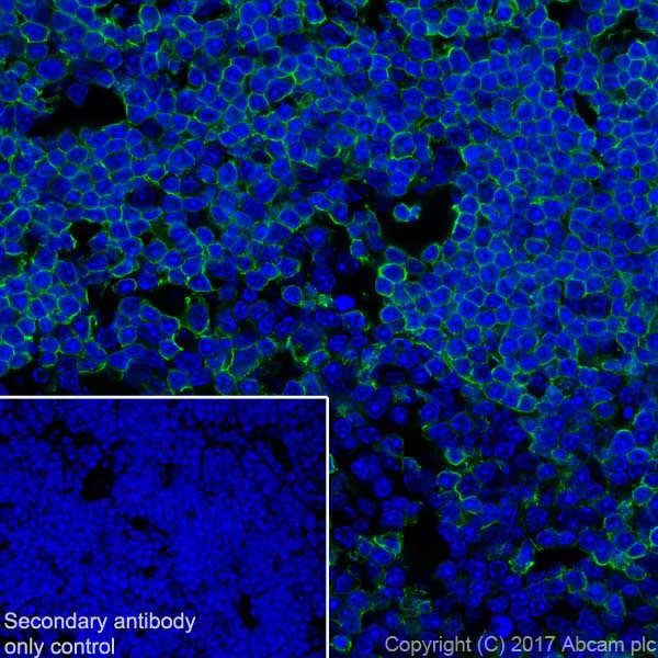
Immunohistochemistry (Frozen sections) - Anti-CD8 alpha antibody [EPR21769] (ab217344)
Immunohistochemical analysis of 4% paraformaldehyde-fixed, 0.2% Triton X-100 permeabilized frozen mouse thymus tissue labeling CD8 alpha with ab217344 at 1/500 dilution, followed by Goat Anti-Rabbit IgG H&L (Alexa Fluor® 488) (ab150077) secondary antibody at 1/1000 dilution (green). Positive membrane staining on mouse thymus tissue section (PMID: 25616911).
The nuclear counter stain is DAPI (blue).
Secondary antibody only control: Used PBS instead of primary antibody, secondary antibody is Goat Anti-Rabbit IgG H&L (Alexa Fluor® 488) (ab150077) at 1/1000 dilution.
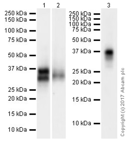
Western blot - Anti-CD8 alpha antibody [EPR21769] (ab217344)
Exposure time : Lane 1: 3 sconds; Lane 2: 40 seconds; Lane 3: 15 seconds.
Blocking/Dilution buffer: 5% NFDM/TBST.
The two bands are different isoforms that are consistent with the literature (PMID 3085089).
The blot was developed on a BIO-RAD® ChemiDoc™ MP instrument.
All lanes:
Western blot - Anti-CD8 alpha antibody [EPR21769] (ab217344) at 1/1000 dilution
Lane 1:
Mouse thymus lysate at 20 µg
Lane 2:
Mouse spleen lysate at 20 µg
Lane 3:
Mouse lymph node lysate at 10 µg
Secondary
All lanes:
Western blot - Goat Anti-Rabbit IgG H&L (HRP) (ab97051) at 1/100000 dilution
Developed using the ECL technique.
Predicted band size: 26 kDa
Observed band size: 35 kDa
