Flow Cytometry - APC Conjugation Kit - Lightning-Link® (ab201807)
Flow Cytometry - APC Conjugation Kit;- Lightning-Link.
Robinson, Andrew P., et al used APC Conjugation Kit - Lightning-Link® (ab201807) as part of characterizing oligodendroglial populations. They used the kit to conjugate APC to Mouse anti-O4 antibody, clone 81, for use in flow cytometry.
SJL/J mice were immunized with PLP139–151 and scored daily for clinical disease. A cohort of SJL/J mice was sacrificed, and spinal cords were analyzed by flow cytometry (n = 5). (A) Cells were distinguished from debris by forward and side scatter then singlet cells were gated. Live cells were gated by dead cell exclusion, and CNS resident cells were identified as CD45− or CD45low. (B) Oligodendroglial cells were defined by double positive staining: A2B5+PDGFRα+ early OPCs, A2B5+NG2+ intermediate OPCs, NG2+O4+ late OPCs, O4+MOG+ pre-myelinating oligodendrocytes, and GALC+MOG+ mature oligodendrocytes.
Image from Robinson, Andrew P., et al., PloS one, 9(9):e107649. doi: 10.1371/journal.pone.0107649. Reproduced under the Creative Commons license https://creativecommons.org/licenses/by/4.0/

Flow Cytometry - APC Conjugation Kit - Lightning-Link® (ab201807)
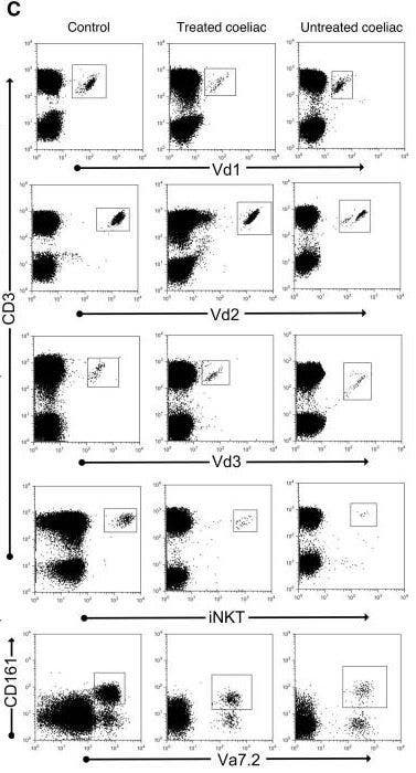
Dunne, Margaret R., et al used APC Conjugation Kit - Lightning-Link® (ab201807) as part of examining coeliac disease. They used the kit to conjugate APC to Vd3 antibody for use in flow cytometry.
Dotplots show flow cytometry data for representative control, treated and untreated coeliac donors.
Image from Dunne, Margaret R., et al., PLoS One, 8(10): e76008, doi: 10.1371/journal.pone.0076008. Reproduced under the Creative Commons license https://creativecommons.org/licenses/by/4.0/
Fluorescence Microscopy - APC Conjugation Kit - Lightning-Link® (ab201807)
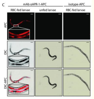
Bouchery, Tiffany, et al used APC Conjugation Kit - Lightning-Link® (ab201807) as part of examining methods for arresting hookworm development. They used the kit to conjugate APC to anti-Na-APR-1 antibody for use with live hookworms in fluorescence microscopy.
Infective L3 stage hookworms were fed in vitro for 24 hr with RBC and treated with 10 μg of 11F3-APC monoclonal antibody against Na-APR-1 or with an isotype matched-RELM-APC control antibody. Larvae were allowed to empty their digestive contents for 2 hours in fresh DMEM before internal labelling was evaluated by confocal microscopy. Data representative of 50 larvae cultured in 3 independent experiments.
Image from Bouchery, Tiffany, et al., PLoS Pathog., 14(3): e1006931; doi: 10.1371/journal.ppat.1006931. Reproduced under the Creative Commons license https://creativecommons.org/licenses/by/4.0/
Flow Cytometry - APC Conjugation Kit - Lightning-Link® (ab201807)
Flow Cytometry - APC Conjugation Kit- Lightning-Link.
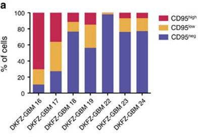
Drachsler, Moritz, et al used APC Conjugation Kit - Lightning-Link® (ab201807) as part of examining expression of CD95. They used the kit to conjugate APC to anti-human CD95 for use in flow cytometry.
The graph shows the CD95 expression in seven patient tumor samples.
Image from Drachsler, Moritz, et al., Cell death & disease 7.4 (2016): e2209-e2209. Reproduced under the Creative Commons license
Fluorescence Microscopy - APC Conjugation Kit - Lightning-Link® (ab201807)
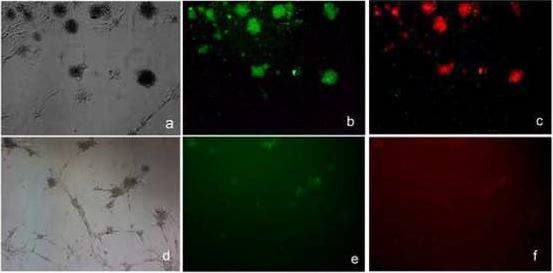
Haque, Nasreen S., Akaash Tuteja, and Niloufar Haque used APC Conjugation Kit - Lightning-Link® (ab201807) as part of examining as part of examining embryoid body formation. They used the kit to conjugate APC to anti-CCR8 antibody for use in immunofluorescence.
Anti CCL1 inhibits the expression of CCR8 and FoxP3 in mouse mesenchymal stem cells (mMSCs). Untreated (5a,-c) and anti-CCL1 treated (5d,-,f) cells were subjected to immunoflourescence with antibodies against FoxP3 (green;5b and e) or CCR8 (red;5c and f).
Image from Haque, Nasreen S., Akaash Tuteja, and Niloufar Haque., PloS one 14.7 (2019): e0218944. Reproduced under the Creative Commons license https://creativecommons.org/licenses/by/4.0/
Fluorescence Microscopy - APC Conjugation Kit - Lightning-Link® (ab201807)
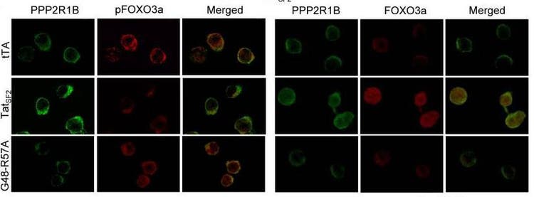
Kim, Nayoung, et al used APC Conjugation Kit - Lightning-Link® (ab201807) as part of examining apoptosis in HIV-1-infected CD4+ primary T cells. They used the kit to conjugate APC to anti-pFOXO3a antibody for use in confocal microscopy.
Jurkat T cells expressing tTA alone, TatSF2+tTA, or TatSF2G48-R57A +tTA were stained with antibodies against PPP2R1B (first and forth columns of panels, green), pFOXO3a (second column, red), and FOXO3a (forth column, red).
Image from Kim, Nayoung, et al., PLoS Pathog., 6(9): e1001103. doi: 10.1371/journal.ppat.1001103. Reproduced under the Creative Commons license https://creativecommons.org/licenses/by/4.0/
