PKC Kinase Activity Assay Kit(AB139437)
$undefined


Key facts
| Detection method | Colorimetric |
| Sample types | Purified protein, Suspension cells, Tissue Extracts, Adherent cells |
| Assay type | Enzyme activity |
| Assay time | 4h 30m |
| Assay Platform | Microplate reader |
Product details - PKC Kinase Activity Assay Kit (ab139437)
PKC Kinase Activity Assay Kit (ab139437) is a non-radioactive assay providing a safe, rapid and reliable method for the screening of inhibitors or activators of PKC and for quantitating the activity of PKC in purified or partially purified enzyme preparations. This kit is based on a solid phase enzyme-linked immuno-absorbent assay (ELISA) that utilizes a specific synthetic peptide as a substrate for PKC and a polyclonal antibody that recognizes the phosphorylated form of the substrate.
PKA activity assay principle
In this assay, a synthetic peptide is used as PKC specific substrate. The substrate, which has pre-coated on the wells on the microplate well provided, is phosphorylated by the PKC present in the sample after addition of ATP.
The phosphorylated substrate is recognized by a polyclonal antibody that recognizes only the phospho-substrate. The phospho-specific antibody is subsequently recognized a HRPconjugated secondary antibody.
The assay is finally developed with TMB substrate and color develops in proportion to the PKC activityexisting in the sample. The color development is stopped with acid stop solution and the intensity of the color is measured in a microplate reader at OD 450 nm.
The assay is designed for the analysis of PKC activity in the solution phase. For the measurement of PKC in partially purified, purified, or crude enzyme preparations from any species.
The kit offers the following advantages:
1. Safe - non-radioactive measurement of kinase activity.
2. Flexible - kinetic and end-point options available.
3. Fast - results in < 4.5 hours.
4. Efficient - only 30 μl diluted sample needed per well.
Functional Studies - PKC Kinase Activity Assay Kit (AB139437)
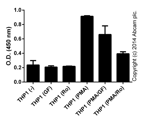
1.5 x e7 THP-1 cells were incubated with 100 nM GF109203X (GF; ab144264) or Ro31-8220 mesylate (Ro; ab120374) for 30 minutes prior to activation with 10 μg x mL-1 PMA (Sigma) for 4 hours. Control cells were left without inhibitors or PMA. Cells were lysed in 1 mL of lysis buffer, and 30 μL were tested for PKC activity (duplicates; +/- SD).
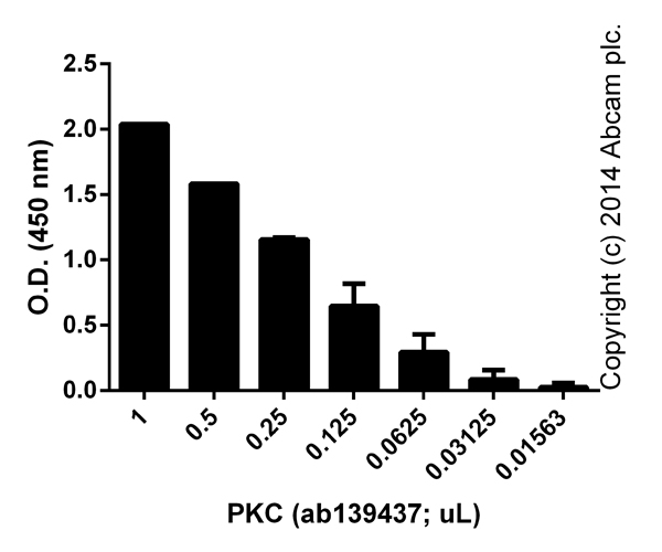
Functional Studies - PKC Kinase Activity Assay Kit (AB139437)
Signal from active PKC with background signal subtracted (duplicates; +/- SD).
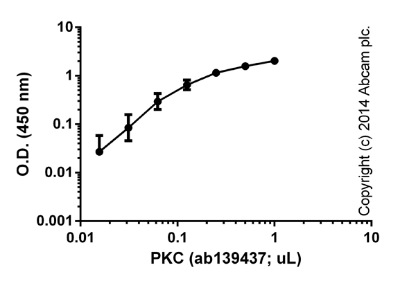
Functional Studies - PKC Kinase Activity Assay Kit (AB139437)
Titration of ab139437 (duplicates; +/- SD).
Schematic Diagram - PKC Kinase Activity Assay Kit (AB139437)
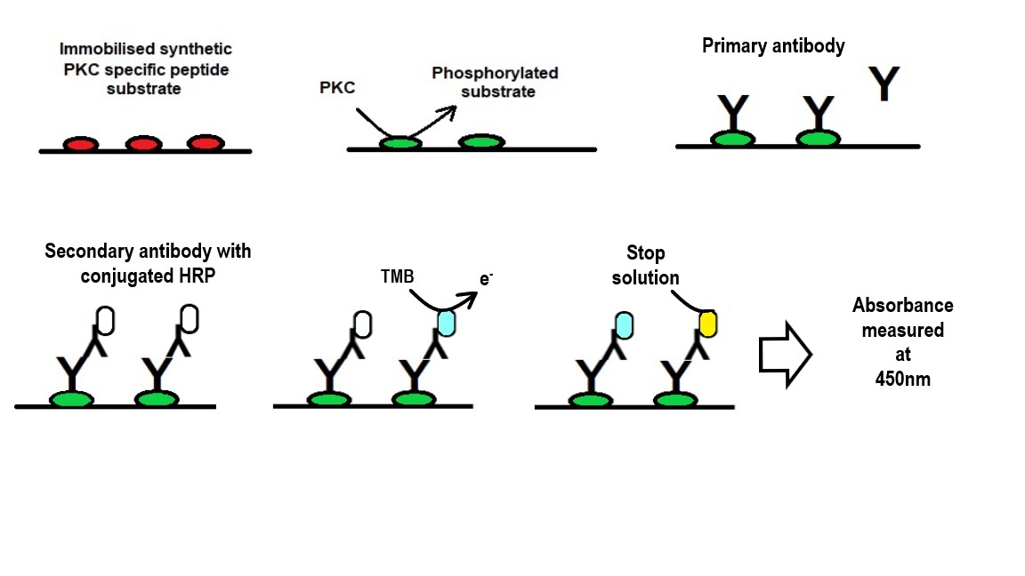
schematic diagram of the assay.
Fitzpatrick, Daniel P. (2020) "A Mechanistic Study on the Non-genotoxic Carcinogenicity of the Food Contaminant Semicarbazide," : Vol. 2 : Iss. 1, Article 2.
Available at : https : //arrow.tudublin.ie/sure_j/vol2/iss1/2
This image is courtesy of Fitzpatrick, Daniel P. (2020) "A Mechanistic Study on the Non-genotoxic Carcinogenicity of the Food Contaminant Semicarbazide," Sure-J: Science Undergraduate Research Journal: Vol. 2: Iss. 1, Article 2
REACH authorisation
Abcam has not and does not intend to apply for the REACH Authorisation of customers' uses of products that contain European Authorisation list (Annex XIV) substances.
It is the responsibility of our customers to check the necessity of application of REACH Authorisation, and any other relevant authorisations, for their intended uses.
Precision
Intra assay
| Sample | n | C.V. |
| Sample | 0 | < 10 |
Inter assay
| Sample | n | C.V. |
| Sample | 0 | < 10 |
What's included?
1 x 96 Tests
| Components | Unit & Qty |
| 20X Wash Buffer | 1 x 30 mL |
| Active PKC | 1 x 30 µL |
| Anti-Rabbit IgG: HRP Conjugate | 1 x 20 µL |
| Antibody Dilution Buffer | 1 x 10 mL |
| ATP | 1 x 2 mg |
| Kinase Assay Dilution Buffer | 1 x 10 mL |
| PKC Phosphospecific Substrate Antibody | 1 x 5 mL |
| PKC Substrate Microtiter Plate | 1 x 1 Unit |
| Stop Solution 2 | 1 x 10 mL |
| TMB Substrate | 1 x 10 mL |
Properties and storage information
| Shipped at conditions | Dry Ice |
| Appropriate short-term storage conditions | Multi |
| Appropriate long-term storage conditions | Multi |
| Storage information | Please refer to protocols |
Supplementary information
This supplementary information is collated from multiple sources and compiled automatically.
Protein kinase C (PKC) comprises a family of serine/threonine kinases with several isoforms such as PKC alpha beta and gamma among others. PKC enzymes play key roles in intracellular signaling particularly in mediating responses to growth factors hormones and other signals. PKC exhibits a molecular weight generally ranging around 77 to 97 kDa depending on the isoform. These enzymes show expression in many tissues but have high concentrations in the brain heart and lung. PKC activation often involves translocation to cellular membranes which is important for their signaling roles.
Biological function summary
PKC influences various cellular processes such as cell proliferation differentiation apoptosis and immune responses. It acts within larger protein complexes serving as a modulator of cellular functions. The activation of PKC leads to its association with different scaffolding proteins impacting a wide array of cell activities. PKC notably affects the function of other kinases and transcription factors demonstrating its integral role in managing cellular behavior and homeostasis.
Pathways
PKC participates in the phosphoinositide signaling pathway and the mitogen-activated protein kinase (MAPK) pathway. In these contexts PKC activation impacts proteins like Ras and Raf kinases which play roles in cellular growth and differentiation. By modulating these pathways PKC becomes an important component in signal transduction cascades influencing cell fate decisions. Understanding the interaction between PKC and these pathways helps in elucidating its role in maintaining normal cellular activities and responses to external stimuli.
PKC is linked to cancer and cardiovascular diseases. In cancer dysregulation of PKC activity often contributes to tumor growth and progression by altering pathways that control cell division and survival. PKC abnormally interacts with proteins like Bcl-2 promoting cell survival. In cardiovascular diseases changes in PKC signaling can impact heart function contributing to conditions like heart failure through interactions with proteins such as troponin. Research on PKC and these diseases can provide insights into developing targeted therapies.
| Shipped At Conditions | Dry Ice |
| Appropriate Long term Storage Conditions | Store at Multi. |