PE / R-Phycoerythrin Conjugation Kit - Lightning-Link®(AB102918)
$undefined


PE conjugation / labeling in < 4 hrs with 30 secs hands-on time using PE Conjugation Kit ab102918. 100% antibody recovery.
Rapid, reproducible PE antibody conjugation kit (also compatible with other proteins-and-peptides):
Fast, easy protocol - add modifier to antibody and incubate for 3 hrs, then add quencher for 30 mins
PE labeled antibody is ready for use in applications like WB, ELISA, and IHC, with no need for further purification
Compatible with most standard antibody formulations
Scalable; use the same conjugation method from 10µg-100mg of antibody
Use Lightning-Link® conjugation with over 45 labels, including Alexa Fluor®, DyLight®, tandem dyes, enzymes, oligonucleotides, and gold nanoparticles
- Proven performance: cited in over 135 publications.
Product details - PE / R-Phycoerythrin Conjugation Kit - Lightning-Link®(AB102918)
R-PE Conjugation Kit/ R-PE Labeling Kit ab102918 uses a simple and quick process for PE labeling / conjugation of antibodies. It can also be used to conjugate other proteins or peptides. To conjugate an antibody to R-PE using this kit:
- add modifier to antibody and incubate for 3 hrs
- add quencher and incubate for 30 mins
The PE conjugated antibody can be used immediately in WB, ELISA, IHC etc. No further purification is required and 100% of the antibody is recovered for use. Learn about buffer compatibility below. Custom size conjugation kits up to 100 mg are available on demand. Please contact us to discuss your requirements.
This product is manufactured by Expedeon, an Abcam company, and was previously called Lightning-Link® R-PE Labeling Kit. 703-0004 is the same as the 3 mg size. 703-0003 is the same as the 5 x 600 ug size. 703-0010 is the same as the 3 x 60 ug size. 703-0030 is the same as the 3 x 10 ug size. 703-0015 is the same as the 600 μg size. 703-0005 is the same as the 60 μg size.
Buffer Requirements for Conjugation
Buffer should be pH 6.5-8.5.
Storing and handling conjugation kits
Lyophilized Lightning-Link® components are hygroscopic.
Kits are intentionally shipped at ambient temperature with silica gel to avoid exposure to moisture. Upon receipt, store the kit frozen and protect from moisture. Before opening the outer container, allow the lyophilized components to reach room temperature to minimize condensation.
Flow Cytometry - PE / R-Phycoerythrin Conjugation Kit - Lightning-Link® (AB102918)

Flow Cytometry - PE / R-Phycoerythrin Conjugation Kit Lightning-Link.
Robinson, Andrew P., et al used PE / R-Phycoerythrin Conjugation Kit - Lightning-Link® (ab102918) as part of characterizing oligodendroglial populations. They used the kit to conjugate PE / R-Phycoerythrin to Mouse monoclonal anti-GALC antibody for use in flow cytometry.
SJL/J mice were immunized with PLP139-151 and scored daily for clinical disease. A cohort of SJL/J mice was sacrificed, and spinal cords were analyzed by flow cytometry (n=5). (A) Cells were distinguished from debris by forward and side scatter then singlet cells were gated. Live cells were gated by dead cell exclusion, and CNS resident cells were identified as CD45- or CD45low. (B) Oligodendroglial cells were defined by double positive staining : A2B5+PDGFRalpha+ early OPCs, A2B5+NG2+ intermediate OPCs, NG2+O4+ late OPCs, O4+MOG+ pre-myelinating oligodendrocytes, and GALC+MOG+ mature oligodendrocytes.
Schematic Diagram - PE / R-Phycoerythrin Conjugation Kit - Lightning-Link® (AB102918)
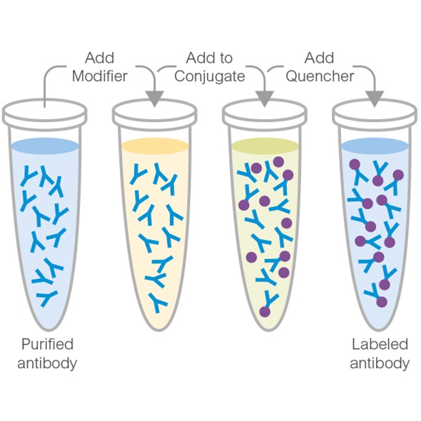
This illustration demonstrates a general procedure of how Lightning-Link® labeling technology enables the direct labeling of antibodies or proteins.
Simply pipette your antibody or biomolecule of choice into the vial of a lyophilized mixture containing the label of interest and incubate for around 3 hours. Please see the ab102918 protocol booklet for more details.
Flow Cytometry - PE / R-Phycoerythrin Conjugation Kit - Lightning-Link® (AB102918)

Flow cytometry using antibody conjugated using EasyLink R-Phycoerythrin conjugation kit (ab102918).
Flow cytometry histogram showing integrin beta-3 positive population of platelets from wildtype and knockout mice. Integrin beta-3 antibody was conjugated using EasyLink R-Phycoerythrin conjugation kit (ab102918). Flow cytometry was performed using platelets from wild type and integrin beta-3 knockout mice. Mice that expressed beta-3 (shown in red) had a clear shift in FL-2 fluorescence over beta-3 knockout mice (shown in black).
Conjugation - PE / R-Phycoerythrin Conjugation Kit - Lightning-Link® (AB102918)

PE / R-Phycoerythrin Conjugation Kit - Lightning-Link® labeling a panel of antibodies for Microsphere immunoassay based on Luminex.
Charlermroj R et al. used ab102918 as part of an experiment studying four different plant pathogens : a fruit blotch bacterium Acidovorax avenae subsp. citrulli (Aac), chilli vein-banding mottle virus (CVbMV, potyvirus), watermelon silver mottle virus (WSMoV, tospovirus serogroup IV) and melon yellow spot virus (MYSV, tospovirus).
(A-G) X-axis is antibody-coated microsphere and y-axis is median fluorescent intensity (MFI) from each RPE-labeled antibody. (H) Summary of selected antibody pair sets for the detection of the four plant pathogens.
Conjugation - PE / R-Phycoerythrin Conjugation Kit - Lightning-Link® (AB102918)

Ronnberg E et al. conjugated PE to an anti-chymase antibody with ab102918 PE Conjugation Kit as part of investigating the role of IL-33 in mast cells.
The mean fluorescence intensity of chymase expression in cells was measured by intra-cellular flow cytometry staining and normalized to the respective isotype control.
This data shows that there was no significant change in the expression on chymase in cord blood-derived mast cells (CBMC) when treated with different combinations of IL-33 and TSLP.
Coagulation - PE / R-Phycoerythrin Conjugation Kit - Lightning-Link® (AB102918)

PE / R-Phycoerythrin Conjugation Kit - Lightning-Link® labeling anti-MUC1 SP antibodies FACS.
Kovjazin R et al. used ab102918 to evaluate the cell surface presence of the MUC1 SP domain by FACS.
The cell surface presence of the MUC1 SP domain was evaluated by gated FACS analysis on PC cells in fresh BM aspirates obtained from MM patients (A–E) and normal naive sample (F–H).
Conjugation - PE / R-Phycoerythrin Conjugation Kit - Lightning-Link® (AB102918)

PE / R-Phycoerythrin Conjugation Kit - Lightning-Link® labeling SPmAb-6 antibody for Imagestream analysis.
Kovjazin R et al. used ab102918 for detection of MUC1 SP on the membrane of MUC1-positive tumor cells.
Conjugation - PE / R-Phycoerythrin Conjugation Kit - Lightning-Link® (AB102918)

PE / R-Phycoerythrin Conjugation Kit - Lightning-Link® labeling A20 antibody for FACS.
Hjelmeland AB et al. used ab102918 as part of an experiment studying the mechanisms that cause A20 regulator to have differential effects on tumor growth and cancer cell behaviors.
They used the PE labelling kit to conjugate phycoerythrin to A20 antibody for use in FACS.
FACS plots are shown for T08-837 (A) and CW468 (B).
Conjugation - PE / R-Phycoerythrin Conjugation Kit - Lightning-Link® (AB102918)

Tan H-X et al. labeled recombinant influenza A H1N1 NP protein with PE or APC fluorochromes using ab102918 PE / R-Phycoerythrin Conjugation Kit and ab201807 APC Conjugation Kit.
Representative HA and NP probe staining in flow cytometry of GC B cells (B220+ IgD- CD38lo GL7+) isolated from lung inducible bronchus-associated tissues (iBALT), mediastinal LN (MLN), and spleen of mice at d35 post-infection with A/Puerto Rico/8/34 (PR8).
Conjugation - PE / R-Phycoerythrin Conjugation Kit - Lightning-Link® (AB102918)

PE / R-Phycoerythrin Conjugation Kit - Lightning-Link® labeling F(ab')2 antibody for Bead array.
Ayoglu B et al. used ab102918 to detect complement activation driven C3 deposition on beads.
For measuring the background, classical, lectin & alternative and only alternative pathway activation, a serially diluted serum sample (1∶1–1∶160) was applied to empty, human IgG. The assay buffer for serum dilution contained either Ca2+-Mg2+, which promotes complement activation, or EDTA, which blocks complement activation. Plot displays for each serum dilution the respective median fluorescence intensity (MFI) value against varying concentrations of human IgG.
Conjugation - PE / R-Phycoerythrin Conjugation Kit - Lightning-Link® (AB102918)

PE conjugation kit used to label DDR2 antibody for Flow Cytometry.
RA Bagchi et al used PE conjugation kit / PE labeling kit ab102918 as part of examining the conversion of fibroblasts to myofibroblasts. They used the PE labeling kit to conjugate phycoerythrin to a DDR2 antibody for use in flow cytometry.
Charts are forward-scatter plots illustrating flow cytometry analysis of cardiac cells from WT and scleraxis KO mice. Left column, unstained cells; center column, stained cells from WT tissue; right column, stained cells from scleraxis KO tissue. Results are representative of assessments from n = 3 independent tissue samples. Purple outline denotes labeled cells, and is derived from unstained plots.
Conjugation - PE / R-Phycoerythrin Conjugation Kit - Lightning-Link® (AB102918)

PE / R-Phycoerythrin Conjugation Kit - Lightning-Link® labeling anti-mouse insulin antibody for Flow Cytometry.
Kalis M et al. used ab102918 as part of examining beta-cells deletion of Dicer1.
They used the kit to conjugate PE to anti-mouse insulin antibody for use in flow cytometry.
Islet cells suspension from 50 islets from 8 weeks old littermates and RIP-Cre Dicer1Δ/Δ mice were stained for insulin and glucagon and analyzed by flow cytometry. Side scatter (SSC) and forward scatter (FSC) analysis of islet cells and the respective gate used to identify live cells. Littermate control islet cells contained significantly more insulin-positive cells (P<0.001) and less glucagon-positive cells (P<0.05) than the RIP-Cre Dicer1Δ/Δ islet cells. Representative of 3 independent experiments.
Conjugation - PE / R-Phycoerythrin Conjugation Kit - Lightning-Link® (AB102918)

PE / R-Phycoerythrin Conjugation Kit - Lightning-Link® labeling ADRB2 antibody for Flow Cytometry.
Bigler MB et al. used ab102918 to assess expression of the epinephrine receptor (ADRB2) on CD3- CD56+ lymphocytes by flow cytometry.
| Shipped At Conditions | Ambient - Cannot Ship with Ice |
| Appropriate Long term Storage Conditions | Store at -20°C. |